. 2014 Nov;94(4):756-70.
doi: 10.1111/mmi.12794. Epub 2014 Oct 7.

Bacillithiol is a major buffer of the labile zinc pool in Bacillus subtilis

Affiliations

Bacillithiol is a major buffer of the labile zinc pool in Bacillus subtilis

Zhen Ma et al. Mol Microbiol. 2014 Nov.

Abstract

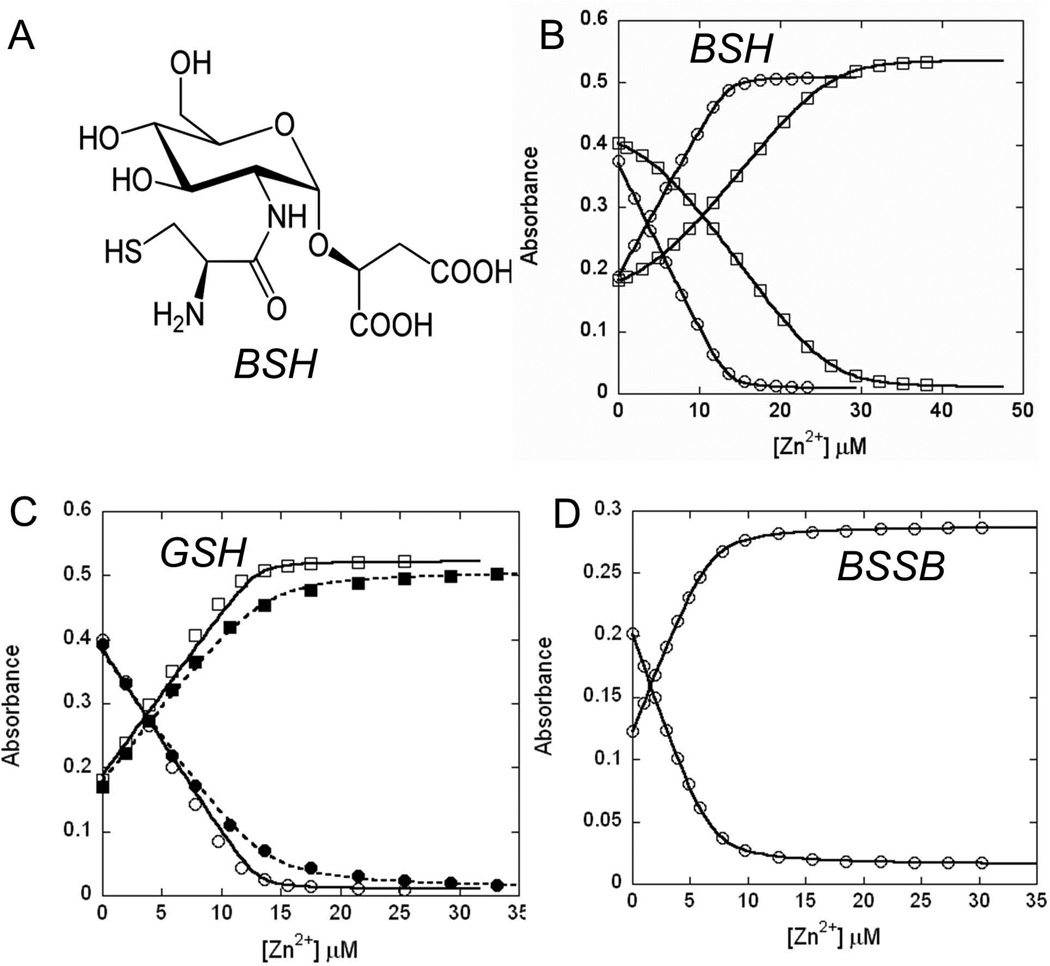
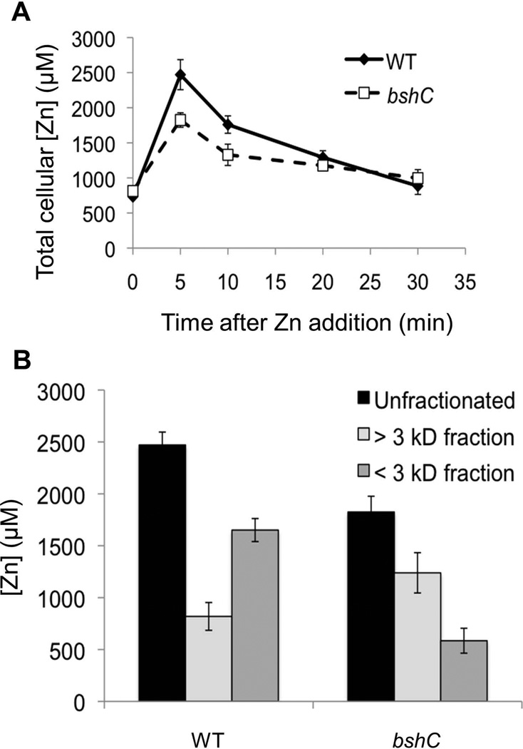
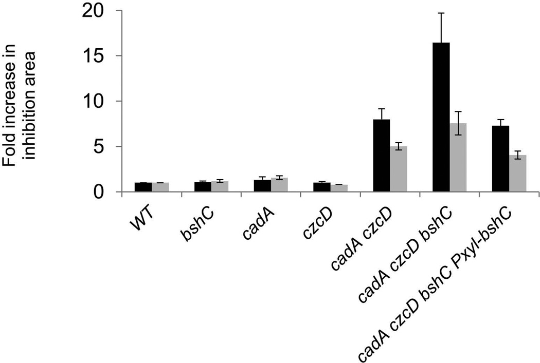
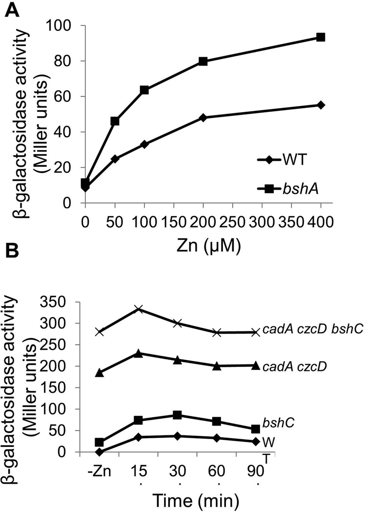
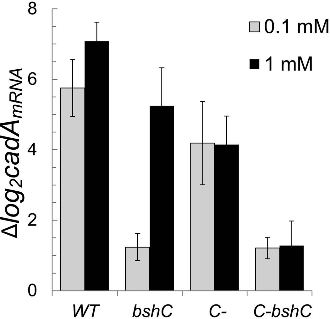
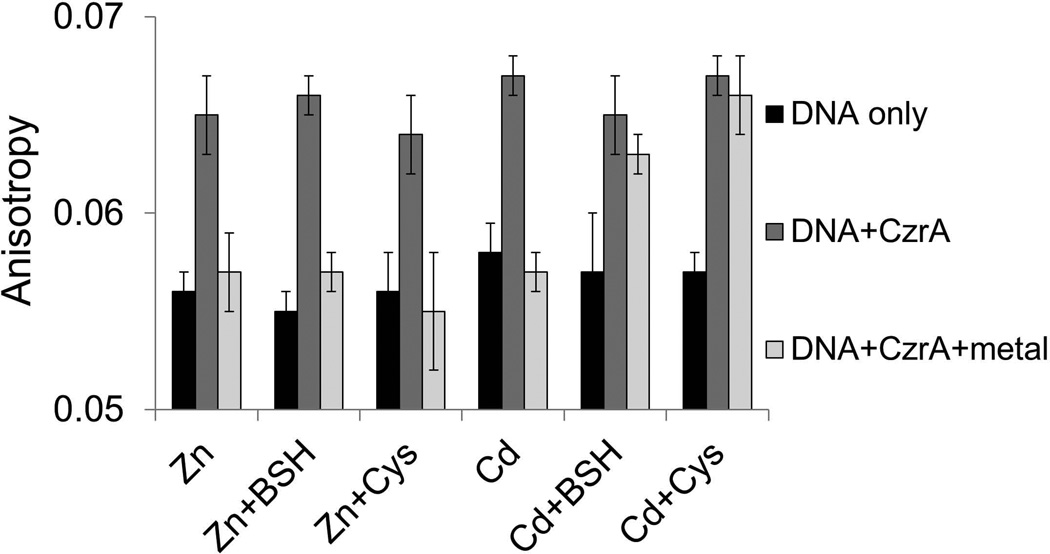
Intracellular zinc levels are tightly regulated since zinc is an essential cofactor for numerous enzymes, yet can be toxic when present in excess. The majority of intracellular zinc is tightly associated with proteins and is incorporated during synthesis from a poorly defined pool of kinetically labile zinc. In Bacillus subtilis, this labile pool is sensed by equilibration with the metalloregulator Zur, as an indication of zinc sufficiency, and by CzrA, as an indication of zinc excess. Here, we demonstrate that the low-molecular-weight thiol bacillithiol (BSH) serves as a major buffer of the labile zinc pool. Upon shift to conditions of zinc excess, cells transiently accumulate zinc in a low-molecular-weight pool, and this accumulation is largely dependent on BSH. Cells lacking BSH are more sensitive to zinc stress, and they induce zinc efflux at lower external zinc concentrations. Thiol reactive agents such as diamide and cadmium induce zinc efflux by interfering with the Zn-buffering function of BSH. Our data provide new insights into intracellular zinc buffering and may have broad relevance given the presence of BSH in pathogens and the proposed role of zinc sequestration in innate immunity.


Figures

Fig 1
Fig 1. BSH binds to Zn(II) with high affinity
(A) Structure of BSH. (B) Zn(II) was titrated into a mixture of 13.5 µM Magfura-2 without (circles) and with 19.2 µM BSH (squares). Absorbance at 325 nm (increasing values) and 366 nm (decreasing values) were plotted. In the absence of BSH, magfura-2 (MF2) binds one Zn(II) with an affinity of 1.7(±0.1) × 107 M−1 (see SI Fig. 1a for comparison with theoretical curves at 10-fold higher and lower affinity), a value similar to the reported affinity of 5.0 × 10⁷ M−1 (VanZile et al., 2000, Simons, 1993). The data from the competitive binding experiment with BSH and Magfura-2 are best fit to a model where BSH forms a 2:1 complex with Zn(II) with stepwise affinity constants of K1= 7.5 (±0.0) × 106 M−1, K2= 2.5 (±0.0) × 105 M−1 (or the accumulative binding constant β2= 1.9 × 10¹² M−2) (see SI Fig. 1b for comparison with theoretical curves at 10-fold higher and lower values for K1 and K2). (C) Zn(II) was titrated into a mixture of 13.5 µM Magfura-2 and 18.8 µM (open symbols, solid line) or 188 µM GSH (filled symbols, dashed line). The data acquired with 188 µM GSH allows an estimation of the GSH-Zn binding constant of ~3×10⁴ M−1. Conditions: 20 mM HEPES, pH 7.7, 0.15 M NaCl, 0.1 mM TCEP. (D) BSSB binds Zn(II) with low affinity. Zn(II) was titrated into a mixture of 6.8 µM Magfura-2 and 200 µM BSSB. Only minor competition was observed suggesting an affinity of ~104 M−1 (see SI Fig. 1c for comparison with theoretical curves at 10-fold higher and lower affinity) Conditions: 20 mM HEPES, pH 7.7, 0.15 M NaCl.
Fig 2
Fig 2. BSH function to buffer Zn(II) under zinc stress conditions
(A) Change of total cellular Zn(II) content in WT (CU1065) and bshC (HB11079) mutant cells following treatment of 250 µM Zn(II) for different time periods. (B) Fraction of total Zn(II) content of WT and bshC cell lysate fractionated through an Amicon ultracentrifugation filter (3 kD molecular weight cutoff) after exposure to 200 µM Zn(II) for 5 min. The data shown represent the mean and standard deviation of three biological replicates.
Fig 3
Fig 3. BSH plays a protective role in cells deficient in Zn(II) efflux
Susceptibility of wild-type and mutant strains (WT, CU1065; bshC, HB11212; cadA, HB11393; czcD, HB11394; cadA czcD, HB11395; cadA czcD bshC, HB11396; cadA czcD bshC Pxyl-bshC, HB11427) to Zn(II) (black bars) or Cd(II) (grey bars) was determined by disk diffusion assay. The data is expressed as the average fold-increase (± standard deviation) of the area of the zone of inhibition relative to wild-type.
Fig 4
Fig 4. Induction of cadA in response to Zn(II) is increased in cells lacking BSH
(A) Induction of PcadA-cat-lacZ as a function of Zn(II) concentration. Wild-type (HB11058) cells (diamonds) and an isogenic bshA null (HB11061) mutant (squares) carrying a cadA-cat-lacZ reporter fusion were grown to mid-logarithmic phase in LB medium with the indicated concentration of Zn(II) and assayed for β-galactosidase. (B) PcadA-lacZ activity in wild-type and mutant strains was monitored as a function of time after 50 µM Zn(II) addition. Representative results are shown for experiments repeated at least three times.
Fig 5
Fig 5. Induction of cadA in response to diamide is reduced in cells lacking mobilizable Zn(II) pools
Expression of cadA was monitored by qRT-PCR in wild-type (CU1065), bshC (HB11212), C- (rpmGA rpmGB rpmE mutant; HB6916), and C- bshC (HB15912) mutant strains after the treatment with 0.1 or 1 mM diamide for 5 minutes. 23S rRNA was used as an internal control and the fold-change between treated and untreated samples were plotted. Results are the mean and standard deviation of 3 biological replicates.
Fig 6
Fig 6. BSH null mutants are affected in metalloregulation
(A) CzrA and ArsR are both members of the ArsR/SmtB family of repressors, but they differ in the location and composition of their sensing sites. The crystal structure of S. aureus CzrA dimer (PDB: 1r1v; 49% identical to the B. subtilis CzrA ortholog) is shown with the two protomers colored green and pink, respectively (Eicken et al., 2003). In CzrA, Zn is coordinated at the α5 site by D84, H86 along with H97’ and H100’ from the opposite protomer (adapted from Pennella et al., 2006). In ArsR, As(III) is coordinated by three Cys residues in the α3 site (putative location highlighted in yellow) (Numbering based on B. subtilis ArsR by alignment with the E. coli ArsR sensing site; Shi et al., 1996). (B) Wild-type cells (blue diamonds) and an isogenic bshC null mutant (red squares) carrying the indicated reporter fusion (PczcD-lacZ WT, HB11423; bshC, HB11424; PcadA-lacZ WT, HB8125; bshC, HB16680; ParsR-lacZ WT, HB5015; bshC, HB16666) were grown to mid-log phase in LB medium with the indicated concentration of metal ions and assayed for β-galactosidase. Results are shown as the mean and standard deviation of triplicate cultures.
Fig 7
Fig 7. CzrA responds directly to Zn(II), but not to Cd(II), in the presence of physiological levels of thiols
The association and dissociation of CzrA-DNA complexes was monitored by fluorescence anisotropy. Anisotropy was determined with 10 nM DNA only (white bars) and after addition of 250 nM CzrA monomer (light grey bars), 1 µM indicated metal ions (deep grey bars). 300 µM thiols were added as indicated. Conditions: 20 mM Tris, pH 8.0, 0.4 M NaCl. Anisotropy values are mean and standard deviations of >4 technical replicates and are representative of experiments repeated at least 3 times with similar magnitude changes, but different absolute anisotropy values.
Fig 8
Fig 8. Bacillithiol (BSH) facilitates Zn-dissociation from CzrA
10 nM FAM-labeled cadA promoter DNA was mixed with 300 nM CzrA and 1 µM Zn in 20 mM Tris, pH 8.0. 0.4 M NaCl. 100 µM TPEN was then added with 300 µM BSH. DNA binding was monitored by fluorescence anisotropy with normalized fractional saturation plotted on the y-axis. x-axis represents the time after TPEN addition. All data were fit to a simple first-order decay model with the half-life (t1/2) of CzrA-Zn complex under these conditions being 1633 s (triangles; TPEN only) and 278 s (squares; TPEN and BSH).

Comment in

References

    1. Akanuma G, Nanamiya H, Natori Y, Nomura N, Kawamura F. Liberation of zinc-containing L31 (RpmE) from ribosomes by its paralogous gene product, YtiA, in Bacillus subtilis. J Bacteriol. 2006;188:2715–2720. - PMC - PubMed
    1. Arguello JM, Gonzalez-Guerrero M, Raimunda D. Bacterial transition metal P(1B)-ATPases: transport mechanism and roles in virulence. Biochemistry. 2011;50:9940–9949. - PMC - PubMed
    1. Arunkumar AI, Campanello GC, Giedroc DP. Solution structure of a paradigm ArsR family zinc sensor in the DNA-bound state. Proc Natl Acad Sci U S A. 2009;106:18177–18182. - PMC - PubMed
    1. Atkinson A, Khalimonchuk O, Smith P, Sabic H, Eide D, Winge DR. Mzm1 influences a labile pool of mitochondrial zinc important for respiratory function. J Biol Chem. 2010;285:19450–19459. - PMC - PubMed
    1. Busenlehner LS, Pennella MA, Giedroc DP. The SmtB/ArsR family of metalloregulatory transcriptional repressors: Structural insights into prokaryotic metal resistance. FEMS microbiology reviews. 2003;27:131–143. - PubMed

Publication types

LinkOut - more resources