

Relationship between Systemic and Cerebral Vascular Disease and Brain Structure Integrity in Normal Elderly Individuals
- PMID: 25213770
- PMCID: PMC4297227
- DOI: 10.3233/JAD-141077
Relationship between Systemic and Cerebral Vascular Disease and Brain Structure Integrity in Normal Elderly Individuals
Abstract
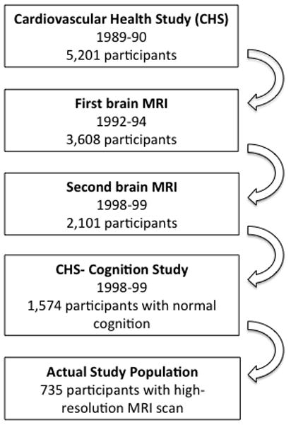
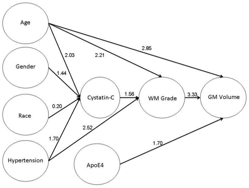
Cerebral white matter lesions (WMLs) are considered a reflection of cerebral and systemic small vessel disease (SVD), and are associated with reductions in brain volume. Like the brain, the kidney is also sensitive to factors that affect vasculature. Glomerular dysfunction due to renal vascular damage can be measured with different biochemical parameters, such as creatinine or cystatin C, although cystatin C is considered to be more accurate than creatinine in the elderly. The purpose of the study was to determine whether manifestations of SVD in the kidney can predict SVD-based damage to the brain. We examined the relationship between glomerular dysfunction as a measure of SVD on WMLs, gray matter (GM) volume, and cognition in 735 cognitively normal participants from the Cardiovascular Health Study Cognition Study. The multivariate analyses controlled for demographic characteristics, hypertension, heart disease, diabetes, Apolipoprotein 4 allele, C reactive protein, lipids, physical activity, smoking, and body mass index (BMI). Elevated cystatin C levels were associated with lower neuropsychological test scores, the presence of MRI-identified brain infarcts, the severity of WMLs, and GM atrophy five years later. In adjusted models, GM volume was significantly associated with cystatin-C only until BMI and severity of WMLs were added to the model, meaning that the effect of SVD on GM volume is mediated by these two variables. These findings suggest that age-related SVD is a process that leads to altered brain structure, and creates a vulnerability state for cognitive decline.
Keywords: Cognitive impairment; cystatin C; gray matter volume; white matter lesions.
Conflict of interest statement
The authors declare no competing interests.
Figures
References
-
- Longstreth WT, Arnold AM, Manolio T, Burke G, Bryan N, Jungreis CA, O'Laery D. Clinical correlates of ventricular and sulcal size on cranial magnetic resonance imaging of 3301 elderly people. The cardiovascular health study. Neuroepidemiology. 2000;19:30–42. - PubMed
-
- Longstreth WT, Arnold AM, Beauchamp NJ, Manolino TA, Lefkowitz D, Jungreis C, Hirsch CH, O'Leary DH, Furberg CD. Incidence, manifestations, and predictors of working white matter on serial cranial magnetic resonance imaging in teh elderly: the Cardiovascular Health Study. Stroke. 2005;36:56–61. - PubMed
-
- Vermeer SE, Hollander M, van Dijk EJ, Hofman A, Koudstaal PJ, Breteler M. Silent brain infarcts and white matter lesions increase stroke risk in the general population: the Rotterdam scan study. Stroke. 2003;34:1126–1129. - PubMed
-
- van Dijk EJ, Prins ND, Vrooman HA, Hofman A, Koudstaal PJ, Breteler MM. Progression of cerebral small vessel disease in relation to risk factors and cognitive consequences: Rotterdam Scan study. Stroke. 2008;39:2712–2719. - PubMed
-
- Manolio TA, Burke GL, O'Leary DH, Evans G, Beauchamp N, Knepper L, Ward B. Relationships of cerebral MRI findings to ultrasonographic carotid atherosclerosis in older adults : the Cardiovascular Health Study. CHS Collaborative Research Group. Arterioscler Thromb Vasc Biol. 1999;19:356–365. - PubMed
Publication types
MeSH terms
Grants and funding
- N01 HC085086/HC/NHLBI NIH HHS/United States
- N01 HC075150/HC/NHLBI NIH HHS/United States
- R56 AG020098/AG/NIA NIH HHS/United States
- N01-HC-85086/HC/NHLBI NIH HHS/United States
- AG05133/AG/NIA NIH HHS/United States
- N01 HC055222/HL/NHLBI NIH HHS/United States
- R01 HL080295/HL/NHLBI NIH HHS/United States
- R01 AG027002/AG/NIA NIH HHS/United States
- R01 AG020098/AG/NIA NIH HHS/United States
- N01-HC-85079/HC/NHLBI NIH HHS/United States
- HL080295/HL/NHLBI NIH HHS/United States
- AG027058/AG/NIA NIH HHS/United States
- AG15928/AG/NIA NIH HHS/United States
- RF1 AG041915/AG/NIA NIH HHS/United States
- R01 AG015928/AG/NIA NIH HHS/United States
- U01 HL080295/HL/NHLBI NIH HHS/United States
- N01 HC015103/HC/NHLBI NIH HHS/United States
- N01 HC085086/HL/NHLBI NIH HHS/United States
- AG027002/AG/NIA NIH HHS/United States
- U54 EB020403/EB/NIBIB NIH HHS/United States
- N01 HC-55222/HC/NHLBI NIH HHS/United States
- N01 HC085079/HL/NHLBI NIH HHS/United States
- N01-HC-75150/HC/NHLBI NIH HHS/United States
- P50 AG005133/AG/NIA NIH HHS/United States
- N01-HC-85239/HC/NHLBI NIH HHS/United States
- AG20098/AG/NIA NIH HHS/United States
- N01 HC075150/HL/NHLBI NIH HHS/United States
- R01 AG023629/AG/NIA NIH HHS/United States
- R01 AG027058/AG/NIA NIH HHS/United States
- N01 HC045133/HC/NHLBI NIH HHS/United States
- AG023629/AG/NIA NIH HHS/United States
- N01 HC035129/HC/NHLBI NIH HHS/United States
- R56 AG023629/AG/NIA NIH HHS/United States
LinkOut - more resources
Full Text Sources
Medical
Research Materials
