. 2014 Oct 31;289(44):30772-30784.
doi: 10.1074/jbc.M114.563270. Epub 2014 Sep 11.

Mouse macrophages completely lacking Rho subfamily GTPases (RhoA, RhoB, and RhoC) have severe lamellipodial retraction defects, but robust chemotactic navigation and altered motility

Affiliations

Mouse macrophages completely lacking Rho subfamily GTPases (RhoA, RhoB, and RhoC) have severe lamellipodial retraction defects, but robust chemotactic navigation and altered motility

Volker Königs et al. J Biol Chem. .

Abstract

RhoA is thought to be essential for coordination of the membrane protrusions and retractions required for immune cell motility and directed migration. Whether the subfamily of Rho (Ras homolog) GTPases (RhoA, RhoB, and RhoC) is actually required for the directed migration of primary cells is difficult to predict. Macrophages isolated from myeloid-restricted RhoA/RhoB (conditional) double knock-out (dKO) mice did not express RhoC and were essentially "pan-Rho"-deficient. Using real-time chemotaxis assays, we found that retraction of the trailing edge was dissociated from the advance of the cell body in dKO cells, which developed extremely elongated tails. Surprisingly, velocity (of the cell body) was increased, whereas chemotactic efficiency was preserved, when compared with WT macrophages. Randomly migrating RhoA/RhoB dKO macrophages exhibited multiple small protrusions and developed large "branches" due to impaired lamellipodial retraction. A mouse model of peritonitis indicated that monocyte/macrophage recruitment was, surprisingly, more rapid in RhoA/RhoB dKO mice than in WT mice. In comparison with dKO cells, the phenotypes of single RhoA- or RhoB-deficient macrophages were mild due to mutual compensation. Furthermore, genetic deletion of RhoB partially reversed the motility defect of macrophages lacking the RhoGAP (Rho GTPase-activating protein) myosin IXb (Myo9b). In conclusion, the Rho subfamily is not required for "front end" functions (motility and chemotaxis), although both RhoA and RhoB are involved in pulling up the "back end" and resorbing lamellipodial membrane protrusions. Macrophages lacking Rho proteins migrate faster in vitro, which, in the case of the peritoneum, translates to more rapid in vivo monocyte/macrophage recruitment.

Keywords: Cell Migration; Cell Motility; Cell Polarity; Chemotaxis; Macrophage; Mouse; Myosin; Ras Homolog Gene Family, Member A (RhoA); Rho (Rho GTPase); Rho GTPases.


Figures

FIGURE 1.
FIGURE 1.
Working model and conditional deletion of RhoA in macrophages. A, time-lapse phase-contrast images (100 × 100 μm, except for t = 2 min) of a migrating mouse resident peritoneal macrophage. White arrows indicate the projected direction of prominent membrane protrusions. B, schematic working model of spatially coordinated Rho signaling in a migrating macrophage. C, gene analysis by RT-PCR. Peritoneal F4/80+ cells (macrophages) purified by cell sorting express mRNA for RhoA and RhoB, but not RhoC. D, Western blot analysis. RhoA and RhoB, but not RhoC, protein could be detected in macrophages. Note that primary antibodies against RhoA, RhoB, or RhoC were applied after cutting the membrane into strips. Lysates from HEK293T cells expressing mouse RhoC were used as positive control. E, Western blot analysis of RhoA and RhoB in macrophages derived from conditional RhoA knock-out mice, obtained by crossing floxed RhoA (RhoAfl/fl) and LysM-Cre mice. A very weak RhoA signal could be detected in macrophages isolated from RhoAfl/fl/LysM-Cre mice, and RhoB levels were markedly increased in these cells. F, high expression levels of RhoB in RhoA−/− macrophages relative to WT cells (n = 5). Error bars indicate means ± S.E. G, flow cytometry analysis of macrophages isolated from LysM-EGFP mice and labeled with phycoerythrin-conjugated anti-F4/80 antibodies. H, DIC and fluorescence images of macrophages isolated from a transgenic mouse expressing EGFP (rather than Cre) under the control of the M lysozyme gene. Note that one of the F4/80+ cells (indicated by a white arrow) does not appear to express EGFP. Images are 100 × 100 μm.
FIGURE 2.
FIGURE 2.
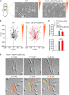
Phenotype and chemotactic behavior of RhoA-deficient macrophages. A, schematic diagram of a chemotaxis μ-slide and 200 × 300-μm snapshots of WT and RhoA-deficient (RhoAfl/fl/LysM-Cre) macrophages in a chemotactic complement component C5a gradient. B, migration plots of WT and RhoA-deficient (RhoAfl/fl/LysM-Cre) macrophages in a C5a gradient. C, summary plots of chemotactic efficiency (chemotaxis index) and mean velocity. The summary bar labeled WT without C5a refers to WT cells migrating in the absence of chemoattractant. Error bars indicate means ± S.E. n.s., not significant. D, high resolution DIC images of spontaneously migrating RhoA-deficient macrophages. Images are 100 × 100 μm. In the lower panel, two migrating cells have been pseudocolored to distinguish them from neighboring cells.
FIGURE 3.
FIGURE 3.
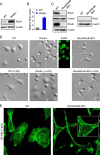
Striking phenotype and chemotactic behavior of pan-Rho-deficient (RhoAfl/fl/LysM-Cre/RhoB−/−) macrophages. A, Western blot analysis of RhoA in macrophages derived from WT and RhoB−/− mice. B, high expression levels of RhoA in RhoB−/− macrophages relative to wild-type cells (n = 3). Error bars indicate means ± S.E. C, Western blot analysis of RhoA, RhoB, and RhoC in macrophages derived from WT and RhoA/RhoB dKO (double knock-out; RhoAfl/fl/LysM-Cre/RhoB−/−) mice. D, morphology of WT, RhoB−/− and RhoA/RhoB dKO macrophages under conditions of no stimulation (upper panel) or activation (lower panel) with 100 ng/ml lipopolysaccharide (+LPS). The DIC images are 100 × 100 μm. F4/80 staining (green) was used to confirm that the unstimulated and rounded up RhoB−/− cells shown are macrophages. E, superresolution structured illumination microscopy (SR-SIM) images of fixed WT and RhoA/RhoB dKO macrophages. F-actin was labeled using Alexa Fluor 488-conjugated phalloidin. The inset (right image) provides an enlarged view of the actin cytoskeleton.
FIGURE 4.
FIGURE 4.
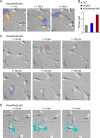
Mechanism of branch formation in spontaneously migrating pan-Rho-deficient (RhoAfl/fl/LysM-Cre/RhoB−/−) macrophages. A, time-lapse DIC images (100 × 100 μm) of a spontaneously migrating RhoA/RhoB dKO (RhoAfl/fl/LysM-Cre/RhoB−/−) macrophage. The dominant lamellipodium, as well as its remnants after incomplete retraction and relocation, have been pseudocolored blue. The rest of the cell has been pseudocolored pale brown (beige). B, mean number of primary branches arising from the cell body of WT, RhoB−/−, and RhoA/RhoB dKO macrophages. *, p < 0.05 (Kruskal-Wallis test and post hoc Dunn test). Error bars indicate means ± S.E. C, further examples of branch formation arising from incomplete membrane retraction following relocation of the dominant lamellipodium. Note that when the lamellipodium (pseudocolored) shifts location, it leaves behind a branch (correspondingly pseudocolored). The DIC images are 100 × 100 μm. D, time-lapse DIC images (100 × 100 μm) showing erratic multifocal membrane protrusions (pseudocolored light blue) in a RhoA/RhoB dKO macrophage.
FIGURE 5.
FIGURE 5.
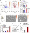
Pan-Rho-deficient (RhoAfl/fl/LysM-Cre/RhoB−/−) macrophages develop exceedingly long trailing ends during chemotaxis. A, migration plots of RhoB−/− and RhoA/RhoB dKO macrophages in a complement component C5a gradient. B, summary plots of chemotactic efficiency (chemotaxis index) and mean velocity, comparing WT, RhoB−/−, and RhoA/RhoB dKO (double knock-out; RhoAfl/fl/LysM-Cre/RhoB−/−) macrophages. Data were obtained after tracking the cell body, except in the fourth column (lower plot), which shows mean velocity of the trailing end of RhoA/RhoB (A/B) dKO macrophages. C, schematic diagram of a chemotaxis slide and 200 × 300-μm snapshots of RhoB−/− and RhoA/RhoB dKO macrophages in a chemotactic C5a gradient. D, plot of maximal tail length observed in WT, RhoB−/−, RhoAfl/fl/LysM-Cre (RhoA−/−), and RhoA/RhoB dKO macrophages migrating in a chemotactic gradient during a 6-h period. E, Western blot analysis of p-MLC2 levels in WT, RhoB−/−, and RhoA/RhoB dKO macrophages. F, plot of the total number of F4/80+ cells that accumulated in the peritoneal cavity of WT (RhoAfl/fl) and RhoA/RhoB dKO mice 48 h after intraperitoneal injection of thioglycollate. *, p < 0.05 (Kruskal-Wallis test and post hoc Dunn test or, in the case of panel F, unpaired Student's t test). n.s., not significant. Error bars indicate means ± S.E.
FIGURE 6.
FIGURE 6.
Phenotype of Myo9b/RhoB double knock-out macrophages. A, bright-field and extended focus images (120 × 120 μm) of living WT and Myo9b/RhoB dKO macrophages, labeled with Alexa Fluor 488-conjugated anti-F4/80 antibodies and imaged by spinning disk confocal microscopy. Three-dimensional views of the same cells are shown on the right. B, Western blot analysis of RhoB in macrophages derived from WT and Myo9b−/− mice. C, low expression levels of RhoB in Myo9b−/− macrophages relative to WT cells (n = 3). Error bars indicate means ± S.E.
FIGURE 7.
FIGURE 7.
Genetic deletion of RhoB in Myo9b−/− macrophages partially rescues velocity. A, schematic diagram of a chemotaxis μ-slide and 200 × 300-μm snapshots of Myo9b−/− and Myo9b/RhoB dKO macrophages in a chemotactic complement component C5a gradient. B, migration plots of Myo9b−/− and Myo9b/RhoB dKO macrophages in a C5a gradient. C, summary plots of chemotactic efficiency (chemotaxis index) and mean velocity, comparing WT, Myo9b−/−, and Myo9b/RhoB dKO macrophages. *, p < 0.05 (Kruskal-Wallis test and post hoc Dunn test). Error bars indicate means ± S.E.
FIGURE 8.
FIGURE 8.
Schematic diagrams indicating the roles of Rho proteins and the negative regulator Myo9b in directed macrophage motility. In wild-type macrophages, both active RhoA and RhoB are involved in the retraction (red arrows) of the trailing end and membrane protrusions, whereas the RhoGAP Myo9b acts at the front to inactivate both RhoA and RhoB. In the absence of the Rho subfamily, there is severely impaired lamellipodial membrane and tail retraction at the back end, but increased motility and robust chemotaxis of the front end (cell body). In addition, macrophages lacking the Rho subfamily exhibit erratic multifocal membrane protrusions.

Similar articles

Cited by

References

    1. Hanley P. J., Xu Y., Kronlage M., Grobe K., Schön P., Song J., Sorokin L., Schwab A., Bähler M. (2010) Motorized RhoGAP myosin IXb (Myo9b) controls cell shape and motility. Proc. Natl. Acad. Sci. U.S.A. 107, 12145–12150 - PMC - PubMed
    1. Ridley A. J., Schwartz M. A., Burridge K., Firtel R. A., Ginsberg M. H., Borisy G., Parsons J. T., Horwitz A. R. (2003) Cell migration: integrating signals from front to back. Science 302, 1704–1709 - PubMed
    1. Abercrombie M., Heaysman J. E., Pegrum S. M. (1971) The locomotion of fibroblasts in culture. IV. Electron microscopy of the leading lamella. Exp. Cell Res. 67, 359–367 - PubMed
    1. Reig G., Pulgar E., Concha M. L. (2014) Cell migration: from tissue culture to embryos. Development 141, 1999–2013 - PubMed
    1. Swaney K. F., Huang C. H., Devreotes P. N. (2010) Eukaryotic chemotaxis: a network of signaling pathways controls motility, directional sensing, and polarity. Annu. Rev. Biophys. 39, 265–289 - PMC - PubMed

Publication types

MeSH terms

LinkOut - more resources