. 2014 Nov 15;307(10):G1002-12.
doi: 10.1152/ajpgi.00164.2014. Epub 2014 Sep 11.

Presence of intramucosal neuroglial cells in normal and aganglionic human colon

Affiliations

Presence of intramucosal neuroglial cells in normal and aganglionic human colon

Kamran Badizadegan et al. Am J Physiol Gastrointest Liver Physiol. .

Abstract

The enteric nervous system (ENS) is composed of neural crest-derived neurons (also known as ganglion cells) the cell bodies of which are located in the submucosal and myenteric plexuses of the intestinal wall. Intramucosal ganglion cells are known to exist but are rare and often considered ectopic. Also derived from the neural crest are enteric glial cells that populate the ganglia and the associated nerves, as well as the lamina propria of the intestinal mucosa. In Hirschsprung disease (HSCR), ganglion cells are absent from the distal gut because of a failure of neural crest-derived progenitor cells to complete their rostrocaudal migration during embryogenesis. The fate of intramucosal glial cells in human HSCR is essentially unknown. We demonstrate a network of intramucosal cells that exhibit dendritic morphology typical of neurons and glial cells. These dendritic cells are present throughout the human gut and express Tuj1, S100, glial fibrillary acidic protein, CD56, synaptophysin, and calretinin, consistent with mixed or overlapping neuroglial differentiation. The cells are present in aganglionic colon from patients with HSCR, but with an altered immunophenotype. Coexpression of Tuj1 and HNK1 in this cell population supports a neural crest origin. These findings extend and challenge the current understanding of ENS microanatomy and suggest the existence of an intramucosal population of neural crest-derived cells, present in HSCR, with overlapping immunophenotype of neurons and glia. Intramucosal neuroglial cells have not been previously recognized, and their presence in HSCR poses new questions about ENS development and the pathobiology of HSCR that merit further investigation.

Keywords: Hirschsprung disease; enteric glial cells; enteric nervous system.


Figures

Fig. 1.
Fig. 1.
Characteristic morphology of normal (a–c) and aganglionic (d–f) human colon. a: Routine hematoxylin-eosin staining of normal colon mucosa after formalin fixation and paraffin embedding shows inconspicuous submucosal nerves (arrowhead) and occasional ganglion cells (arrow, expanded in inset). b: By immunohistochemistry of formalin-fixed, paraffin-embedded tissue, delicate calretinin-positive dendritic cellular processes are seen in the lamina propria, with cell nuclei present in the calretinin-positive regions (arrows, inset). c: By enzymatic histochemistry on a corresponding frozen section of fresh (unfixed) tissue, acetylcholinesterase (ACE)-positive neurites are not seen in the normal colon mucosa. d: Aganglionic colon shows marked neural hypertrophy (arrow) and no submucosal ganglion cells. e and f: calretinin-immunoreactive cellular processes are absent in the aganglionic colon from a patient with Hirschsprung disease (HSCR), while prominent ACE-positive dendritic processes are seen in the lamina propria, some associated with cell nuclei (f, arrows, inset). Morphological features of formalin-fixed, paraffin-embedded tissue (a, b, d, and e) differ slightly, as expected, from frozen sections of fresh tissue (c and f). Scale bar = 100 μm (a and d) and 25 μm (b, c, e, and f).
Fig. 2.
Fig. 2.
Nucleated cells with dendritic cellular processes and neural and/or glial differentiation are present in the mucosa of normal gastrointestinal tract. a: Calretinin-immunoreactive dendritic processes and cell bodies are present in formalin-fixed, paraffin-embedded tissue sections of normal colon mucosa. Note cell-cell connection between the 2 nuclei marked by arrows. b–d: CD56-positive (b), synaptophysin-positive (c), and S100-positive (d) cells with similar morphology are also present in the colon mucosa. e: Rare delicate VIP-positive processes are identified. f–h: Calretinin-positive dendritic cells are also seen in the mucosa of the appendix (f), small intestine (g), and stomach (h). Scale bar = 50 μm (a–h).
Fig. 3.
Fig. 3.
Immunohistochemical coexpression of markers suggests a single population of intramucosal dendritic cells (arrows point to cell bodies). a: Tandem immunohistochemical staining of formalin-fixed, paraffin-embedded tissue sections of normal colon mucosa for calretinin (brown) and CD56 (red) shows coexpression of 2 markers (mixed red and brown pigments) in the same cells. b: Tandem staining with calretinin (brown) and synaptophysin (red) shows similar findings. Inset: coexpression of calretinin and synaptophysin in an intramucosal ganglion cell from a nearby region. While immunohistochemical expression patterns are similar (i.e., cellular coexpression without complete subcellular colocalization), the intramucosal ganglion cell is morphologically distinct from the intramucosal dendritic cells by virtue of its prominent cell body, including a round vesicular nucleus and a prominent nucleolus, neither of which resembles the cell body of dendritic cells (a and b, arrows). c: Staining specificity, spatial resolution, and red-brown color contrast of the method are highlighted in a submucosal ganglion from the same tissue (calretinin in brown, CD56 in red). Scale bar = 10 μm (a–c).
Fig. 4.
Fig. 4.
Intramucosal nucleated dendritic cells expressing neural markers are present in HSCR. a–c: Formalin-fixed, paraffin-embedded tissue sections of colon mucosa from the aganglionic segment of patients with HSCR show no expression of calretinin (a), but intact expression of CD56 (b) and synaptophysin (c) in nucleated cells (arrows) with dendritic processes. Scale bar = 50 μm (a–c).
Fig. 5.
Fig. 5.
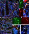
Intramucosal dendritic cells coexpress neural and glial markers. a: Double immunofluorescence performed with a neural marker (Tuj1, green) and an enteric glial marker (S100, red) on frozen sections of normal colon mucosa shows cellular coexpression. Nuclear stain (4′,6-diamidino-2-phenylindole dihydrochloride, blue) depicts the crypt and lamina propria anatomy. b–d: High-magnification image of a double-labeled cell. e–h: Immunofluorescence with Tuj1 and glial fibrillary acidic protein (GFAP), also on frozen sections of normal colon mucosa, shows cellular coexpression (e), highlighted in the high-magnification images (f–h). i: Staining with Tuj1 (red) and a neural crest cell marker [human natural killer-1 (HNK1), green] supports coexpression in the same lamina propria cells (arrows), providing evidence in favor of a neural crest origin. Scale bar = 40 μm (a), 20 μm (b and c), 15 μm (d and f–h), 50 μm (e), and 30 μm (i).
Fig. 6.
Fig. 6.
Mucosal neuroglial cells are present in HSCR. a–c: Immunofluorescence microscopy and confocal imaging of colon mucosa in formalin-fixed, paraffin-embedded tissue sections from the proximal ganglionated colon of a patient with HSCR shows Tuj1 (a) and S100 (b) coexpression (c, arrows). Tuj1 is also expressed by intraepithelial neuroendocrine cells (a, arrows). d–f: Aganglionic mucosa from the same patient with HSCR reveals persistence of these double-expressing cells, albeit with a reduction in Tuj1 (d) and S100 (e) expression (merged in f). Merged images (c and f) are magnified (relative to a–d) to highlight areas of interest. Scale bar = 70 μm (a, b, d, and e) and 45 μm (c and f).
Fig. 7.
Fig. 7.
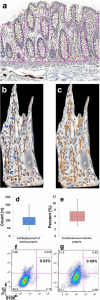
Quantitative analysis and flow cytometry of intramucosal dendritic cells. a: Immunostained sections were scanned, and well-oriented regions where crypts reached the muscularis mucosae were selected for analysis. In each region, 5 separate foci were manually selected by following the epithelial basement membrane (magenta lines within rectangular boxes). b and c: Each masked focus was subjected to a manual cell count (b, blue arrows) and automated measurement of brown diaminobenzidine (DAB) pigment (c, orange mask over brown pigment). d and e: Box-and-whisker plots were generated for the number of cell bodies per unit area (d) and for DAB-positive fractional area (e) using data from 10 patients, including 5 analysis foci in 2 separate regions per patient. f: Representative scatter plot of intestinal cells incubated with anti-rabbit Alexa Fluor 488-conjugated and anti-mouse Alexa Fluor 647-conjugated secondary antibodies (1:500 dilution) without primary antibodies. g: Representative scatter plot of cells incubated with anti-Tuj1 and anti-S100, each at 1:100 dilution, and visualized with the same secondary antibodies (1:500 dilution). Axes represent fluorescence intensity for Tuj1 (y-axis) and S100 (x-axis) on a logarithmic scale. Percentage of cells that costain for Tuj1 and S100 [0.02% in the negative control (f) and 0.40% in the presence of primary antibodies (g)] confirms coexpression in a distinct population of cells. Data are representative of triplicate samples.
Fig. 8.
Fig. 8.
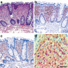
Intestinal pathology supports the presence of an intramucosal cell with neuroglial differentiation. a–c: Formalin-fixed, paraffin-embedded colonic lesion diagnosed as an “intramucosal neuroma” (surgical pathology diagnosis) demonstrates benign proliferation of lamina propria spindle and dendritic cells (a) that express calretinin (b, brown) and S100 (c, brown) by immunohistochemistry. d: Another abnormal spindle cell proliferation with similar morphology and immunohistochemical expression of S100 (red) and calretinin (brown) was observed in the appendiceal lamina propria of a child who underwent appendectomy for abdominal pain. Scale bar = 75 μm (a–c) and 25 μm (d).

References

    1. Akbar A, Yiangou Y, Facer P, Brydon WG, Walters JR, Anand P, Ghosh S. Expression of the TRPV1 receptor differs in quiescent inflammatory bowel disease with or without abdominal pain. Gut : 767–774, 2010. - PubMed
    1. Akbar A, Yiangou Y, Facer P, Walters JR, Anand P, Ghosh S. Increased capsaicin receptor TRPV1-expressing sensory fibres in irritable bowel syndrome and their correlation with abdominal pain. Gut : 923–929, 2008. - PMC - PubMed
    1. Balemba OB, Grondahl ML, Mbassa GK, Semuguruka WD, Hay-Smith A, Skadhauge E, Dantzer V. The organisation of the enteric nervous system in the submucous and mucous layers of the small intestine of the pig studied by VIP and neurofilament protein immunohistochemistry. J Anat : 257–267, 1998. - PMC - PubMed
    1. Barshack I, Fridman E, Goldberg I, Chowers Y, Kopolovic J. The loss of calretinin expression indicates aganglionosis in Hirschsprung's disease. J Clin Pathol : 712–716, 2004. - PMC - PubMed
    1. Burns AJ, Douarin NM. The sacral neural crest contributes neurons and glia to the post-umbilical gut: spatiotemporal analysis of the development of the enteric nervous system. Development : 4335–4347, 1998. - PubMed

MeSH terms