

Prolonged exposure of cholestatic rats to complete dark inhibits biliary hyperplasia and liver fibrosis
- PMID: 25214401
- PMCID: PMC4216989
- DOI: 10.1152/ajpgi.00288.2014
Prolonged exposure of cholestatic rats to complete dark inhibits biliary hyperplasia and liver fibrosis
Abstract
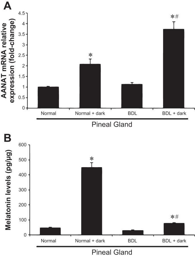
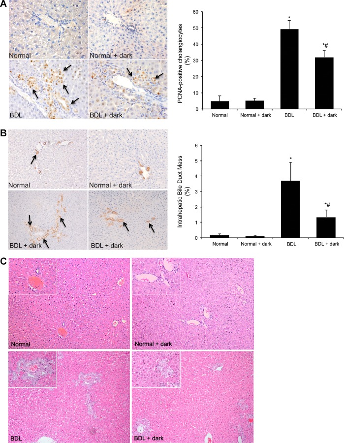
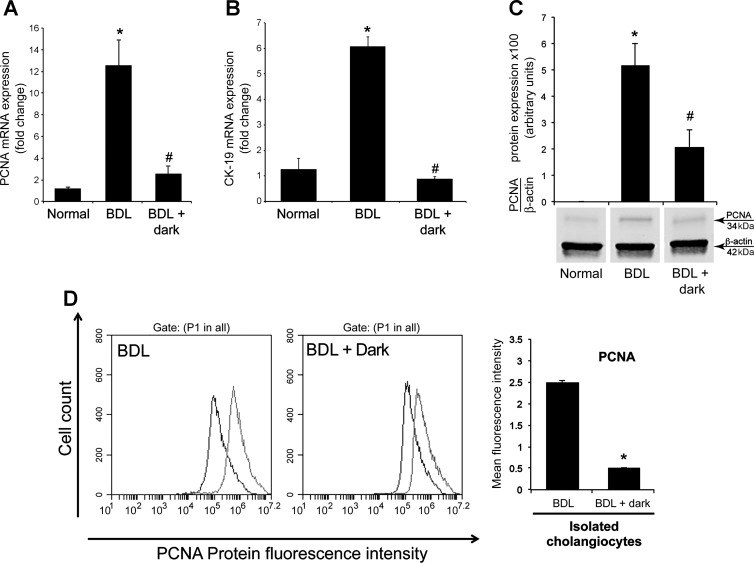
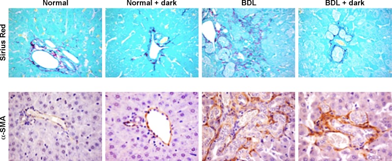
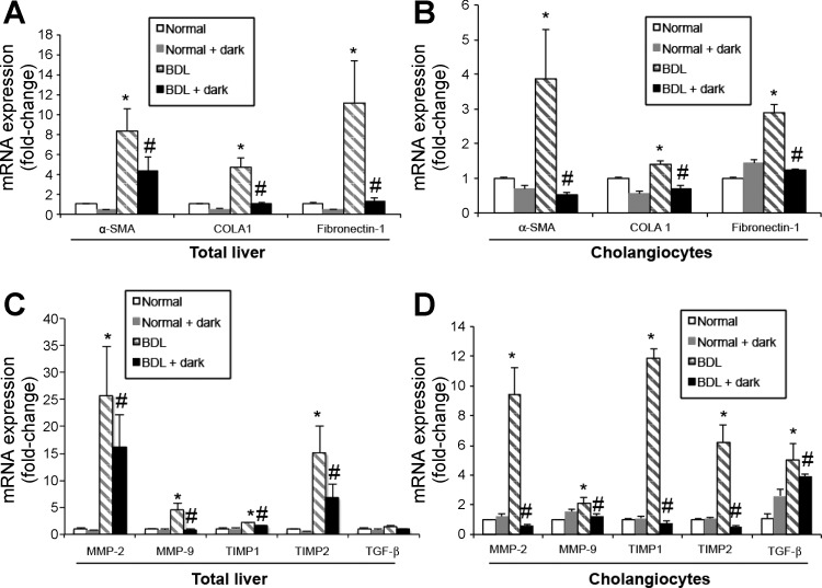
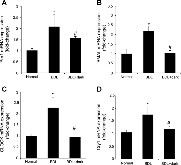
Biliary hyperplasia and liver fibrosis are common features in cholestatic liver disease. Melatonin is synthesized by the pineal gland as well as the liver. Melatonin inhibits biliary hyperplasia of bile duct-ligated (BDL) rats. Since melatonin synthesis (by the enzyme serotonin N-acetyltransferase, AANAT) from the pineal gland increases after dark exposure, we hypothesized that biliary hyperplasia and liver fibrosis are diminished by continuous darkness via increased melatonin synthesis from the pineal gland. Normal or BDL rats (immediately after surgery) were housed with light-dark cycles or complete dark for 1 wk before evaluation of 1) the expression of AANAT in the pineal gland and melatonin levels in pineal gland tissue supernatants and serum; 2) biliary proliferation and intrahepatic bile duct mass, liver histology, and serum chemistry; 3) secretin-stimulated ductal secretion (functional index of biliary growth); 4) collagen deposition, liver fibrosis markers in liver sections, total liver, and cholangiocytes; and 5) expression of clock genes in cholangiocytes. In BDL rats exposed to dark there was 1) enhanced AANAT expression/melatonin secretion in pineal gland and melatonin serum levels; 2) improved liver morphology, serum chemistry and decreased biliary proliferation and secretin-stimulated choleresis; and 4) decreased fibrosis and expression of fibrosis markers in liver sections, total liver and cholangiocytes and reduced biliary expression of the clock genes PER1, BMAL1, CLOCK, and Cry1. Thus prolonged dark exposure may be a beneficial noninvasive therapeutic approach for the management of biliary disorders.
Keywords: biliary epithelium; cholestasis; clock genes; melatonin; secretin.
Figures

Similar articles
-
Pinealectomy or light exposure exacerbates biliary damage and liver fibrosis in cholestatic rats through decreased melatonin synthesis.Biochim Biophys Acta Mol Basis Dis. 2019 Jun 1;1865(6):1525-1539. doi: 10.1016/j.bbadis.2019.03.002. Epub 2019 Mar 16. Biochim Biophys Acta Mol Basis Dis. 2019. PMID: 30890428 Free PMC article.
-
Modulation of the biliary expression of arylalkylamine N-acetyltransferase alters the autocrine proliferative responses of cholangiocytes in rats.Hepatology. 2013 Mar;57(3):1130-41. doi: 10.1002/hep.26105. Epub 2013 Feb 7. Hepatology. 2013. PMID: 23080076 Free PMC article.
-
Melatonin inhibits cholangiocyte hyperplasia in cholestatic rats by interaction with MT1 but not MT2 melatonin receptors.Am J Physiol Gastrointest Liver Physiol. 2011 Oct;301(4):G634-43. doi: 10.1152/ajpgi.00206.2011. Epub 2011 Jul 14. Am J Physiol Gastrointest Liver Physiol. 2011. PMID: 21757639 Free PMC article.
-
Melatonin synthesis and clock gene regulation in the pineal organ of teleost fish compared to mammals: Similarities and differences.Gen Comp Endocrinol. 2019 Aug 1;279:27-34. doi: 10.1016/j.ygcen.2018.07.010. Epub 2018 Jul 17. Gen Comp Endocrinol. 2019. PMID: 30026020 Review.
-
Avian biological clock - Immune system relationship.Dev Comp Immunol. 2017 Jan;66:130-138. doi: 10.1016/j.dci.2016.05.017. Epub 2016 May 26. Dev Comp Immunol. 2017. PMID: 27235884 Review.
Cited by
-
Knockout of microRNA-21 reduces biliary hyperplasia and liver fibrosis in cholestatic bile duct ligated mice.Lab Invest. 2016 Dec;96(12):1256-1267. doi: 10.1038/labinvest.2016.112. Epub 2016 Oct 24. Lab Invest. 2016. PMID: 27775690 Free PMC article.
-
Melatonin Protects Cholangiocytes from Oxidative Stress-Induced Proapoptotic and Proinflammatory Stimuli via miR-132 and miR-34.Int J Mol Sci. 2020 Dec 18;21(24):9667. doi: 10.3390/ijms21249667. Int J Mol Sci. 2020. PMID: 33352965 Free PMC article.
-
Functional and structural features of cholangiocytes in health and disease.Cell Mol Gastroenterol Hepatol. 2015 Jul 1;1(4):368-380. doi: 10.1016/j.jcmgh.2015.05.005. Cell Mol Gastroenterol Hepatol. 2015. PMID: 26273695 Free PMC article.
-
miR-24 Inhibition Increases Menin Expression and Decreases Cholangiocarcinoma Proliferation.Am J Pathol. 2017 Mar;187(3):570-580. doi: 10.1016/j.ajpath.2016.10.021. Epub 2017 Jan 11. Am J Pathol. 2017. PMID: 28087162 Free PMC article.
-
Circadian rhythms of liver physiology and disease: experimental and clinical evidence.Nat Rev Gastroenterol Hepatol. 2016 Apr;13(4):217-26. doi: 10.1038/nrgastro.2016.8. Epub 2016 Feb 24. Nat Rev Gastroenterol Hepatol. 2016. PMID: 26907879 Review.
References
-
- Alpini G, Glaser S, Robertson W, Rodgers RE, Phinizy JL, Lasater J, LeSage GD. Large but not small intrahepatic bile ducts are involved in secretin-regulated ductal bile secretion. Am J Physiol Gastrointest Liver Physiol 272: G1064–G1074, 1997. - PubMed
-
- Alpini G, Glaser S, Ueno Y, Pham L, Podila PV, Caligiuri A, LeSage G, LaRusso NF. Heterogeneity of the proliferative capacity of rat cholangiocytes after bile duct ligation. Am J Physiol Gastrointest Liver Physiol 274: G767–G775, 1998. - PubMed
-
- Alpini G, Roberts S, Kuntz SM, Ueno Y, Gubba S, Podila PV, LeSage G, LaRusso NF. Morphological, molecular, and functional heterogeneity of cholangiocytes from normal rat liver. Gastroenterology 110: 1636–1643, 1996. - PubMed
-
- Alpini G, Ulrich C, Roberts S, Phillips JO, Ueno Y, Podila PV, Colegio O, LeSage G, Miller LJ, LaRusso NF. Molecular and functional heterogeneity of cholangiocytes from rat liver after bile duct ligation. Am J Physiol Gastrointest Liver Physiol 272: G289–G297, 1997. - PubMed
Publication types
MeSH terms
Substances
Grants and funding
LinkOut - more resources
Full Text Sources
Other Literature Sources