. 2015 Jan 15;24(2):480-91.
doi: 10.1093/hmg/ddu468. Epub 2014 Sep 11.

Tissue-specific responses to the LRPPRC founder mutation in French Canadian Leigh Syndrome

Affiliations

Tissue-specific responses to the LRPPRC founder mutation in French Canadian Leigh Syndrome

Florin Sasarman et al. Hum Mol Genet. .

Abstract

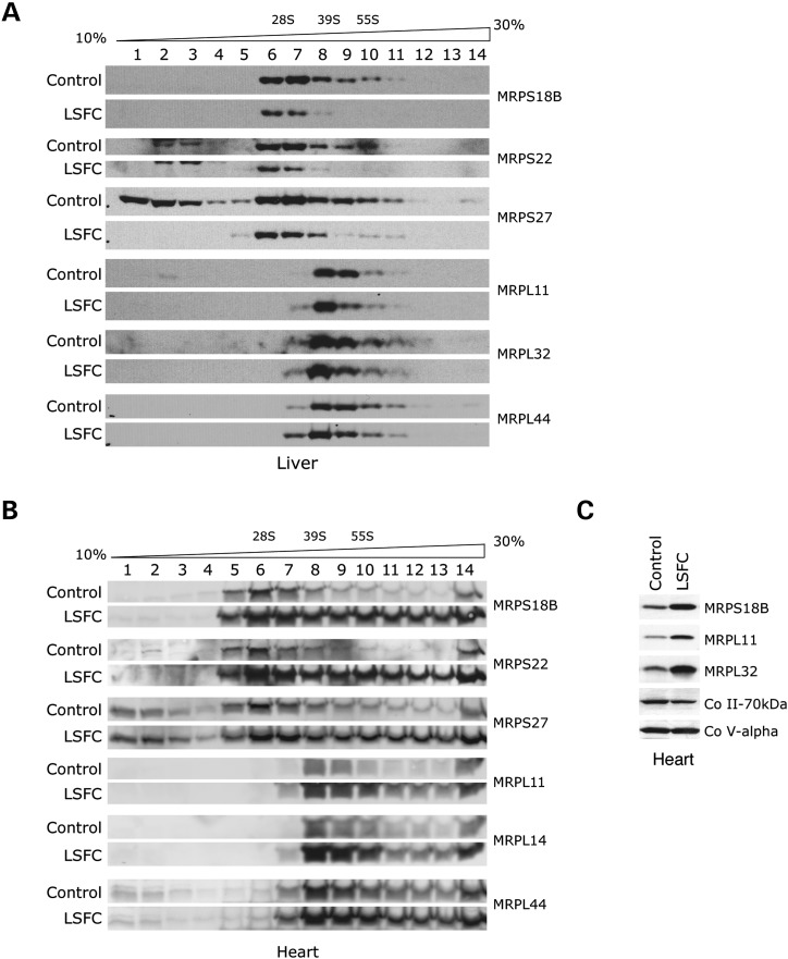
French Canadian Leigh Syndrome (LSFC) is an early-onset, progressive neurodegenerative disorder with a distinct pattern of tissue involvement. Most cases are caused by a founder missense mutation in LRPPRC. LRPPRC forms a ribonucleoprotein complex with SLIRP, another RNA-binding protein, and this stabilizes polyadenylated mitochondrial mRNAs. LSFC fibroblasts have reduced levels of LRPPRC and a specific complex IV assembly defect; however, further depletion of mutant LRPPRC results in a complete failure to assemble a functional oxidative phosphorylation system, suggesting that LRPPRC levels determine the nature of the biochemical phenotype. We tested this hypothesis in cultured muscle cells and tissues from LSFC patients. LRPPRC levels were reduced in LSFC muscle cells, resulting in combined complex I and IV deficiencies. A similar combined deficiency was observed in skeletal muscle. Complex IV was only moderately reduced in LSFC heart, but was almost undetectable in liver. Both of these tissues showed elevated levels of complexes I and III. Despite the marked biochemical differences, the steady-state levels of LRPPRC and mitochondrial mRNAs were extremely low, LRPPRC was largely detergent-insoluble, and SLIRP was undetectable in all LSFC tissues. The level of the LRPPRC/SLIRP complex appeared much reduced in control tissues by the first dimension blue-native polyacrylamide gel electrophoresis (BN-PAGE) analysis compared with fibroblasts, and even by second dimension analysis it was virtually undetectable in control heart. These results point to tissue-specific pathways for the post-transcriptional handling of mitochondrial mRNAs and suggest that the biochemical defects in LSFC reflect the differential ability of tissues to adapt to the mutation.


Figures

Figure 1.
Figure 1.
Knockdown of SLIRP in control fibroblasts phenocopies the mitochondrial translation defect in LSFC fibroblasts. (A) Control fibroblasts were transiently transfected with two different siRNA constructs specific to SLIRP, or with a fluorescent control siRNA (Alexa). On the sixth day post-transfection, the mitochondrial translation products were pulse-labeled with a mix of [35S]-methionine and cysteine in the presence of emetine, an inhibitor of cytoplasmic translation, and analyzed by PAGE. The 13 mitochondrial translation products are indicated to the left of the figure: seven subunits of complex I (ND), one subunit of complex III, three subunits of complex IV (COX) and two subunits of complex V (ATP). The asterisk indicates an anomalous, unidentified translation product typically detected in LSFC fibroblasts. (B) The knockdown of SLIRP and the concomitant reduction in steady-state levels of LRPPRC were confirmed by immunoblotting with specific antibodies. The 70 kDa subunit of complex II was used as a loading control.
Figure 2.
Figure 2.
Mitochondrial translation defect and combined OXPHOS deficiency in LSFC cultured muscle cells. (A) Immunoblot analysis showing reduced levels of LRPPRC and SLIRP in extracts from fetal and post-natal cultured muscle cells from LSFC subjects. Prohibitin was used as loading control. The sample for LSFC fetal myotubes was slightly under loaded. (B) Mitochondrial translation products in fetal and post-natal myoblasts and myotubes from LSFC subjects were pulse-labeled with a mix of [35S]-methionine and cysteine in the presence of emetine, an inhibitor of cytoplasmic translation and analyzed by PAGE. The 13 mitochondrial translation products are indicated to the left of the figure: seven subunits of complex I (ND), one subunit of complex III, three subunits of complex IV (COX) and two subunits of complex V (ATP). The asterisk indicates a translation product with anomalous migration also detected in LSFC fibroblasts. (C) Blue-native PAGE analysis of the five OXPHOS complexes revealed reduced levels of assembled complex IV (COX) and complex I in cultured muscle cells from LSFC subjects. Individual complexes were detected with subunit-specific antibodies.
Figure 3.
Figure 3.
Generalized OXPHOS defect following knockdown of LRPPRC in LSFC cultured muscle cells. LSFC and control myoblasts or myotubes were transiently transfected either with an siRNA construct specific to LRPPRC, or with a fluorescent control siRNA (Alexa), and then (A) analyzed by immunoblotting with antibodies against LRPPRC, SLIRP or subunits of the OXPHOS complexes, (B) pulse-labeled with a mix of [35S]-methionine and cysteine in the presence of emetine and (C) analyzed by BN-PAGE (as described in Fig. 2).
Figure 4.
Figure 4.
Generalized reduction of steady-state levels of mitochondrial mRNAs in LSFC tissues. Total RNA was extracted from muscle, heart and liver of controls and an LSFC subject, followed by either standard northern blotting analysis with probes specific for the mitochondrial mRNAs or rRNAs indicated at the left of the panel (A), or by urea-PAGE and hybridization with oligonucleotide probes complementary to the mitochondrial and cytoplasmic tRNAs indicated at the right of the panel (B).
Figure 5.
Figure 5.
Tissue-specific OXPHOS deficiencies in LSFC tissues. Mitochondrial extracts from muscle, heart and liver of one LSFC subject, several controls and four disease controls were analyzed by BN-PAGE (A) and by western blotting (B) with antibodies against subunits of the OXPHOS complexes or against LRPPRC and SLIRP, as indicated. In (A), ‘Co I deficiency’ refers to two subjects with assembly defects of complex I; in (B), ‘COX15’ and ‘EFG1’ refer to two subjects with mutations in an assembly factor of COX and a mitochondrial translation factor, respectively. (C) Cleared mitochondrial extracts from control liver, heart and muscle were analyzed by immunoblotting with antibodies against LRPPRC, SLIRP or subunits of the OXPHOS complexes.
Figure 6.
Figure 6.
Altered detergent solubility of LRPPRC and of other mitochondrial translation proteins in LSFC liver. (A) Mitochondria from muscle, heart and liver samples of the LSFC subject and controls were extracted in dodecyl maltoside, then mixed with loading buffer containing SDS without extract clearing by centrifugation, followed by immunoblot analysis with antibodies against LRPPRC or the 70 kDa subunit of complex II. (B, C) Mitochondria isolated from liver samples of one LSFC subject and controls were extracted in dodecyl maltoside or taurodeoxycholate (for TACO1 detection), then loading buffer containing SDS was added to the extracts without extract clearing by centrifugation (‘Input’). Duplicate samples were centrifuged following extraction in dodecyl maltoside or taurodeoxycholate, and each soluble (‘Supernatant’) and insoluble (‘Pellet’) fraction was then mixed with loading buffer containing SDS. All samples were analyzed by immunoblotting with antibodies against LRPPRC, SLIRP, mitochondrial ribosomal proteins (MRPS27, MRPL44), factors or enzymes involved in mitochondrial translation (EFG1, TACO1, MTFMT, PDF, CCDC56) and other mitochondrial proteins, as detailed in the text.
Figure 7.
Figure 7.
LRPPRC and SLIRP are partly associated with the mitochondrial membrane fraction. Alkaline carbonate extracts of mitochondria from HEK293 cells (A) or from control and LSFC fibroblasts (B) were analyzed by immunoblotting with antibodies against LRPPRC, SLIRP and the following protein controls: COX I (multiple pass, integral inner membrane), SCO1 (single pass, inner membrane), VDAC (outer membrane), Co II-70 kDa (soluble, inner membrane-associated), HSP 60 and 75 (soluble).
Figure 8.
Figure 8.
Analysis of ribosome assembly in LSFC liver and heart. Cleared mitochondrial lysates from control and LSFC liver (A) and heart (B) were loaded on a discontinuous sucrose gradient and centrifuged, after which fractions were collected for immunoblot analysis with antibodies against subunits of the large (mitochondrial ribosomal protein, large) and small (mitochondrial ribosomal protein, small) mitochondrial ribosomal subunits. The approximate migration of the small (28S) and of the large (39S) mitochondrial ribosomal subunits and the monosome (55S) are indicated for reference. (C) Western blotting analysis of cleared mitochondrial extracts from control and LSFC heart with antibodies against small and large mitochondrial ribosomal subunits. One subunit of complex II and one of complex V were used as loading controls.
Figure 9.
Figure 9.
The LRPPRC–SLIRP complex is undetectable in normal heart tissue by the first and second dimension BN/SDS-PAGE analysis. (A) First dimension BN-PAGE analysis of the LRPPRC–SLIRP complex with an antibody against LRPPRC, and of complexes II and IV (COX) with subunit-specific antibodies, in control and LSFC liver. (B) Comparison of the levels of LRPPRC–SLIRP complex in control tissues and cells by the first dimension BN-PAGE analysis. The LRPPRC–SLIRP complex was detected with an antibody against LRPPRC and complex IV was detected with an antibody against its nuclear subunit COX IV. (C) Extracts from control tissues and cells were run in the first dimension on a non-denaturing, blue native gel (BN-PAGE), after which individual lanes were cut out and run on the second dimension on a denaturing gel (SDS-PAGE). Antibodies were then used to detect LRPPRC and SLIRP, as indicated.

Similar articles

Cited by

References

    1. Koopman W.J., Willems P.H., Smeitink J.A. Monogenic mitochondrial disorders. N. Engl. J. Med. 2012;366:1132–1141. - PubMed
    1. Nunnari J., Suomalainen A. Mitochondria: in sickness and in health. Cell. 2012;148:1145–1159. - PMC - PubMed
    1. Schon E.A., Przedborski S. Mitochondria: the next (neurode)generation. Neuron. 2011;70:1033–1053. - PMC - PubMed
    1. Leigh D. Subacute necrotizing encephalomyelopathy in an infant. J. Neurochem. 1951;14:216–221. - PMC - PubMed
    1. Zhu Z., Yao J., Johns T., Fu K., De Bie I., Macmillan C., Cuthbert A.P., Newbold R.F., Wang J., Chevrette M., et al. SURF1, encoding a factor involved in the biogenesis of cytochrome c oxidase, is mutated in Leigh syndrome. Nat. Genet. 1998;20:337–343. - PubMed

Publication types