. 2015 Jan 15;24(2):492-505.
doi: 10.1093/hmg/ddu469. Epub 2014 Sep 11.

Loss of nNOS inhibits compensatory muscle hypertrophy and exacerbates inflammation and eccentric contraction-induced damage in mdx mice

Affiliations

Loss of nNOS inhibits compensatory muscle hypertrophy and exacerbates inflammation and eccentric contraction-induced damage in mdx mice

Stanley C Froehner et al. Hum Mol Genet. .

Abstract

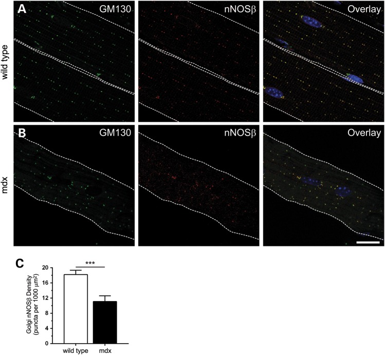
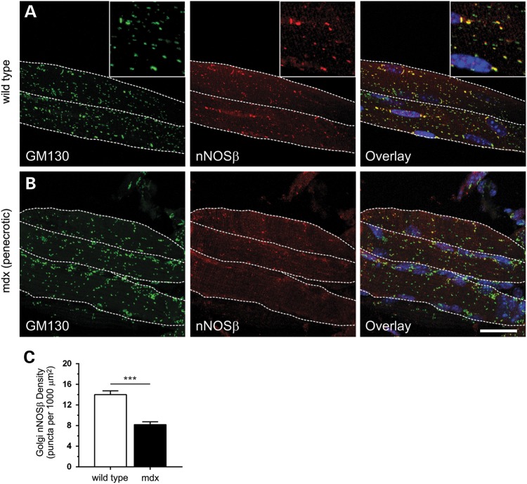
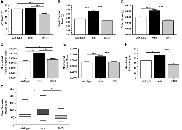
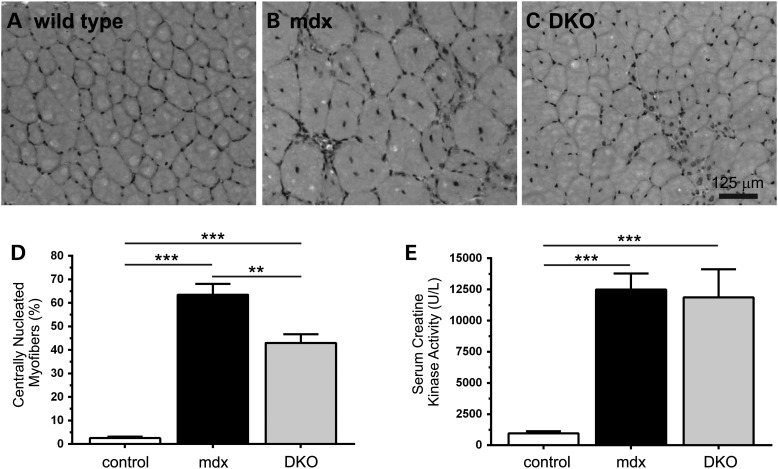
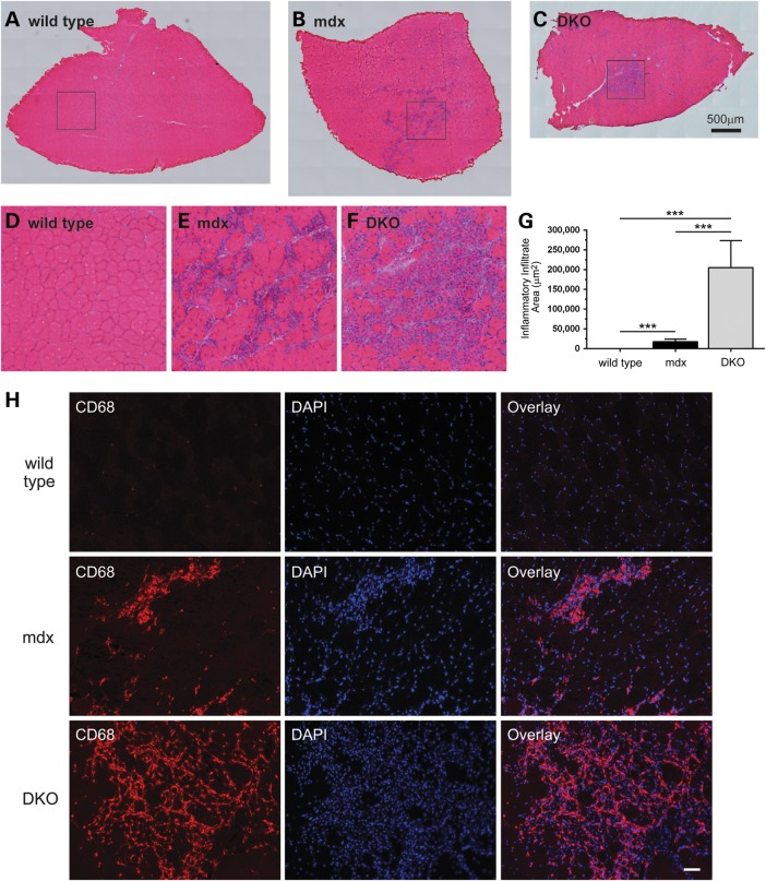
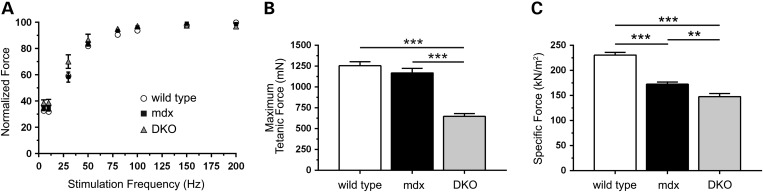
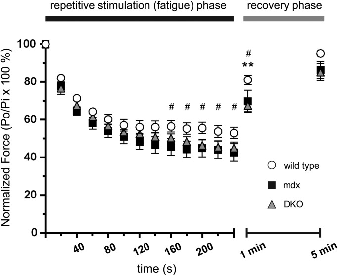
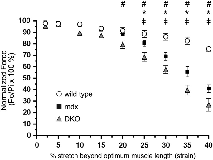
Approaches targeting nitric oxide (NO) signaling show promise as therapies for Duchenne and Becker muscular dystrophies. However, the mechanisms by which NO benefits dystrophin-deficient muscle remain unclear, but may involve nNOSβ, a newly discovered enzymatic source of NO in skeletal muscle. Here we investigate the impact of dystrophin deficiency on nNOSβ and use mdx mice engineered to lack nNOSμ and nNOSβ to discern how the loss of nNOS impacts dystrophic skeletal muscle pathology. In mdx muscle, nNOSβ was mislocalized and its association with the Golgi complex was reduced. nNOS depletion from mdx mice prevented compensatory skeletal muscle cell hypertrophy, decreased myofiber central nucleation and increased focal macrophage cell infiltration, indicating exacerbated dystrophic muscle damage. Reductions in muscle integrity in nNOS-null mdx mice were accompanied by decreases in specific force and increased susceptibility to eccentric contraction-induced muscle damage compared with mdx controls. Unexpectedly, muscle fatigue was unaffected by nNOS depletion, revealing a novel latent compensatory mechanism for the loss of nNOS in mdx mice. Together with previous studies, these data suggest that localization of both nNOSμ and nNOSβ is disrupted by dystrophin deficiency. They also indicate that nNOS has a more complex role as a modifier of dystrophic pathology and broader therapeutic potential than previously recognized. Importantly, these findings also suggest nNOSβ as a new drug target and provide a new conceptual framework for understanding nNOS signaling and the benefits of NO therapies in dystrophinopathies.


Figures

Figure 1.
Figure 1.
Loss of dystrophin causes mislocalization of Golgi-targeted nNOSβ. To determine the impact of dystrophin deficiency on nNOSβ localization, gastrocnemius myofibers from 8-week-old wild-type and mdx mice were immunolabeled with anti-GM130 and anti-nNOS antibodies. GM130 marks the cis-Golgi compartment. Nuclei (blue) were counterstained with DAPI. The sarcolemma is marked by a dashed white line. Single optical sections obtained by confocal microscopy are shown. (A) In wild-type muscle, nNOSβ exhibited stereotypic ‘beads on a string’ localization to the cis-Golgi compartment of the subsarcolemmal Golgi complex indicated by extensive colocalization with GM130. nNOSβ is also localized to the Golgi adjacent to myonuclei. (B) In mdx muscle, loss of dystrophin caused mislocalization of both the Golgi and nNOSβ. (C) Dystrophin deficiency caused a 39% reduction (P < 0.001) in subsarcolemmal Golgi-nNOSβ density (the number Golgi puncta positive for both GM130 and nNOSβ per 1000 μm2) compared with wild-type controls. ***P < 0.001. n = 6 and 8 for wild-type and mdx groups, respectively. Scale bar: 20 microns.
Figure 2.
Figure 2.
Reductions in Golgi-nNOSβ density in prenecrotic 2-week-old mdx muscle. To determine if nNOSβ association with the Golgi was decreased in mdx muscles, nNOSβ densities were determined in 2-week-old gastrocnemius myofibers from wild-type and mdx mice. Gastrocnemius myofibers were immunolabeled with anti-GM130 and anti-nNOS antibodies and nuclei counterstained with DAPI. Differences in myofiber geometry at 2 weeks necessitated the use of maximum projection images of myofibers which were obtained by confocal and two photon microscopy. The sarcolemma is marked by a dashed white line. (A) In 2-week-old wild-type muscle, the Golgi complex is non-uniformly distributed compared with adult muscle (left panel). But nNOSβ still colocalizes extensively with GM130 as in 8-week-old muscle. Yellow puncta in the overlay inset image highlight the high degree of association of nNOSβ with subsarcolemmal Golgi stacks, while green puncta represent intermyofibrillar Golgi stacks found deeper inside the muscle that lack nNOSβ. (B) In 2-week-old prenecrotic mdx muscle, loss of dystrophin has no impact on Golgi complex distribution relative to wild-type controls (left panel). However, prenecrotic muscles exhibit a marked reduction in Golgi-targeted nNOSβ (middle panel) highlighted in the overlay (right panel). (C) Dystrophin deficiency significantly reduced subsarcolemmal Golgi-nNOSβ density by 42% compared with wild-type controls. ***P < 0.001. n = 5 for wild-type and mdx groups. Inset image is 2.25× magnification. Scale bar: 20 microns.
Figure 3.
Figure 3.
nNOSβ and nNOSμ depletion prevents compensatory skeletal muscle hypertrophy in mdx mice. (A) mdx mice lacking nNOSμ and nNOSβ (DKO) had decreased body mass compared with wild-type and mdx controls. (B and C) mdx mice exhibited a stereotypical increase in tibialis anterior (TA) and soleus muscle mass that was prevented by loss of nNOS. (D and E) Normalization of TA and soleus mass to body mass revealed that reductions in muscle mass resulting from the loss of nNOS were not solely due to decreased body mass. (F) Mean myofiber Feret diameter from wild-type, mdx and DKO TA muscles. As expected, the mean mdx myofiber Feret diameter was larger than wild-type controls reflecting bigger muscle cells; however this increase was prevented in DKO muscles. (G) The range of Feret diameters represented in box and whisker plots in wild-type, mdx and DKO groups. Boxes (25th–75th percentile) contain median (horizontal bar) values. The whiskers represent minimum and maximum Feret diameters and the medians of each group are compared. The median and range of myofiber Feret diameters were increased in dystrophin-null TA muscles indicating compensatory hypertrophy in mdx mice. Decreases Feret diameter spread as well as median Feret diameter showed that the hypertrophic growth of mdx muscle was prevented by nNOSμ and nNOSβ depletion. *P < 0.05, ***P < 0.001. (A–E) n = 19–24 for each group. F, n = 4 per group.
Figure 4.
Figure 4.
nNOS depletion reduces mdx muscle central nucleation without impacting hyperCKemia. (A and B) Dystrophin-deficient muscle exhibit a stereotypical increase in the fraction of muscle cells with centrally localized nuclei indicating that those myofibers have undergone degeneration and/or regeneration. Representative hematoxylin (nuclei stain) and eosin (cytoplasm stain) labeled muscle sections from mdx tibialis anterior muscles show a stereotypical elevation of centrally localized nuclei compared with wild-type controls. (C) Loss of both nNOSβ and nNOSμ decreased the fraction of myofibers with centrally localized nuclei suggesting perturbation of mdx muscle cell degeneration/regeneration. (D) Quantitation of the decrease in central nucleation in DKO mice. (E) Persistent elevated serum creatine kinase activities (hyperCKemia) in mdx mice marking extensive muscle damage and turnover. HyperCKemia in DKO mice was not different from mdx controls. **P < 0.01, ***P < 0.001. (A–D), n ≥ 4 per group. E, n = 15, 17 and 11 for wild-type, mdx and DKO groups, respectively.
Figure 5.
Figure 5.
Elimination of nNOSβ and nNOSμ promotes macrophage infiltration in mdx skeletal muscle. We investigated the extent of inflammatory cell influx in wild-type, mdx and DKO tibialis anterior skeletal muscles using hematoxylin and eosin staining. (A–C) Representative images constructed by automated image tiling of whole sections from the midbelly of tibialis anterior muscles are shown. (A) Control muscles exhibit no significant inflammation. (B) As expected mdx muscles show pronounced inflammatory cell infiltration. (C) DKO tibialis muscles exhibited large areas of focal inflammatory cell accumulation marking exacerbated inflammation. (D–F) These images represent the magnified areas within the boxes of A, B and C, respectively. (G) Quantitation of inflammatory cell infiltration areas reveals an extensive increase in focal inflammatory cell infiltration in DKO muscle compared with mdx and wild-type controls. (H) To provide evidence that these infiltrating cells were macrophages, muscle sections were labeled with anti-CD68 antibody and nuclei counterstained with DAPI. Very few CD68-positive cells could be observed in wild-type muscle (H, top row). However, large numbers of CD68-positive macrophages were observed in mdx muscles as expected (H, middle row). nNOS depletion from mdx muscles increased numbers of CD68-positive macrophages in DKO muscles indicating greater focal inflammation (H, bottom row). These data suggest an important anti-inflammatory role for nNOS splice variants in dystrophin-deficient skeletal muscle. ***P < 0.001 from one-way ANOVA analysis and Bonferrroni multiple comparison tests. For wild-type, mdx and DKO groups, n ≥ 4. Scale bar in H, 50 μm.
Figure 6.
Figure 6.
nNOSβ and nNOSμ depletion exacerbates mdx muscle weakness. The impact of nNOS depletion on the isometric tetanic contractile properties of mdx tibialis anterior (TA) muscles was determined in situ. (A) wild-type, mdx and nNOS-deficient mdx muscles exhibited similar normalized tetanic force outputs across a wide range of stimulation frequencies. (B) Maximal tetanic force generation capacity in mdx TA muscles was comparable to wild-type. In contrast, DKO muscles displayed a 45 and 48% reduction in maximal tetanic force generating capacity compared with mdx and wild-type controls, respectively. (C) As expected, mdx TA muscles exhibited a significant 25% decrease in specific force (maximal tetanic force normalized to cross-sectional area) compared with wild-type controls. mdx muscle-specific force deficits were further exacerbated by the loss of nNOS. Thus, force deficits in DKO muscle reflect intrinsic muscle weakness and are not due to decreased muscle mass. **P < 0.01, ***P < 0.001. For wild-type, mdx and DKO groups: n = 15, 15 and 10 in A; n = 12, 13 and 10 in B; n = 12, 15 and 11 in C, respectively.
Figure 7.
Figure 7.
mdx muscles are protected from exaggerated muscle fatigue caused by nNOS deficiency. The fatigue resistance of tibialis anterior muscles in wild-type, mdx, and DKO mice was determined in situ. After 160 s of repetitive stimulation, mdx muscles exhibited mild but significant force deficits compared with wild-type controls, indicating reduced fatigue resistance. Surprisingly, DKO muscles exhibited similar force deficits to mdx muscle. At 1 min into the recovery phase, both mdx and DKO TA muscles exhibited significant postexercise weakness (14 and 17% respectively) compared with wild-type TA controls. However, full force recovery was observed after 5 min of rest confirming that force defects were reversible and not due to contraction-induced damage. #P < 0.05 wild-type versus mdx. *P < 0.05 wild-type versus DKO. n = 10, 6 and 7, for wild-type, mdx and DKO groups, respectively.
Figure 8.
Figure 8.
Loss of nNOS from dystrophin-null muscle exacerbates eccentric contraction-induced muscle damage. To determine whether nNOS elimination from mdx muscle impacted eccentric contraction-induced muscle damage, TA muscles from wild-type, mdx and DKO mice were subjected to a series of lengthening contractions of progressively increasing lengths (stretch) in situ. At stretches beyond 25%, mdx muscle exhibited reduced force output compared with wild-type controls. Force deficits were exacerbated in DKO muscles at strains beyond 20%. This suggested DKO mice had an increased susceptibility to eccentric contraction-induced damage. *P < 0.05 wild-type versus mdx. #P < 0.05 wild-type versus DKO. P < 0.05 mdx versus DKO. n = 7, 8 and 7, for wild-type, mdx and DKO groups, respectively.

Similar articles

Cited by

References

    1. Koenig M., Hoffman E.P., Bertelson C.J., Monaco A.P., Feener C., Kunkel L.M. Complete cloning of the Duchenne muscular dystrophy (DMD) cDNA and preliminary genomic organization of the DMD gene in normal and affected individuals. Cell. 1987;50:509–517. - PubMed
    1. Hoffman E.P., Brown R.H., Jr, Kunkel L.M. Dystrophin: the protein product of the Duchenne muscular dystrophy locus. Cell. 1987;51:919–928. - PubMed
    1. Brenman J.E., Chao D.S., Xia H., Aldape K., Bredt D.S. Nitric oxide synthase complexed with dystrophin and absent from skeletal muscle sarcolemma in Duchenne muscular dystrophy. Cell. 1995;82:743–752. - PubMed
    1. Crosbie R.H., Barresi R., Campbell K.P. Loss of sarcolemma nNOS in sarcoglycan-deficient muscle. FASEB J. 2002;16:1786–1791. - PubMed
    1. Kobayashi Y.M., Rader E.P., Crawford R.W., Iyengar N.K., Thedens D.R., Faulkner J.A., Parikh S.V., Weiss R.M., Chamberlain J.S., Moore S.A., et al. Sarcolemma-localized nNOS is required to maintain activity after mild exercise. Nature. 2008;456:511–515. - PMC - PubMed

Publication types

MeSH terms

LinkOut - more resources