. 2014 Sep 23;111(38):13948-53.
doi: 10.1073/pnas.1400821111. Epub 2014 Sep 11.

Deep, noninvasive imaging and surgical guidance of submillimeter tumors using targeted M13-stabilized single-walled carbon nanotubes

Affiliations

Deep, noninvasive imaging and surgical guidance of submillimeter tumors using targeted M13-stabilized single-walled carbon nanotubes

Debadyuti Ghosh et al. Proc Natl Acad Sci U S A. .

Abstract

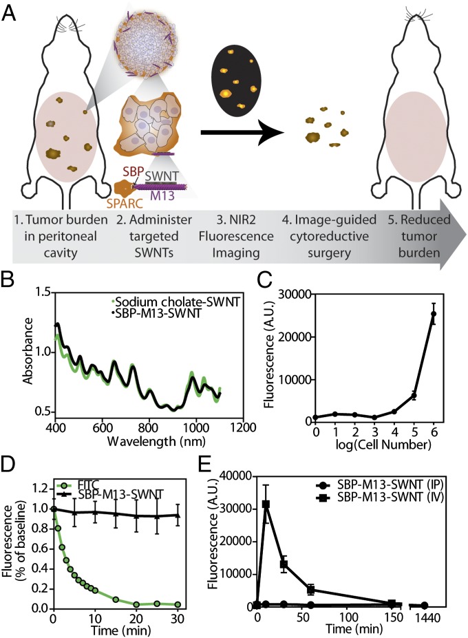
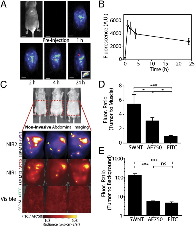
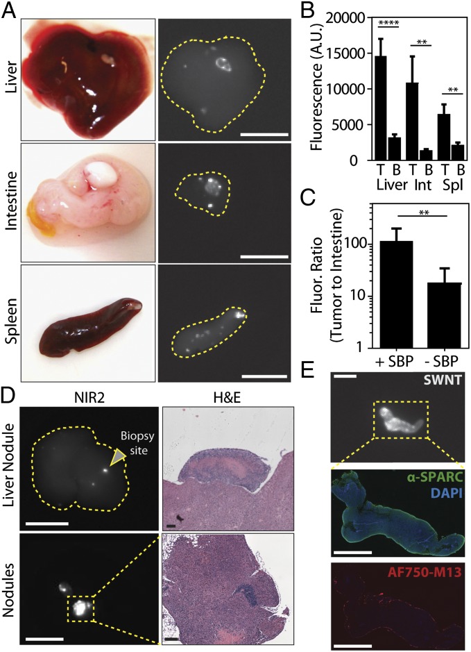
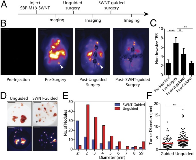
Highly sensitive detection of small, deep tumors for early diagnosis and surgical interventions remains a challenge for conventional imaging modalities. Second-window near-infrared light (NIR2, 950-1,400 nm) is promising for in vivo fluorescence imaging due to deep tissue penetration and low tissue autofluorescence. With their intrinsic fluorescence in the NIR2 regime and lack of photobleaching, single-walled carbon nanotubes (SWNTs) are potentially attractive contrast agents to detect tumors. Here, targeted M13 virus-stabilized SWNTs are used to visualize deep, disseminated tumors in vivo. This targeted nanoprobe, which uses M13 to stably display both tumor-targeting peptides and an SWNT imaging probe, demonstrates excellent tumor-to-background uptake and exhibits higher signal-to-noise performance compared with visible and near-infrared (NIR1) dyes for delineating tumor nodules. Detection and excision of tumors by a gynecological surgeon improved with SWNT image guidance and led to the identification of submillimeter tumors. Collectively, these findings demonstrate the promise of targeted SWNT nanoprobes for noninvasive disease monitoring and guided surgery.

Keywords: M13 bacteriophage; cancer imaging; fluorescence-guided surgery.


Conflict of interest statement

The authors declare no conflict of interest.

Figures

Fig. 1.
Fig. 1.
Characterization of tumor-targeting SBP–M13–SWNT probe. (A) Schematic illustrating association with ovarian tumor nodules for noninvasive detection by NIR2 fluorescence and surgical excision. (B) Absorbance spectra of SWNTs in sodium cholate and as SBP–M13–SWNT probe. (C) In vitro sensitivity of SBP–M13–SWNT fluorescence in ovarian cancer cell culture. n = 3 per group. Error bars represent SE. (D) Photobleaching fluorescence decay of FITC and SBP–M13–SWNTs under continuous excitation. Error bars represent SD. (E) Pharmacokinetic circulation study of SBP–M13–SWNT administered i.v. (IV) and intraperitoneally (IP). n = 3 per timepoint. Errors bars represent SE.
Fig. 2.
Fig. 2.
Noninvasive tumor imaging with SBP–M13–SWNTs. (A) Representative whole-abdomen NIR2 imaging series following i.p. administration of SBP–M13–SWNTs. (Inset) Surgically excised tumor nodule (denoted by red arrow) observed 24 h postinjection of SBP–M13–SWNTs. (B) NIR2 fluorescence intensity in the abdomen of tumor-bearing animals following IP administration of SBP–M13–SWNTs up to 24 h postinjection (n = 6; error bars represent SE). (C) Noninvasive imaging of ovarian tumors using SBP–M13 conjugated to SWNTs (NIR2), AlexaFluor750 (NIR1), and FITC (Visible) (top to bottom). Arrows in the SWNT panel denote nodules visible only by SWNTs (n = 3 animals). (D and E) Tumor-to-muscle ratio and TBR from noninvasive images obtained with SWNTs, AF750, and FITC (n = 3 per group; ***P < 0.001; *P < 0.05; one-way ANOVA and Tukey posttests). Error bars, s.d. [Scale bar, 1 cm (A).]
Fig. 3.
Fig. 3.
Specificity of SBP–M13–SWNTs for OVCAR8 tumor nodules in the abdominal cavity. (A) Photographs and NIR2 fluorescence (10–50-ms exposure) of tumor nodules implanted on several peritoneal organs. (B) Quantification of nodule and organ-specific background for nodules present on the liver, intestine, and spleen (n = 8–11 nodules per organ; **P < 0.01; ****P < 0.0001; two-tailed t tests). (C) Tumor-to-intestine ratio for targeted and untargeted M13–SWNT probes. Intestinal tissue used for background intensity (+SBP, n = 6; –SBP, n = 13; **P < 0.01; two-tailed t test). (D) Representative NIR2 fluorescence and H&E staining of a positive nodule revealing characteristic tumor histology. (E) Immunofluorescence staining reveals colocalization of M13–SBP–SWNTs conjugated to AlexaFluor750 dye with SPARC expression in an NIR2-positive nodule. [Scale bars, 10 mm (A), 10 mm (D, NIR2), 250 μm (D, H&E liver nodule), 125 μm (D, H&E nodules), 5 mm (E, NIR2), and 2.5 mm (E, SPARC, AF750–M13).]
Fig. 4.
Fig. 4.
Surgical removal of tumors with SBP–M13–SWNT guidance. (A) Schematic of serial surgical procedure. (B) Representative whole-abdomen NIR2 images before injection of SBP–M13–SWNT, before surgery, after an initial unguided surgery, and after subsequent SBP–M13–SWNT-guided surgery. White arrow indicates an SWNT-positive nodule detected only during image-guided surgery. (C) TBRs during surgery. Muscle from the hind limb was used for background (**P < 0.01; ****P < 0.0001; two-tailed t tests; error bars represent SE). (D) Photographs and NIR2 images of excised tumor nodules following unguided and SWNT-guided surgery. (E) Histogram of tumor diameters removed with and without guidance. (F) Dot plot of individual tumor nodule diameters excised with and without SWNT-guidance (guided, n = 74; unguided, n = 146; **P < 0.01; two-tailed t test). [Scale bars, 1 cm (B), 1 cm (C, photograph), and 1 cm (C, NIR2).]

References

    1. Weissleder R, Pittet MJ. Imaging in the era of molecular oncology. Nature. 2008;452(7187):580–589. - PMC - PubMed
    1. Urano Y, et al. Rapid cancer detection by topically spraying a γ-glutamyltranspeptidase-activated fluorescent probe. Sci Transl Med. 2011;3(110):ra119. - PMC - PubMed
    1. Nguyen QT, et al. Surgery with molecular fluorescence imaging using activatable cell-penetrating peptides decreases residual cancer and improves survival. Proc Natl Acad Sci USA. 2010;107(9):4317–4322. - PMC - PubMed
    1. van Dam GM, et al. Intraoperative tumor-specific fluorescence imaging in ovarian cancer by folate receptor-α targeting: First in-human results. Nat Med. 2011;17(10):1315–1319. - PubMed
    1. Lim YT, et al. Selection of quantum dot wavelengths for biomedical assays and imaging. Mol Imaging. 2003;2(1):50–64. - PubMed

Publication types