

Retrospective analysis of 18F-FDG PET/CT for staging asymptomatic breast cancer patients younger than 40 years
- PMID: 25214641
- PMCID: PMC4414239
- DOI: 10.2967/jnumed.114.143297
Retrospective analysis of 18F-FDG PET/CT for staging asymptomatic breast cancer patients younger than 40 years
Abstract
National Comprehensive Cancer Network guidelines consider (18)F-FDG PET/CT for only clinical stage III breast cancer patients. However, there is debate whether TNM staging should be the only factor in considering if PET/CT is warranted. Patient age may be an additional consideration, because young breast cancer patients often have more aggressive tumors with potential for earlier metastases. This study assessed PET/CT for staging of asymptomatic breast cancer patients younger than 40 y.
Methods: In this Institutional Review Board-approved retrospective study, our hospital information system was screened for breast cancer patients younger than 40 y who underwent staging PET/CT before any treatment. Patients with symptoms or conventional imaging findings suggestive of distant metastases or with prior malignancy were excluded. Initial stage was based on physical examination, mammography, ultrasound, and breast MR imaging. PET/CT was then evaluated to identify unsuspected extraaxillary regional nodal and distant metastases.
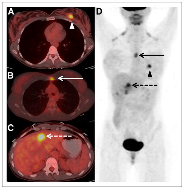
Results: One hundred thirty-four patients with initial breast cancer stage I to IIIC met inclusion criteria. PET/CT findings led to upstaging to stage III or IV in 28 patients (21%). Unsuspected extraaxillary regional nodes were found in 15 of 134 patients (11%) and distant metastases in 20 of 134 (15%), with 7 of 134 (5%) demonstrating both. PET/CT revealed stage IV disease in 1 of 20 (5%) patients with initial clinical stage I, 2 of 44 (5%) stage IIA, 8 of 47 (17%) stage IIB, 4 of 13 (31%) stage IIIA, 4 of 8 (50%) stage IIIB, and 1 of 2 (50%) stage IIIC. All 20 patients upstaged to stage IV were histologically confirmed. Four synchronous thyroid and 1 rectal malignancies were identified.
Conclusion: PET/CT revealed distant metastases in 17% of asymptomatic stage IIB breast cancer patients younger than 40 y. Although guidelines of the National Comprehensive Cancer Network recommend against systemic staging in patients with stage II disease, our data suggest that PET/CT might be valuable in younger patients with stage IIB and III disease. Use of PET/CT in younger patients has the potential to reduce the morbidity and cost of unnecessary therapies in young breast cancer patients.
Keywords: FDG; PET; breast cancer; metastases; staging.
© 2014 by the Society of Nuclear Medicine and Molecular Imaging, Inc.
Figures


Comment in
-
Breast Cancer Staging: To Which Women Should 18F-FDG PET/CT Be Offered?J Nucl Med. 2015 Aug;56(8):1293. doi: 10.2967/jnumed.115.160945. Epub 2015 Jun 4. J Nucl Med. 2015. PMID: 26045310 No abstract available.
-
Reply: Breast Cancer Staging: To Which Women Should 18F-FDG PET/CT Be Offered?J Nucl Med. 2015 Aug;56(8):1293-4. doi: 10.2967/jnumed.115.161042. Epub 2015 Jun 25. J Nucl Med. 2015. PMID: 26112021 No abstract available.
References
-
- Carkaci S, Macapinlac HA, Cristofanilli M, et al. Retrospective study of 18F-FDG PET/CT in the diagnosis of inflammatory breast cancer: preliminary data. J Nucl Med. 2009;50:231–238. - PubMed
-
- Fuster D, Duch J, Paredes P, et al. Preoperative staging of large primary breast cancer with [18F]fluorodeoxyglucose positron emission tomography/computed tomography compared with conventional imaging procedures. J Clin Oncol. 2008;26:4746–4751. - PubMed
-
- Alberini JL, Lerebours F, Wartski M, et al. 18F-fluorodeoxyglucose positron emission tomography/computed tomography (FDG-PET/CT) imaging in the staging and prognosis of inflammatory breast cancer. Cancer. 2009;115:5038–5047. - PubMed
-
- Bernsdorf M, Berthelsen AK, Wielenga VT, et al. Preoperative PET/CT in early-stage breast cancer. Ann Oncol. 2012;23:2277–2282. - PubMed
Publication types
MeSH terms
Substances
Grants and funding
LinkOut - more resources
Full Text Sources
Other Literature Sources
Medical
Miscellaneous