Toll-like receptor 4 in bone marrow-derived cells contributes to the progression of diabetic retinopathy
- PMID: 25214718
- PMCID: PMC4156976
- DOI: 10.1155/2014/858763
Toll-like receptor 4 in bone marrow-derived cells contributes to the progression of diabetic retinopathy
Abstract
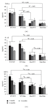
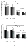
Diabetic retinopathy (DR) is a major microvascular complication in diabetics, and its mechanism is not fully understood. Toll-like receptor 4 (TLR4) plays a pivotal role in the maintenance of the inflammatory state during DR, and the deletion of TLR4 eventually alleviates the diabetic inflammatory state. To further elucidate the mechanism of DR, we used bone marrow transplantation to establish reciprocal chimeric animals of TLR4 mutant mice and TLR4 WT mice combined with diabetes mellitus (DM) induction by streptozotocin (STZ) treatment to identify the role of TLR4 in different cell types in the development of the proinflammatory state during DR. TLR4 mutation did not block the occurrence of high blood glucose after STZ injection compared with WT mice but did alleviate the progression of DR and alter the expression of the small vessel proliferation-related genes, vascular endothelial growth factor (VEGF), and hypoxia inducible factor-1α (HIF-1α). Grafting bone marrow-derived cells from TLR4 WT mice into TLR4 mutant mice increased the levels of TNF-α, IL-1β, and MIP-2 and increased the damage to the retina. Similarly, VEGF and HIF-1α expression were restored by the bone marrow transplantation. These findings identify an essential role for TLR4 in bone marrow-derived cells contributing to the progression of DR.
Figures


Similar articles
-
Attenuation of streptozotocin-induced diabetic retinopathy with low molecular weight fucoidan via inhibition of vascular endothelial growth factor.Exp Eye Res. 2013 Oct;115:96-105. doi: 10.1016/j.exer.2013.06.011. Epub 2013 Jun 28. Exp Eye Res. 2013. PMID: 23810809
-
Association of the TLR4 signaling pathway in the retina of streptozotocin-induced diabetic rats.Graefes Arch Clin Exp Ophthalmol. 2015 Mar;253(3):389-98. doi: 10.1007/s00417-014-2832-y. Epub 2014 Oct 31. Graefes Arch Clin Exp Ophthalmol. 2015. PMID: 25359392
-
Deletion of toll-like receptor 4 ameliorates diabetic retinopathy in mice.Arch Physiol Biochem. 2023 Apr;129(2):519-525. doi: 10.1080/13813455.2020.1841795. Epub 2020 Nov 6. Arch Physiol Biochem. 2023. PMID: 33156707
-
Role of toll-like receptor 4 in diabetic retinopathy.Pharmacol Res. 2022 Jan;175:105960. doi: 10.1016/j.phrs.2021.105960. Epub 2021 Oct 28. Pharmacol Res. 2022. PMID: 34718133 Review.
-
Hypoxia-inducible factor-1α: A promising therapeutic target for vasculopathy in diabetic retinopathy.Pharmacol Res. 2020 Sep;159:104924. doi: 10.1016/j.phrs.2020.104924. Epub 2020 May 25. Pharmacol Res. 2020. PMID: 32464323 Review.
Cited by
-
Administration of glycyrrhetinic acid reinforces therapeutic effects of mesenchymal stem cell-derived exosome against acute liver ischemia-reperfusion injury.J Cell Mol Med. 2020 Oct;24(19):11211-11220. doi: 10.1111/jcmm.15675. Epub 2020 Sep 9. J Cell Mol Med. 2020. PMID: 32902129 Free PMC article.
-
Role of HMGB1 signaling in the inflammatory process in diabetic retinopathy.Cell Signal. 2020 Sep;73:109687. doi: 10.1016/j.cellsig.2020.109687. Epub 2020 Jun 1. Cell Signal. 2020. PMID: 32497617 Free PMC article. Review.
-
Regulation of toll-like receptor signaling pathways in age-related eye disease: from mechanisms to targeted therapeutics.Inflammopharmacology. 2025 Aug 19. doi: 10.1007/s10787-025-01913-9. Online ahead of print. Inflammopharmacology. 2025. PMID: 40828357 Review.
-
RAGE plays key role in diabetic retinopathy: a review.Biomed Eng Online. 2023 Dec 19;22(1):128. doi: 10.1186/s12938-023-01194-9. Biomed Eng Online. 2023. PMID: 38115006 Free PMC article. Review.
-
Friend or Foe? Resident Microglia vs Bone Marrow-Derived Microglia and Their Roles in the Retinal Degeneration.Mol Neurobiol. 2017 Aug;54(6):4094-4112. doi: 10.1007/s12035-016-9960-9. Epub 2016 Jun 18. Mol Neurobiol. 2017. PMID: 27318678 Review.
References
-
- Stitt AW, Lois N, Medina RJ, Adamson P, Curtis TM. Advances in our understanding of diabetic retinopathy. Clinical Science. 2013;125(1):1–17. - PubMed
-
- Antonetti DA, Barber AJ, Bronson SK, et al. Diabetic retinopathy: seeing beyond glucose-induced microvascular disease. Diabetes. 2006;55(9):2401–2411. - PubMed
Publication types
MeSH terms
Substances
LinkOut - more resources
Full Text Sources
Other Literature Sources
Medical

