Comparative evaluation of sealing ability of four different restorative materials used as coronal sealants: an in vitro study
- PMID: 25214726
- PMCID: PMC4148566
Comparative evaluation of sealing ability of four different restorative materials used as coronal sealants: an in vitro study
Abstract
Background: The purpose of the present study was to evaluate and compare the sealing ability of glass ionomer cement (GIC), composite resin, gray mineral trioxide aggregate (GMTA) and white mineral trioxide aggregate (WMTA) when placed coronally as double - sealing material over gutta-percha in root canal treated teeth.
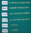
Materials and methods: A sample of 70 freshly extracted human single rooted teeth were cleaned, shaped and obturated with gutta-percha and AH Plus. The gutta-percha was reduced to a depth of 4 mm from the cemento enamel junction using hot plugger and standardized access cavities with 4 mm depth were prepared at the coronal ends of the roots. The specimens were randomly divided into four groups containing 15 teeth each depending on the restorations they received in the coronal cavity. A positive control group of five teeth received no restorative barrier over gutta-percha. All root surfaces were covered with two coats of nail varnish, leaving only the access openings uncovered except teeth in the negative control group, which were completely covered with nail varnish. All teeth were immersed in India ink, cleared and observed under stereomicroscope for the depth of dye penetration.
Results: The results were tabulated and analyzed using Kruskal-Wallis test and multiple comparison between each group was carried out using Mann-Whitney test. The groups sealed with GMTA and WMTA showed least dye penetration than other groups and the difference was statistically significant. Highest dye penetration was seen with groups sealed with GIC and was statistically significant compared with other three groups.
Conclusion: The results showed that the GMTA and WMTA provided significantly better coronal seal when compared to other two restorations. The composite resin also showed significantly better seal than the unsealed group and the group sealed GIC, which showed highest leakage that was equivalent to that of unsealed group.
Keywords: Coronal microleakage; coronal seal; double-seal technique; mineral trioxide aggregate.
Conflict of interest statement
Figures


Similar articles
-
Evaluation of coronal microleakage of intra-orifice barrier materials in endodontically treated teeth: A systematic review.J Conserv Dent. 2022 Nov-Dec;25(6):588-595. doi: 10.4103/jcd.jcd_377_22. Epub 2022 Oct 13. J Conserv Dent. 2022. PMID: 36591578 Free PMC article. Review.
-
Intracoronal sealing comparison of mineral trioxide aggregate and glass ionomer.Quintessence Int. 2005 Jul-Aug;36(7-8):539-45. Quintessence Int. 2005. PMID: 15997934 Clinical Trial.
-
An Evaluation of MTA Cements as Coronal Barrier.Iran Endod J. 2006 Fall;1(3):106-8. Epub 2006 Oct 1. Iran Endod J. 2006. PMID: 24454453 Free PMC article.
-
An in vitro study of apical and coronal microleakage of laterally condensed gutta percha with Ketac-Endo and AH-26.Aust Dent J. 1998 Aug;43(4):262-8. doi: 10.1111/j.1834-7819.1998.tb00175.x. Aust Dent J. 1998. PMID: 9775474
-
Microleakage comparison of glass-ionomer and white mineral trioxide aggregate used as a coronal barrier in nonvital bleaching.Med Oral Patol Oral Cir Bucal. 2011 Nov 1;16(7):e1017-21. doi: 10.4317/medoral.17306. Med Oral Patol Oral Cir Bucal. 2011. PMID: 21743399
Cited by
-
Comparison of Dentinal Microleakage in Three Interim Dental Restorations: An In Vitro Study.J Int Soc Prev Community Dent. 2022 Dec 30;12(6):590-595. doi: 10.4103/jispcd.JISPCD_183_21. eCollection 2022 Nov-Dec. J Int Soc Prev Community Dent. 2022. PMID: 36777014 Free PMC article.
-
Comparative Evaluation of Sealing Ability of Three Different Materials as Barriers to Coronal Microleakage in Root-Filled Teeth: An In Vitro Study.J Pharm Bioallied Sci. 2024 Feb;16(Suppl 1):S659-S662. doi: 10.4103/jpbs.jpbs_921_23. Epub 2023 Dec 20. J Pharm Bioallied Sci. 2024. PMID: 38595523 Free PMC article.
-
Effect of an Intraorifice Barrier on Endodontically Treated Teeth: A Systematic Review and Meta-Analysis of In Vitro Studies.Biomed Res Int. 2022 Jan 20;2022:2789073. doi: 10.1155/2022/2789073. eCollection 2022. Biomed Res Int. 2022. PMID: 35097115 Free PMC article.
-
Evaluation of coronal microleakage of intra-orifice barrier materials in endodontically treated teeth: A systematic review.J Conserv Dent. 2022 Nov-Dec;25(6):588-595. doi: 10.4103/jcd.jcd_377_22. Epub 2022 Oct 13. J Conserv Dent. 2022. PMID: 36591578 Free PMC article. Review.
-
Pulpal responses to mineral trioxide aggregate with and without zinc oxide addition in mature canine teeth after full pulpotomy.Sci Rep. 2025 May 7;15(1):15957. doi: 10.1038/s41598-025-00061-y. Sci Rep. 2025. PMID: 40335528 Free PMC article.
References
-
- Verma MR, Desai VM, Shahani SN. Importance of coronal seal following routine endodontic therapy. Endodontology. 1992;1(2):17–9.
-
- Saunders WP, Saunders EM. Coronal leakage as a cause of failure in root-canal therapy: A review. Endod Dent Traumatol. 1994;10(3):105–8. - PubMed
-
- Ray HA, Trope M. Periapical status of endodontically treated teeth in relation to the technical quality of the root filling and the coronal restoration. Int Endod J. 1995;28(1):12–8. - PubMed
-
- Roghanizad N, Jones JJ. Evaluation of coronal microleakage after endodontic treatment. J Endod. 1996;22(9):471–3. - PubMed
-
- Torabinejad M, Chivian N. Clinical applications of mineral trioxide aggregate. J Endod. 1999;25(3):197–205. - PubMed
LinkOut - more resources
Full Text Sources
