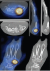
Assessment of Inter-modality Spatial Alignment Accuracy in Hybrid Single Photon Emission Computed Tomography in Patients with Hand and Wrist Pain
- PMID: 25214811
- PMCID: PMC4145159
- DOI: 10.4103/1450-1147.136732
Assessment of Inter-modality Spatial Alignment Accuracy in Hybrid Single Photon Emission Computed Tomography in Patients with Hand and Wrist Pain
Abstract
Single photon emission computed tomography (SPECT) and computed tomography (CT) integrated in one system (SPECT/CT) is an effective co-registration technique that helps to localize and characterize lesions in the hand and wrist. However, patient motion may cause misalignment between the two modalities leading to potential misdiagnosis. The aim of the present study was to evaluate the hardware-based registration accuracy of multislice SPECT/CT of the hand and wrist and to determine the effect of misalignment errors on diagnostic accuracy. A total of 55 patients who had multislice SPECT/CT of the hand and wrist between July 2008 and January 2010 were included. Two reviewers independently evaluated the fused images for any misalignments with six degrees of freedom: Translation and rotation in the X, Y and Z directions. The results were tested against an automated fusion tool (Syntegra). More than half of the patients had moved during SPECT scanning (Reviewer 1: 29 patients; Reviewer 2: 30 patients) and they all originated in the Y-direction translation (vertical hand motion). Five fused images had significant misalignment errors that could have led to misdiagnosis. The Wilcoxon test indicated statistically non-significant difference (P > 0.05) between reviewers and statistically non-significant difference between the reviewers and software registration. The study also showed high inter-reviewer agreement (κ = 0.87). Hand movement during the SPECT scan was common, but significant misalignments and subsequent misdiagnosis were infrequent. Future studies should investigate the use of hand and wrist immobilization devices and reductions of scan time to minimize patient motion.
Keywords: Image fusion; registration; single photon emission computed tomography/computed tomography.
Conflict of interest statement
Figures


Similar articles
-
Imaging non-specific wrist pain: interobserver agreement and diagnostic accuracy of SPECT/CT, MRI, CT, bone scan and plain radiographs.PLoS One. 2013 Dec 30;8(12):e85359. doi: 10.1371/journal.pone.0085359. eCollection 2013. PLoS One. 2013. PMID: 24386468 Free PMC article. Clinical Trial.
-
Acceptance test of a commercially available software for automatic image registration of computed tomography (CT), magnetic resonance imaging (MRI) and 99mTc-methoxyisobutylisonitrile (MIBI) single-photon emission computed tomography (SPECT) brain images.J Digit Imaging. 2008 Sep;21(3):329-37. doi: 10.1007/s10278-007-9042-7. J Digit Imaging. 2008. PMID: 17549564 Free PMC article.
-
Anatomical accuracy of hybrid SPECT/spiral CT in the lower spine.Nucl Med Commun. 2006 Jun;27(6):521-8. doi: 10.1097/00006231-200606000-00008. Nucl Med Commun. 2006. PMID: 16710107
-
Single-photon emission computed tomography/computed tomography in brain tumors.Semin Nucl Med. 2007 Jan;37(1):34-47. doi: 10.1053/j.semnuclmed.2006.08.003. Semin Nucl Med. 2007. PMID: 17161038 Review.
-
Fusion imaging in nuclear medicine--applications of dual-modality systems in oncology.Cancer Biother Radiopharm. 2004 Feb;19(1):1-10. doi: 10.1089/108497804773391621. Cancer Biother Radiopharm. 2004. PMID: 15068606 Review.
Cited by
-
Development of a Protocol for SPECT/CT in the Assessment of Wrist Disorders.J Wrist Surg. 2016 Nov;5(4):297-305. doi: 10.1055/s-0036-1583314. Epub 2016 May 2. J Wrist Surg. 2016. PMID: 27777821 Free PMC article.
References
-
- von Schulthess GK, Pelc NJ. Integrated-modality imaging: The best of both worlds. Acad Radiol. 2002;9:1241–4. - PubMed
-
- Schillaci O. Hybrid SPECT/CT: A new era for SPECT imaging? Eur J Nucl Med Mol Imaging. 2005;32:521–4. - PubMed
-
- Townsend DW, Cherry SR. Combining anatomy and function: The path to true image fusion. Eur Radiol. 2001;11:1968–74. - PubMed
-
- Even-Sapir E, Keidar Z, Bar-Shalom R. Hybrid imaging (SPECT/CT and PET/CT) - Improving the diagnostic accuracy of functional/metabolic and anatomic imaging. Semin Nucl Med. 2009;39:264–75. - PubMed
-
- Fogelman I. Bone scanning. In: Maisey M, Britton K, Gilday D, editors. Clinical Nuclear Medicine. Cambridge: Chapman & Hall; 1991. pp. 131–57.
LinkOut - more resources
Full Text Sources
Other Literature Sources

