. 2014 Sep 12:5:4888.
doi: 10.1038/ncomms5888.

Basal foot MTOC organizes pillar MTs required for coordination of beating cilia

Affiliations

Basal foot MTOC organizes pillar MTs required for coordination of beating cilia

Daniel K Clare et al. Nat Commun. .

Abstract

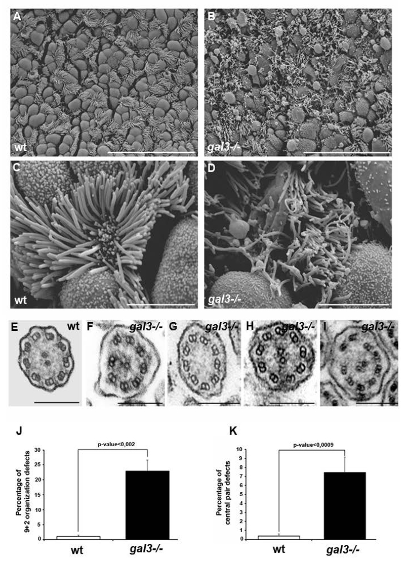
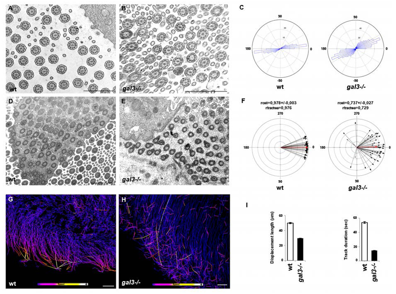
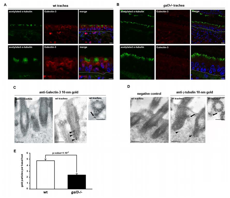
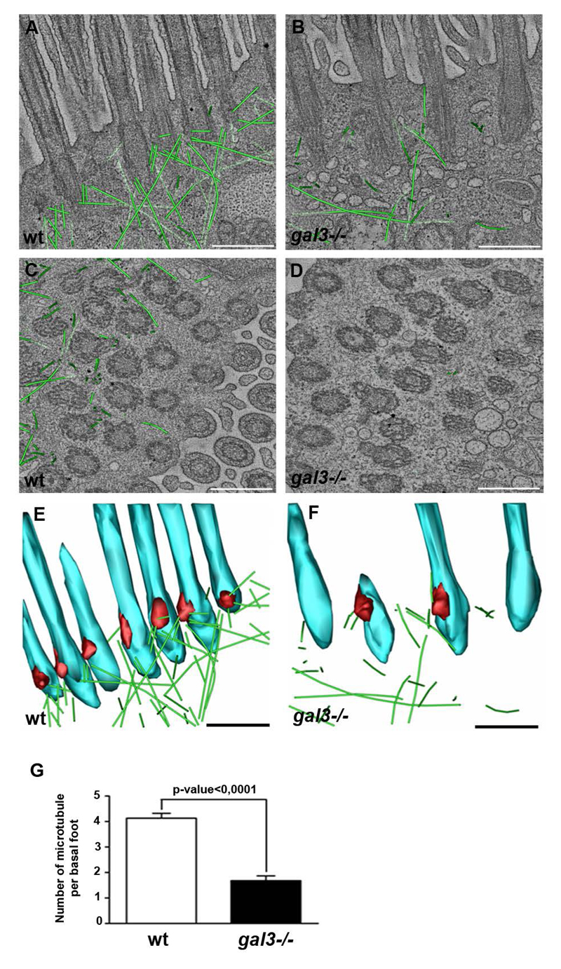
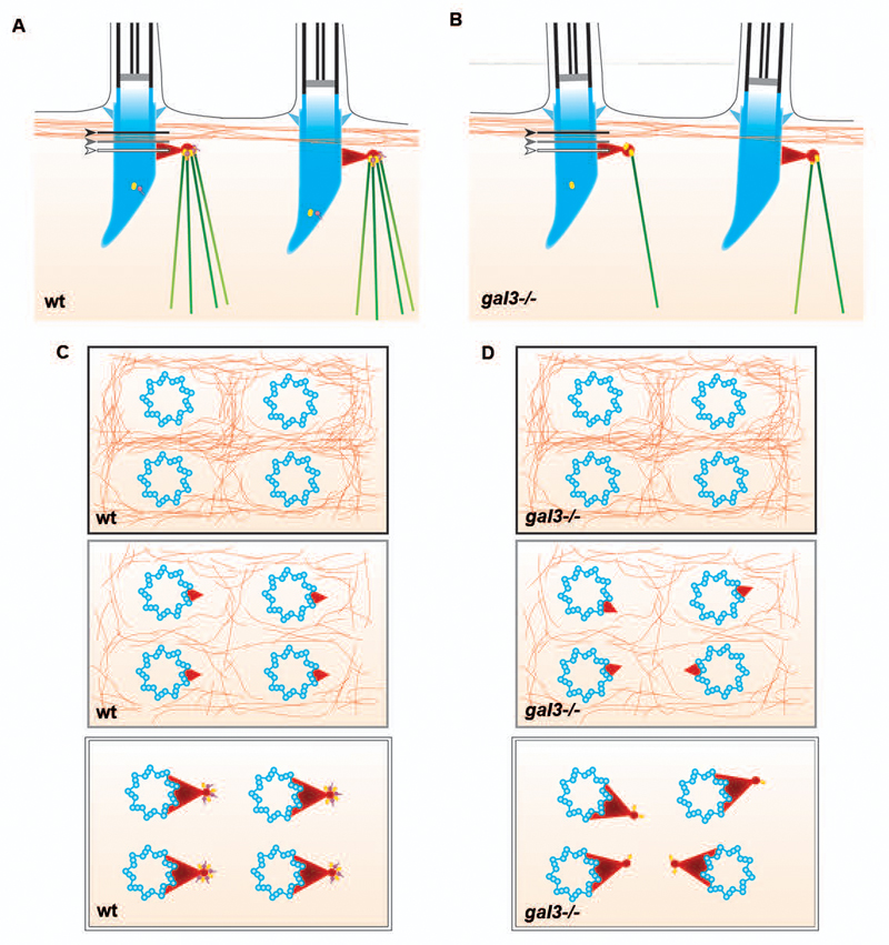
Coordination of ciliary beating is essential to ensure mucus clearance in the airway tract. The orientation and synchronization of ciliary motion responds in part to the organization of the underlying cytoskeletal networks. Using electron tomography on mouse trachea, we show that basal bodies are collectively hooked at the cortex by a regular microtubule array composed of 4-5 microtubules. Removal of galectin-3, one of basal-body components, provokes misrecruitment of γ-tubulin, disorganization of this microtubule framework emanating from the basal-foot cap, together with loss of basal-body alignment and cilium orientation, defects in cilium organization and reduced fluid flow in the tracheal lumen. We conclude that galectin-3 plays a crucial role in the maintenance of the microtubule-organizing centre of the cilium and the 'pillar' microtubules, and that this network is instrumental for the coordinated orientation and stabilization of motile cilia.


Conflict of interest statement

the authors declare no conflict of interest.

Figures

Figure 1
Figure 1. Defective motile cilium organization in mouse gal3-/- tracheas.
A-D, the luminal surface of wt (A and C) (N = 4 wt mice) and gal3-/- (B and D) (N = 4 gal3-/- mice) tracheal epithelium was analyzed using SEM. Low magnifications (A and B) and high magnifications (C and D) are presented. Scale bars, A and B, 20μm and C and D, 5μm. E-I, TEM ultrastructural analyses of axonemal transverse sections in wt (E) and gal3-/- (F-I) tracheas. Scale bars, E-I, 200nm. J, statistical analysis of the 9+2 axonemal organization defects in wt (white) and gal3-/- (black) tracheas. Mann-Whitney test, p-value = 0,0019. N (wt) = 9 mice, n (wt) = 305 cilia; N (gal3-/-) = 9 mice, n (gal3-/-) = 940 cilia. K, statistical analysis of the central pair defects in wt (white) and gal3-/- (black) tracheas. Mann-Whitney test, p-value = 0,00087. N (wt) = 9 mice, n (wt) = 889 cilia; N (gal3-/-) = 9 mice, n (gal3-/-) = 599 cilia. Values are mean ± SEM.
Figure 2
Figure 2. Cilium organization and flow directionality are lost in mouse gal3-/- ciliated cells.
TEM ultrastructural analysis of axonemal (A and B) and BB (D and E) transversal sections in wt and gal3-/- mouse tracheas. Scale bars, A, B, D and E, 1μm. C, statistical analyses of central MT pair orientation in individual cells with MATLAB. wt cilium mean angular deviation: ±23.01°; gal3-/- cilium mean angular deviation: ±39.19°; Watson-Wheeler homogeneity test, p-value = 6,91.10-15. N (wt) =8 mice, n (wt) =338 axonemes; N (gal3-/-) =7 mice, n (gal3-/-) =411 axonemes. F, statistical analyses of BB orientation in individual cells and along the tracheal axis with Oriana2.0. rcell (wt) = 0,978±0,003; rtrachea (wt) = 0,976; rcell (gal3-/-) = 0,737±0,027; rtrachea (gal3-/-) = 0,729. N (wt) = 3 mice, n (wt) = 284 basal feet; N (gal3-/-) = 3 mice, n (gal3-/-) = 341 basal feet. G and H, representative patterns of fluorescent bead dynamics above wt (G) and gal3-/- (H) tracheal explants. Scale bars, 30μm. I, statistical analyses of fluorescent bead behavior on the surface of wt (white) and gal3-/- (black) tracheal epithelia. Fluorescent bead track displacement lengths and durations were automatically measured using Imaris software. N (wt) = 6 mice, one tracheal ring per animal, n (wt) = 3981 tracked beads; N (gal3-/-) = 6 mice, 1 tracheal ring per animal, n (gal3-/-) = 11156 tracked beads. N (wt track displacement length) = 49±0,8μm, n (wt track duration) = 53,84±1,34sec, n (gal3-/- track displacement length) = 29,63±0,29 μm, n (gal3-/- track duration) = 14,57±0,22sec. Values are mean ± SEM.
Figure 3
Figure 3. Galectin-3 is present at the basal foot cap and ciliary rootlet of motile cilium in tracheal epithelial cells.
A, confocal microscopy analysis of Galectin-3 distribution in mouse tracheal cells. Longitudinal paraffin sections were immunostained with monoclonal anti-acetylated α-tubulin (green) and polyclonal anti-Galectin-3 (red) antibodies. B, Gal3-/- mouse tracheas were used as a negative control for Galectin-3 immunofluorescence. Galectin-3 is highly enriched at the cilium base of tracheal epithelial cells. Nuclei were detected by Hoechst 33342 staining (blue). Tracheal epithelium is demarcated by a white dotted line. Scale bars, upper panel 10μm, lower panel 5μm. C-D, TEM ultrastrural analyses of Galectin-3 (C) and γ-tubulin (D) localizations at the BBs. Lowicryl sections were immunostained with monoclonal anti-Galectin-3 or polyclonal anti-γ-tubulin antibodies. Gal3-/- mouse tracheas were used as a negative control for Galectin-3 immunogold staining (C). Incubation of the sections with only the secondary antibody was used as a negative control (D). Scale bars, 300nm. E, statistical analysis of the number of γ-tubulin gold particles per basal foot in wt (white) and gal3-/- (black). Mann-Whitney test, p-value= 1,283.10-8. N (wt) = 3 mice, n (wt) = 67 basal feet; N (gal3-/-) = 3 mice, n (gal3-/-) = 50 basal feet. Values are mean ± SEM.
Figure 4
Figure 4. Association of MTs with basal feet is severely disrupted in gal3-/- mouse tracheas.
Representative EM tomographic slices, generated by averaging 5 z-sections of the reconstructed filtered volume, of the cortical domain of wt (A and C) and gal3-/- (B and D) ciliated cells are presented with superimposition of the contour of the MTs (green). The identification of a MT is based on its shape and diameter (20nm). Longitudinal (A and B) and transversal (C and D) sections are shown. E and F, 3D models of the EM tomographic reconstructions of cilium base (blue), basal foot (red) and MTs (green) were generated for wt (E) and gal3-/- (F) ciliated cells, and representative tilted longitudinal views are presented. Scale bars, 500nm. G, statistical analysis of the number of MTs per basal foot in wt (white) and gal3-/- (black). Mann-Whitney test, p-value=3,18.10-12. N (wt) = 3 mice, n (wt) = 60 basal feet; N (gal3-/-) = 3 mice, n (gal3-/-) = 53 basal feet. Values are mean ± SEM.
Figure 5
Figure 5. Schemes showing the predicted association of the MT network and the actin cytoskeleton with the basal bodies of motile cilia from the acquired tomograms in wt (A and C) and in gal3-/- (B and D) ciliated cells, in side (A and B) or top (C and D) views.
MTs (green), actin cables (orange), Galectin-3 (purple) and γ-tubulin (yellow) are represented at the BBs (blue) and at the basal foot cap (red). The relative position of the actin cytoskeleton to the basal foot in wt (A) and gal3-/- (B) is showed with black (above the basal foot), grey (the top of the basal foot) and white (at the level of the basal foot cap) arrows and lines. C and D, black, grey and white panels correspond to black, grey and white line positions in A and B.

Similar articles

Cited by

References

    1. Knowles MR, Boucher RC. Mucus clearance as a primary innate defense mechanism for mammalian airways. The Journal of clinical investigation. 2002;109:571–577. - PMC - PubMed
    1. Randell SH, Boucher RC. Effective mucus clearance is essential for respiratory health. American journal of respiratory cell and molecular biology. 2006;35:20–28. - PMC - PubMed
    1. Mall MA. Role of cilia, mucus, and airway surface liquid in mucociliary dysfunction: lessons from mouse models. Journal of aerosol medicine and pulmonary drug delivery. 2008;21:13–24. - PubMed
    1. Marshall WF, Kintner C. Cilia orientation and the fluid mechanics of development. Current opinion in cell biology. 2008;20:48–52. - PMC - PubMed
    1. Wallingford JB. Planar cell polarity signaling, cilia and polarized ciliary beating. Current opinion in cell biology. 2010;22:597–604. - PMC - PubMed

Publication types

MeSH terms