. 2014 Sep 11;158(6):1254-1269.
doi: 10.1016/j.cell.2014.08.029.

Resetting transcription factor control circuitry toward ground-state pluripotency in human

Affiliations

Resetting transcription factor control circuitry toward ground-state pluripotency in human

Yasuhiro Takashima et al. Cell. .

Erratum in

Abstract

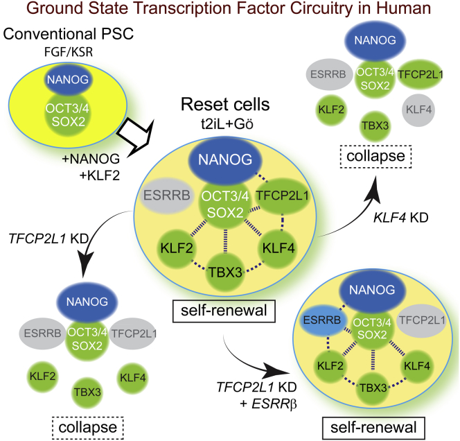
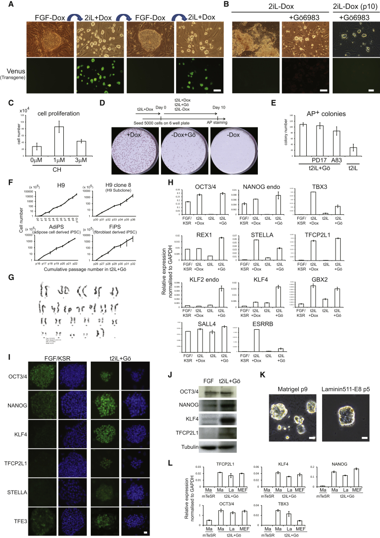
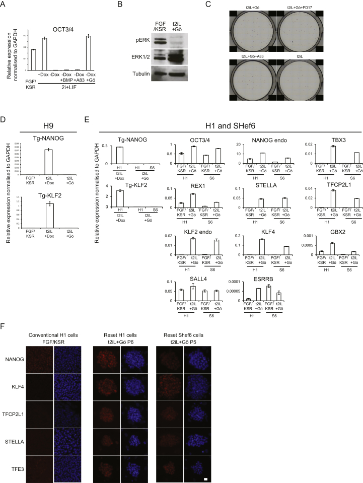
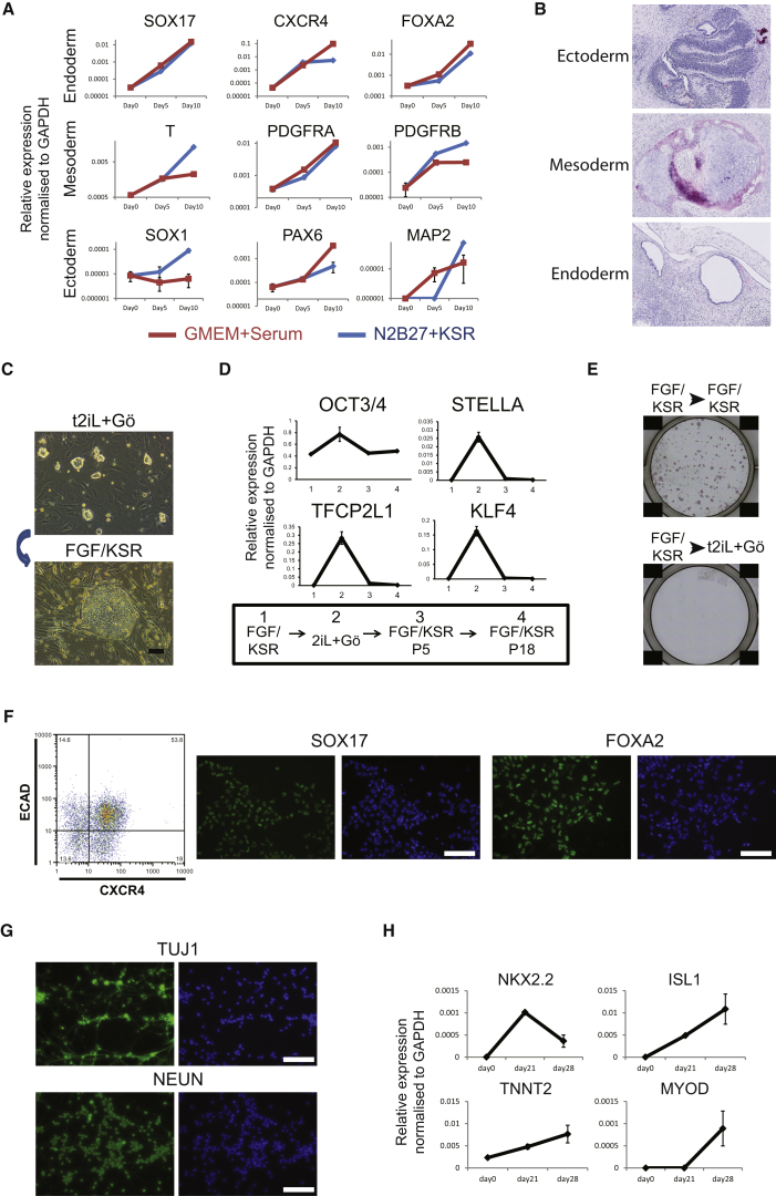
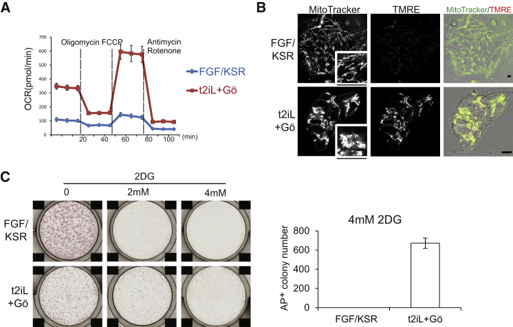
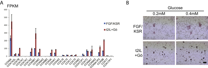
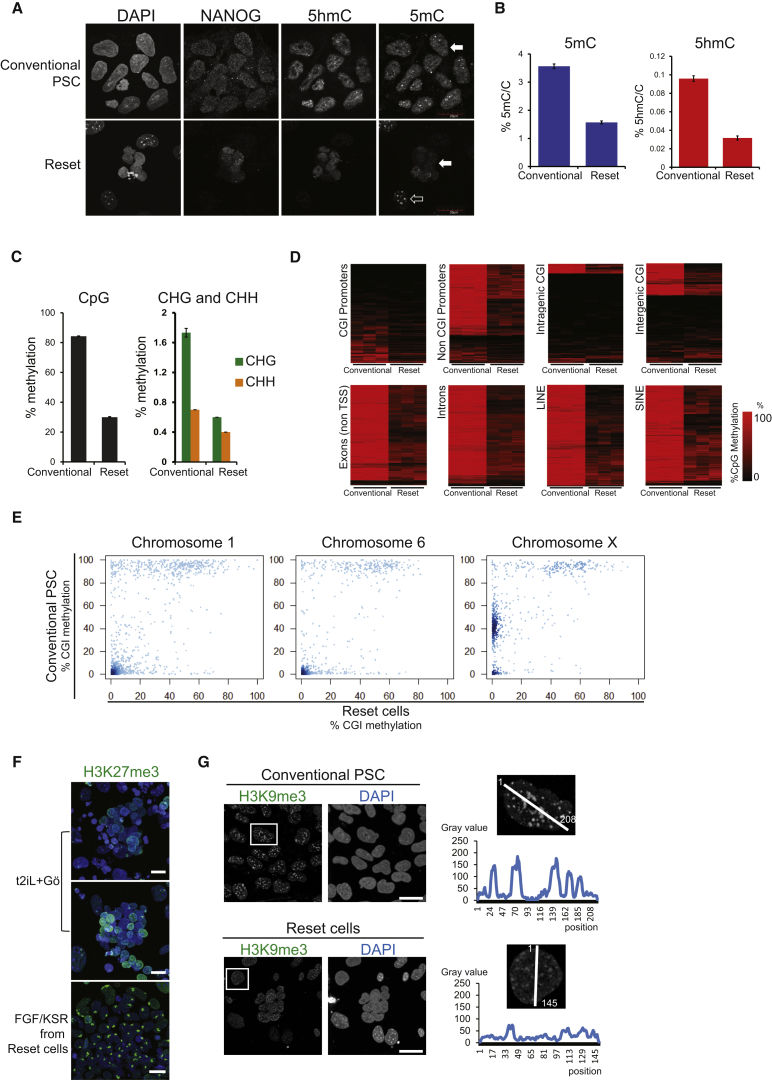
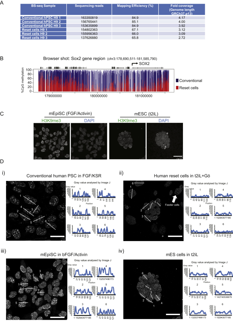
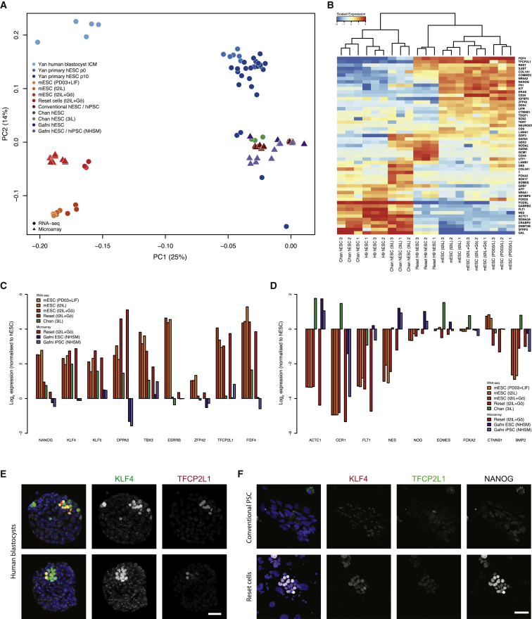
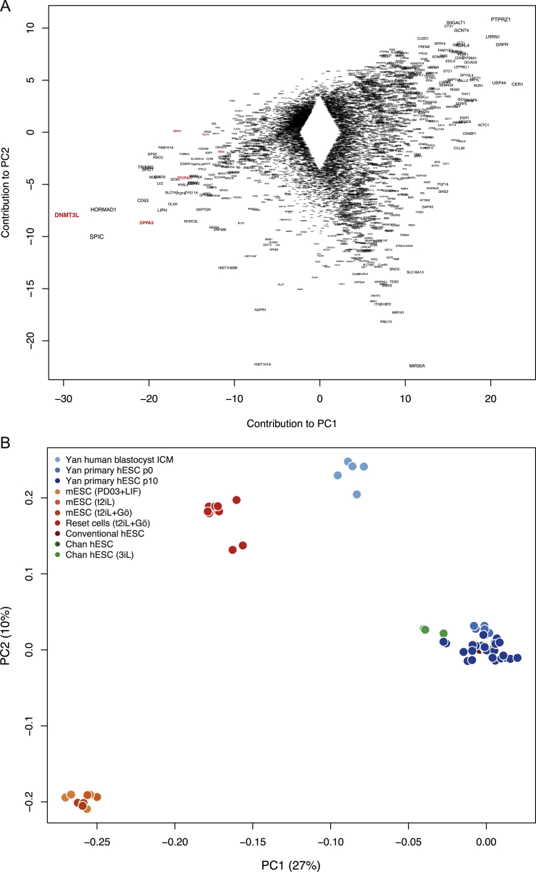
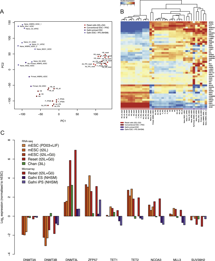
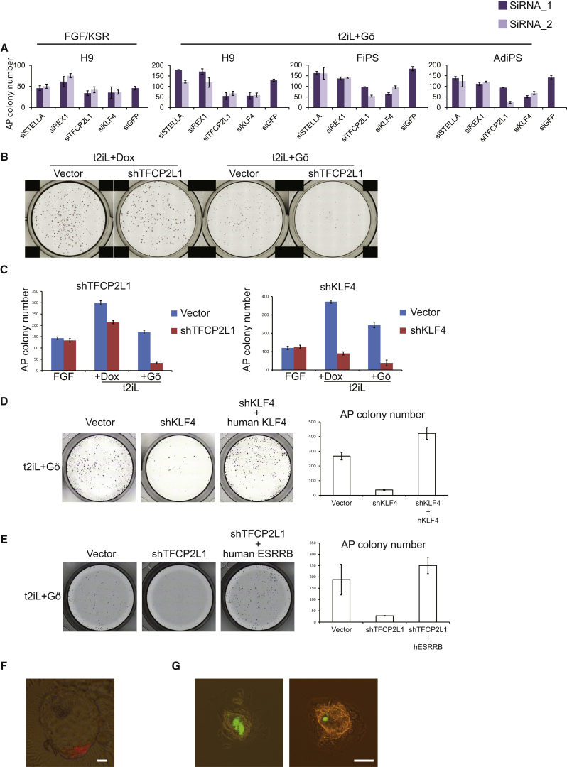
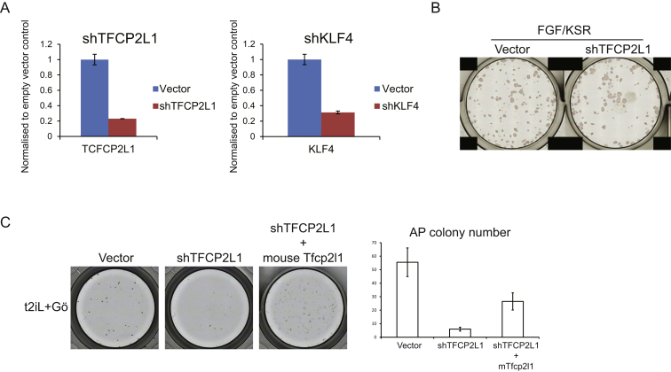
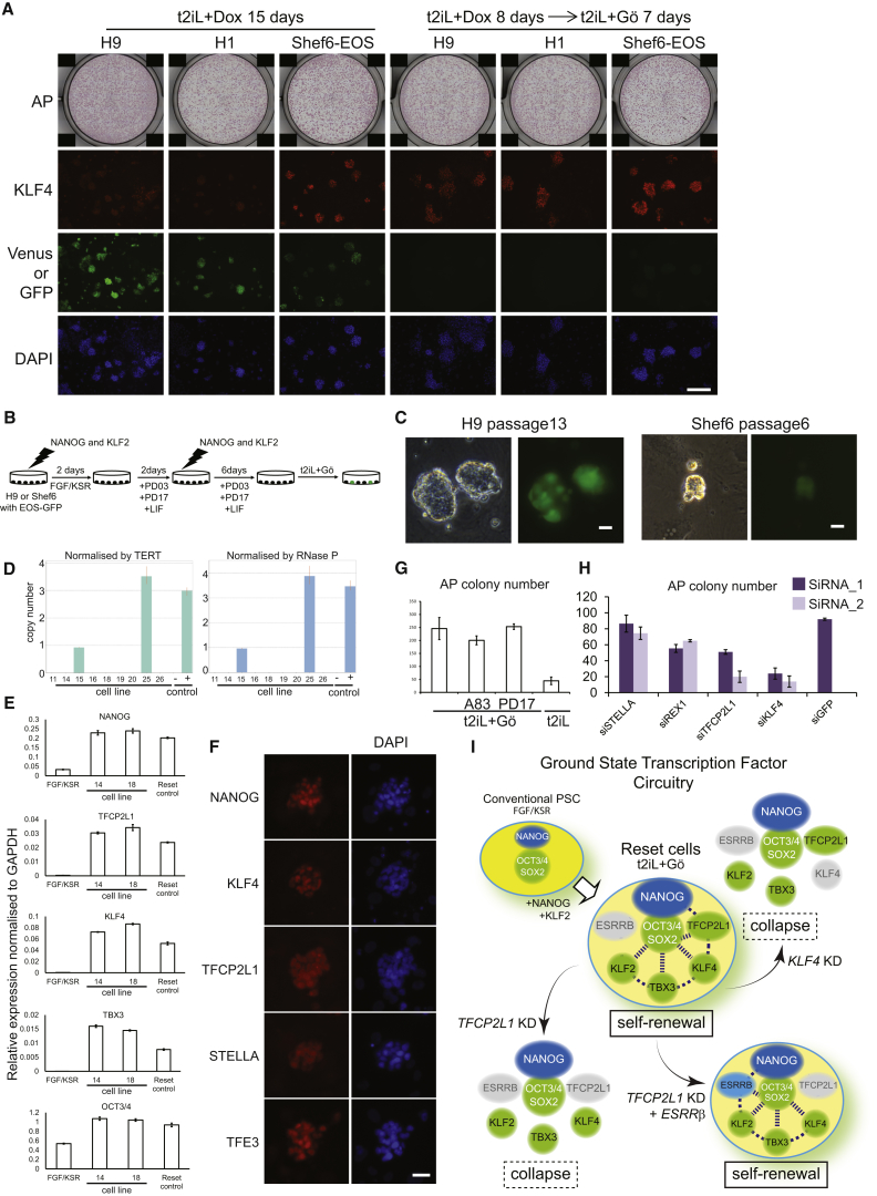
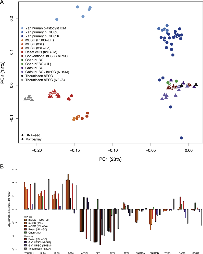
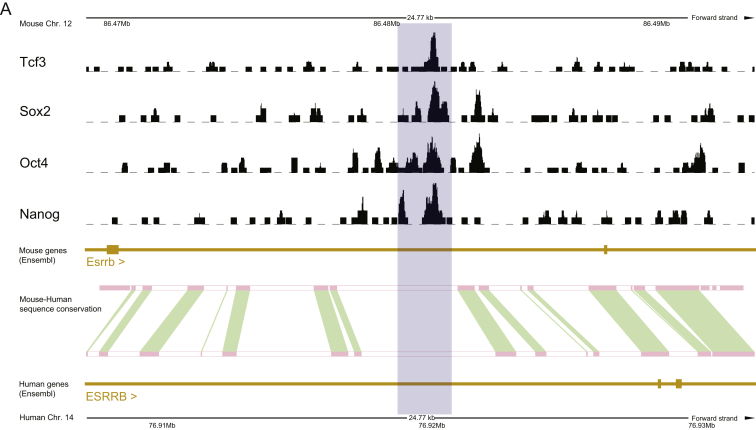
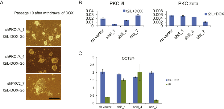
Current human pluripotent stem cells lack the transcription factor circuitry that governs the ground state of mouse embryonic stem cells (ESC). Here, we report that short-term expression of two components, NANOG and KLF2, is sufficient to ignite other elements of the network and reset the human pluripotent state. Inhibition of ERK and protein kinase C sustains a transgene-independent rewired state. Reset cells self-renew continuously without ERK signaling, are phenotypically stable, and are karyotypically intact. They differentiate in vitro and form teratomas in vivo. Metabolism is reprogrammed with activation of mitochondrial respiration as in ESC. DNA methylation is dramatically reduced and transcriptome state is globally realigned across multiple cell lines. Depletion of ground-state transcription factors, TFCP2L1 or KLF4, has marginal impact on conventional human pluripotent stem cells but collapses the reset state. These findings demonstrate feasibility of installing and propagating functional control circuitry for ground-state pluripotency in human cells.


Figures

None
Graphical abstract
Figure 1
Figure 1
Resetting Human PSC Data in this and other figures are from H9 cells unless otherwise indicated, but similar results were obtained from H1 and Shef6 cells and various iPS cell lines (Table S1 and Figure S1). (A) Induction or silencing of transgenes combined with switching between 2iL and FGF/KSR supports expansion of colonies with distinct morphology. Transgene expression indicated by the Venus reporter. (B) PKC inhibitor Gö6983 maintains colony morphology in the absence of transgene expression. Intrinsic fluorescence of Gö produces a faint red background signal. (C) Expansion in different CH concentrations. Cells were plated at 5 × 104 cells per well and were cultured for 4 days in PD03 with LIF and 0, 1, or 3 μM CH. (D) Cells previously cultured in t2iL with DOX were plated in 6-wells without ROCKi in conditions indicated and were stained after 10 days. (E) Cells maintained in t2iL+Gö were seeded in 12-well dishes without ROCKi in conditions indicated. (F) Cell proliferation data. (G) G-banded karyotype of reset cells at passage 16 (converted at parental passage 40). (H) qRT-PCR for pluripotency factor transcripts. (I) Immunostaining for ground-state pluripotency markers. The dot in the TFCP2L1 image of reset cells is due to intrinsic fluorescence of Gö. (J) Immunoblotting for ground-state pluripotency proteins in conventional and reset cells. (K) Reset cells on Matrigel or laminin 511-E8. (L) qRT-PCR for ground-state transcription factor transcripts in reset cells after five passages on Matrigel (Ma) or Laminin511-E8 (La). Cultures on MEF and conventional PSC on Matrigel in mTeSR are controls. Scale bars: (A and B) 100 μM, (I and K) 20 μM. Error bars indicate SD.
Figure S1
Figure S1
Resetting Human PSC, Related to Figure 1 (A) OCT4 expression after withdrawal of DOX. OCT4 mRNA expression was measured by qRT-PCR. (B) Immunoblotting for ERK1/2 and pERK1/2. Protein was extracted from H9 cells cultured in FGF/KSR or reset in t2iL+Gö from a single well of a 6-well plate (1 × 106 cells). One fifth of the sample was fractionated by SDS electrophoresis, electroblotted and probed with indicated antibodies. (C) Image of colony forming assay quantified in Figure 1E. Reset H9 cells maintained in t2iL+Gö were seeded without ROCKi at 2000 cells/well in 12-well plates in t2iL+Gö. FGF receptor inhibitor PD173074 (0.5 μM) or TGF-β/activin receptor inhibitor A83-01 (0.25 μM) were added as indicated. Colonies were stained for alkaline phosphatase after 7 days. (D) Transgene-specific qRT-PCR assay. H9 cells harboring DOX-inducible NANOG and KLF2 transgenes were assayed in the indicated culture conditions. (E) Expression of ground-state transcription factor transcripts. qRT-PCR assay on reset H1 and Shef6 cells. (F) Immunostaining for ground-state pluripotency markers. Conventional and reset H1 and Shef6 cells were stained with antibodies against the indicated markers. Note nuclear localization of TFE3 in reset cells. Error bars indicate SD.
Figure 2
Figure 2
Differentiation (A) Expression of lineage markers in embryoid bodies formed from reset cells in KSR or serum. (B) Teratomas formed from reset cells in three out of ten mice. (C) Reset cells convert to conventional PSC morphology after transfer to FGF/KSR. Bottom panel shows typical colony four passages after transfer. (D) Downregulation of naive markers in FGF/KSR. (E) Colony formation after transfer of reset cells into FGF/KSR for two passages. Cells plated in the presence of ROCKi. (F) Definitive endoderm differentiation in activin and Wnt. Flow cytometry and immunostaining. (G) Neuronal differentiation after dual inhibition of activin and BMP pathways. (H) qRT-PCR assay of cardiac lineage markers in embryoid body outgrowths. Scale bars: (C, F, and G) 100 μM.
Figure 3
Figure 3
Mitochondrial Activity (A) Oxygen consumption rate (OCR) measurements. (B) Mitochondrial staining. MitoTracker is a general stain; TMRE staining is dependent on mitochondrial membrane activity. Scale bar, 10 μM; inset, 15 μM. (C) Colony formation in 2-deoxyglucose. 3 × 104 cells were seeded in 12-well plates and were cultured for 7 days with indicated concentrations of 2-deoxyglucose (2DG). Error bars indicate SD. See also Figure S2.
Figure S2
Figure S2
Mitochondrial Activity, Related to Figure 3 (A) COX gene expression determined from RNA-seq analysis. Data extracted from sample analysis in Figure 5 (B) Proliferation in low glucose. After single-cell dissociation, 3x10⁴ cells were seeded on 12-well plates and cultured for 7 days in the indicated concentrations of glucose. ROCKi was added for seeding conventional PSC. Conventional PSC failed to generate colonies. Reset cells are tolerant against low glucose produce multiple colonies. Scale bars: 200 μM. Error bars indicate SD.
Figure 4
Figure 4
Epigenome Analysis (A) Immunostaining for 5mC, 5hmC, and NANOG. Conventional PSC exhibit pronounced 5mC staining (white arrow). Reset cells display reduced 5mC signal (white arrow) in contrast to feeder cells (unfilled arrow). (B) Quantification by mass spectrometry of global 5mC and 5hmC levels. (C) Quantitative summaries of whole-genome BS-seq data from three biological replicates. (D) Heatmaps of methylation levels in up to 10,000 random samplings of previously classified genomic regions: CpG island (CGI) or non-CGI promoters; intragenic and intergenic CGI; exons; introns; LINEs and SINEs. (E) Scatter plots of CGI methylation percentages on the X chromosome and autosomes. (F) Immunostaining for H3K27me3, counterstained with DAPI. Representative fields of reset cells and after passaging in FGF/KSR. (G) Immunostaining for H3K9me3. Intensity and distribution analysis by Image J. Scale bars: (F and G) 20 μM. Error bars indicate SD. See also Figure S3.
Figure S3
Figure S3
Epigenome Analysis, Related to Figure 4 (A) Sequencing coverage of whole-genome bisulfite sequencing libraries. (B) BS-seq data for methylation at the SOX2 locus in conventional versus reset H9 cultures. (C) Immunofluorescence staining for H3K9me3 in mouse cells. Images of mouse post-implantation epiblast stem cells (EpiSC) in FGF/activin and mouse ESC in t2iL. H3K9me3 appears in green and DAPI staining in blue. (D) Intensity and distribution of H3K9me3. Six cells were selected at random and intensity and distribution of staining were analyzed by Image J. i). Conventional human PSC in FGF/KSR. ii). Reset cells in t2iL+Gö. iii). Mouse EpiSC in bFGF/Activin. iv). Mouse ESC in t2iL. Scale bars, 20 μM.
Figure 5
Figure 5
Comparative Expression Analysis (A) PCA of RNA-seq and microarray data from this study with RNA-seq data from Chan et al. (2013), microarray data from Gafni et al. (2013), and single-cell RNA-seq data from Yan et al. (2013). Samples generated in this study were additionally hybridized to the identical array platform used by Gafni et al. (2013) to facilitate direct comparison. Data were normalized to conventional PSC in each study. Similar clustering is apparent using RNA-seq data alone (Figure S4B), discounting the influence of platform differences. (B) RNA-seq meta-analysis reveals two major groups, with reset cells featuring expression patterns most similar to ESC. Values displayed correspond to the expression level in each sample scaled by the mean expression of each gene across samples. (C) Reset cells display transcription factor hallmarks of ground-state ESC. Data normalized to expression from conventional human PSC as above. (D) Reset cells feature downregulation of lineage markers. (E) Immunostaining of KLF4 and TFCP2L1 in the human ICM. (F) Coexpression of KLF4, TFCP2L1, and NANOG in reset cells. Scale bar: (E and F) 50 μM. See also Figures S4 and S5 and Tables S2, S3 and S4.
Figure S4
Figure S4
Comparative Expression Analysis, Related to Figure 5 (A) Genes contributing to principal components distinguishing reset cells from conventional human PSC. Gene symbols were extracted from the PCA and the labels scaled relative to the magnitude of variance. Pluripotency regulators are present in the leftmost area defining reset cells, whereas numerous lineage-specific genes can be found to the right expressed in conventional human PSC cultures. (B) Platform-specific principal component analysis of RNA-seq data. Data are compared from reset cells, mouse ESC cultured in three different conditions, conventional human PSC and 3iL samples from Chan et al. Clustering of cell types when applied to a single technology recapitulates the integrated analysis in Figure 5A.
Figure S5
Figure S5
Marker Genes Distinguish Reset Cells from Conventional Human PSC and Alternative Protocols, Related to Figure 5 (A) Platform-specific principal component analysis of microarray data from this study and those reported in Gafni et al. Samples were hybridized to the same array platform to allow for direct comparison. Reset cells (light red) occupy a tight cluster to the right and conventional PSC (dark red) toward the bottom. Cells described as naive in Gafni et al. (violet) exhibit wide variation and appear unrelated to ground-state cells. (B) Heatmap comparing the expression of 48 pluripotency and lineage marker genes selected by the International Stem Cell Consortium (Adewumi et al., 2007) between reset cells, conventional PSC cultures and those reported in Gafni et al., based on Affymetrix Human Gene 1.0 ST data. Reset cells form a distinct, relatively uniform population with robust expression of pluripotency genes and repression of lineage markers. In contrast, reportedly naive cells from Gafni et al. display many of the same traits as conventional PSC with mixed expression of lineage markers and significant reduction of key pluripotency regulators. Only genes for which a difference in expression was observed are displayed (i.e., scaled expression > 1 or < −1 in at least one sample). (C) Panel of chromatin modification genes associated with DNA methylation and demethylation, histone methylation and acetylation. Expression trends in ground-state ESC are recapitulated in reset cells, whereas weaker or divergent transcription is evident in PSC cultured in alternative conditions. Expression levels are scaled relative to conventional PSC samples from each study. Data from different platforms are separated by spaces between bars.
Figure 6
Figure 6
Functional Interrogation of the Reset State (A) Colony formation after siRNA knockdown in 4,000 cells in FGF/KSR or 2,000 cells in t2iL+Gö. Colony size is variable for conventional PSC, but numbers are relatively consistent. Histogram shows mean colony counts from duplicate assays. (B) Colony formation after shTFCP2L1 knockdown (KD) in indicated conditions. (C) Quantification of colony formation by shTFCP2L1 or shKLF4 knockdown cells. (D) Rescue of KLF4 knockdown with KLF4 transgene. (E) Colony formation by shTFCP2L1 knockdown cells transfected with ESRRB. (F) Morula aggregation. Six of 42 embryos aggregated with reset cells contained Cherry-positive cells, as shown. Scale bar, 20 μM. (G) Blastocyst injection. After 72 hr, 9 of 32 embryos injected with reset cells showed GFP-positive cells in the ICM/epiblast, as shown. Scale bar, 100 μM. Error bars indicate SD. See also Figure S6.
Figure S6
Figure S6
Functional Interrogation of Transcription Factor Circuitry, Related to Figure 6 (A) shRNA knockdown of TFCP2L1 and KLF4. Knockdown cells and cells transfected with empty vector were maintained by expression of NANOG and KLF2 transgenes in t2iL+DOX. Knockdown was evaluated by qRT-PCR. (B) Colony formation after shTFCP2L1 knockdown (KD) in FGF/KSR. Parental H9 cells were stably transfected with empty vector or shTFCP2L1 construct and selected in puromycin. 4000 cells were plated in FGF/KSR with ROCKi in 12-well plates. (C) Rescue of TFCP2L1 knockdown with mouse Tfcp2l1. Colony formation by shTFCP2L1 knockdown cells transfected with mouse Tfcp2l1 expression vector. Knockdown cells maintained by DOX induction of NANOG and KLF2 were transfected with a piggyBac mTfcp2l1 expression vector and transfectant pools established by selection in hygromycin. Cells were then plated at 2000 cells/well in 24-well plates in t2iL+Gö without DOX and stained for alkaline phosphatase after 7 days. Error bars indicate SD.
Figure 7
Figure 7
Resetting by Transient Transgenesis (A) Time span for resetting with inducible NANOG and KLF2. At day 8, cultures were replated in triplicate with or without DOX. Colonies were analyzed at day 15. Few colonies are obtained with DOX exposure <6 days. Reporter expression in Shef6-EOS cells was assessed by GFP after DOX withdrawal. Exposure time was constant for all images, yielding lower EOS-GFP signal relative to hCMV-Venus. Scale bar, 100 μM. (B) Scheme for generation of reset cells by transient transfection. (C) Phase contrast and fluorescence images of reset cells generated by transient transfection of H9 and Shef6 PSC. (D) Detection of transgene-free cultures by TaqMan copy number assay. Cells with (+) or without (−) a blasticidin transgene provide controls. (E) qRT-PCR assay for transcription factor expression in expanded transgene-free reset cells. (F) Immunofluorescence staining of expanded transgene-free reset cells. (G) Colony-forming assays on transgene-free reset cells seeded in the indicated conditions without ROCKi. (H) Colony formation after siRNA knockdown in transgene-free reset cells. Histogram shows mean colony counts from duplicate assays. (I) ESC express general pluripotency factors Oct4 and Sox2 plus an interconnected transcription factor circuitry that sustains self-renewal. Resetting induces expression of these factors in human PSC apart from ESRRB. Self-renewal is less robust in human, and knockdown of single components, TFCP2L1, or KLF4 causes collapse. Error bars indicate SD.
Figure S7
Figure S7
Transcriptome Meta-Analysis, Related to Discussion (A) Principal component analysis of human and mouse PSC. Expression data were analyzed from conventional and 6i/L/A cultured human PSC (Theunissen et al.), H9 conventional and reset cells (this study), ground-state ESC cultured in three different conditions (this study), conventional and NHSM cultured human PSC (Gafni et al.), conventional and 3iL cultured human PSC (Chan et al.) and single cells from human blastocyst ICMs and primary explant cultures (Yan et al.). 6i/L/A samples share global similarity with reset cells but appear more divergent from mouse ESC. (B) Comparison of individual human and mouse genes expressed in alternate culture conditions. Data are normalized to expression levels from conventional human PSC profiled on each platform. Notably, cells cultured in 6i/L/A largely recapitulate expression trends in reset cells and ground-state ESC, but reduced expression of TET1 and SOX17 and upregulation of DNA methyltransferase DNMT3A and germ cell marker TDRD1 indicate noteworthy differences, potentially reflecting developmental distinctions in pluripotent stage. Expression levels are scaled relative to conventional PSC samples from each study. Data from different platforms are separated by spaces between bars.
Figure S8
Figure S8
Incomplete Conservation between Mouse and Human Esrrb Loci, Related to Discussion Esrrb locus of the mouse genome showing Nanog, Oct4, Sox2, and Tcf3 binding sites determined by ChIP-seq with sequences homologous to the equivalent human locus indicated. Signal tracks for ChIP-seq data from Marson et al. (Marson et al., 2008), were obtained from the the ES cell ChIP-seq compendium (Martello et al., 2012) (A) Gray rectangle delineates the binding site of Tcf3, Sox2, Oct4 and Nanog in mouse where there is no conserved sequence between mouse and human. Conserved sequence is shown in red and green.
Figure S9
Figure S9
Knockdown of aPKC iota/lambda and zeta, Related to Discussion Two different shRNA vectors, shPKC iota_1 and shPKC iota_4, were used for knockdown of PKC iota/lambda. shPKCzeta_8 was used for knockdown of PKC zeta. Scale bars: 100 μM. (A) Brightfield images of shPKC iota KD and shPKC zeta KD cells cultured in t2iL. shPKC iota KD cell retain undifferentiated morphology whereas shPKC zeta cells progressively differentiate. (B) Knockdown efficiency of each shRNA. (C) OCT4 expression at passage 10 (KD lines) or 3 (control). Error bars indicate SD.

References

    1. Adewumi O., Aflatoonian B., Ahrlund-Richter L., Amit M., Andrews P.W., Beighton G., Bello P.A., Benvenisty N., Berry L.S., Bevan S., International Stem Cell Initiative Characterization of human embryonic stem cell lines by the International Stem Cell Initiative. Nat. Biotechnol. 2007;25:803–816. - PubMed
    1. Amit M., Carpenter M.K., Inokuma M.S., Chiu C.P., Harris C.P., Waknitz M.A., Itskovitz-Eldor J., Thomson J.A. Clonally derived human embryonic stem cell lines maintain pluripotency and proliferative potential for prolonged periods of culture. Dev. Biol. 2000;227:271–278. - PubMed
    1. Anders S., Huber W. Differential expression analysis for sequence count data. Genome Biol. 2010;11:R106. - PMC - PubMed
    1. Anders S., Pyl P.T., Huber W. HTSeq — A Python framework to work with high-throughput sequencing data. bioRxiv. 2014 doi: 10.1101/002824. Published online February 20, 2014. - DOI - PMC - PubMed
    1. Bedzhov I., Zernicka-Goetz M. Self-organizing properties of mouse pluripotent cells initiate morphogenesis upon implantation. Cell. 2014;156:1032–1044. - PMC - PubMed

Supplemental References

    1. Aflatoonian B., Ruban L., Shamsuddin S., Baker D., Andrews P., Moore H. Generation of Sheffield (Shef) human embryonic stem cell lines using a microdrop culture system. In Vitro Cell. Dev. Biol. Anim. 2010;46:236–241. - PubMed
    1. Krueger F., Andrews S.R. Bismark: a flexible aligner and methylation caller for Bisulfite-Seq applications. Bioinformatics. 2011;27:1571–1572. - PMC - PubMed
    1. Marson A., Levine S.S., Cole M.F., Frampton G.M., Brambrink T., Johnstone S., Guenther M.G., Johnston W.K., Wernig M., Newman J. Connecting microRNA genes to the core transcriptional regulatory circuitry of embryonic stem cells. Cell. 2008;134:521–533. - PMC - PubMed
    1. Moretti A., Bellin M., Jung C.B., Thies T.M., Takashima Y., Bernshausen A., Schiemann M., Fischer S., Moosmang S., Smith A.G. Mouse and human induced pluripotent stem cells as a source for multipotent Isl1+ cardiovascular progenitors. FASEB J. 2010;24:700–711. - PubMed
    1. Thomson J.A., Itskovitz-Eldor J., Shapiro S.S., Waknitz M.A., Swiergiel J.J., Marshall V.S., Jones J.M. Embryonic stem cell lines derived from human blastocysts. Science. 1998;282:1145–1147. - PubMed

Publication types

Associated data