. 2014 Sep 12;9(9):e107758.
doi: 10.1371/journal.pone.0107758. eCollection 2014.

Lactobacilli inactivate Chlamydia trachomatis through lactic acid but not H2O2

Affiliations

Lactobacilli inactivate Chlamydia trachomatis through lactic acid but not H2O2

Zheng Gong et al. PLoS One. .

Abstract

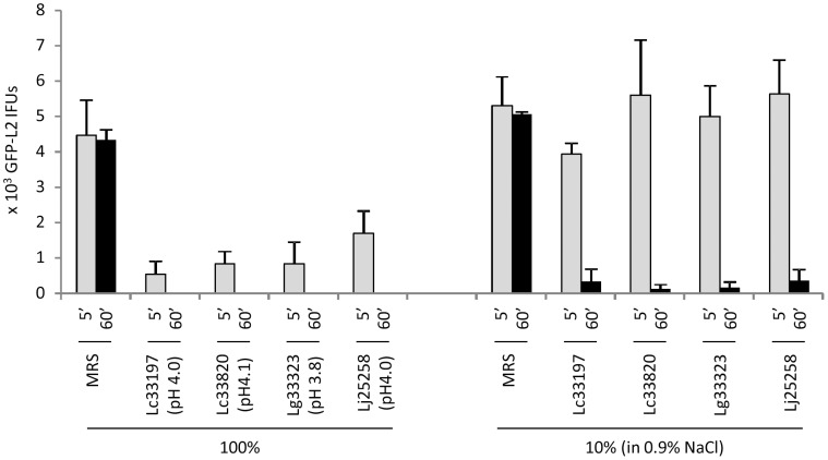
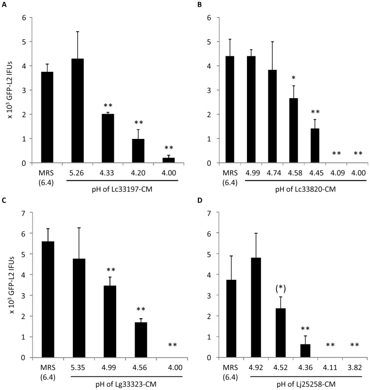
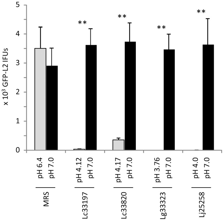
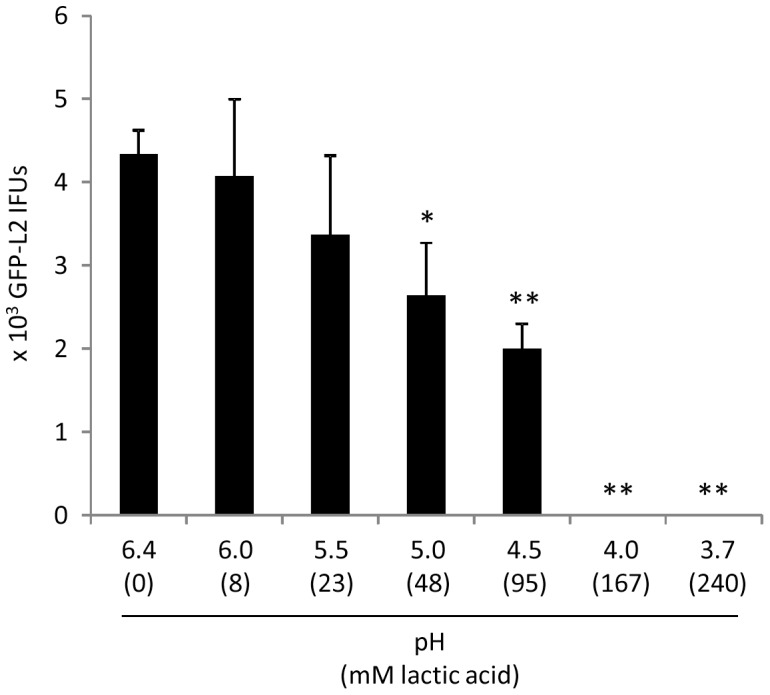
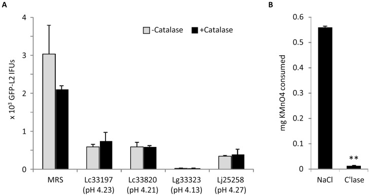
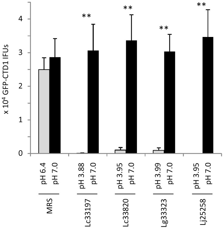
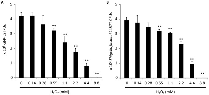
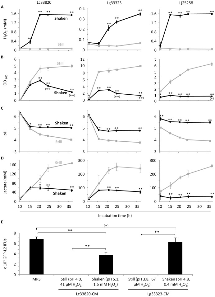
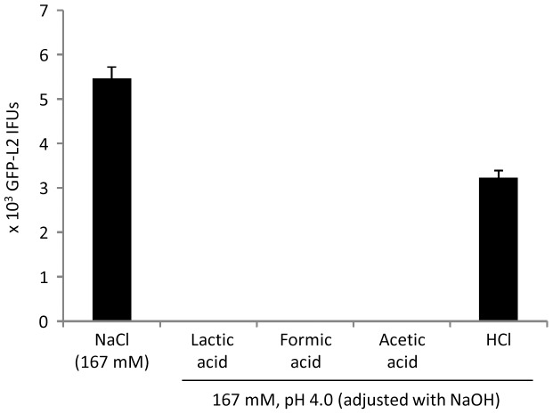
Lactobacillus species dominate the microbiome in the lower genital tract of most reproductive-age women. Producing lactic acid and H2O2, lactobacilli are believed to play an important role in prevention of colonization by and growth of pathogens. However, to date, there have been no reported studies characterizing how lactobacilli interact with Chlamydia trachomatis, a leading sexually transmitted bacterium. In this report, we demonstrate inactivation of C. trachomatis infectivity by culture media conditioned by Lactobacillus crispatus, L. gasseri and L. jensenii, known to be dominating organisms in the human vaginal microbiome. Lactobacillus still cultures produced lactic acid, leading to time- and concentration-dependent killing of C. trachomatis. Neutralization of the acidic media completely reversed chlamydia killing. Addition of lactic acid into Lactobacillus-unconditioned growth medium recapitulated the chlamydiacidal activity of conditioned media. The H2O2 concentrations in the still cultures were found to be comparable to those reported for the cervicovaginal fluid, but insufficient to inactivate chlamydiae. Aeration of Lactobacillus cultures by shaking markedly induced H2O2 production, but strongly inhibited Lactobacillus growth and lactic acid production, and thus severely affected acidification, leading to significantly reduced chlamydiacidal efficiency. These observations indicate lactobacilli inactivate chlamydiae primarily through maintaining acidity in a relatively hypoxic environment in the vaginal lumen with limited H2O2, which is consistent with the notion that women with higher vaginal pH are more prone to sexually transmitted C. trachomatis infection. In addition to lactic acid, formic acid and acetic acid also exhibited potent chlamydiacidal activities. Taken together, our findings imply that lowering the vaginal pH through engineering of the vaginal microbiome and other means will make women less susceptible to C. trachomatis infection.


Conflict of interest statement

Competing Interests: The authors have declared that no competing interests exist.

Figures

Figure 1
Figure 1. Time- and concentration-dependent inactivation of elementary bodies (EBs) of GFP-L2, derived from C. trachomatis serovar L2, by Lactobacillus-conditioned medium (LCM).
LCM was collected from overnight still cultures of L. crispatus 33197 (Lc33197), L. crispatus 33820 (Lc33820), L. gasseri 33323 (Lg33323) and L. jensenii 25258 (Lj25258). pH values of undiluted (100%) LCM are shown. EBs were treated with undiluted LCM or LCM diluted 10 fold with 0.9% NaCl for 5 min or 1 h. Following treatments, surviving EBs were serially diluted and then inoculated onto McCoy monolayers; resulting inclusion-forming units were scored by fluorescence microscopy. Values were averages ± standard deviations of triplicate experiments.
Figure 2
Figure 2. Effects of LCM longitudinally collected from still cultures of Lc33197 (A), Lc33820 (B), Lg33323 (C) and Lj25258 (D).
pH values of LCM are shown on the horizontal axis. GFP-L2 EBs were treated for 1 h. Surviving bacteria were quantified as outlined in Fig. 1 legend. Values were averages ± standard deviations of triplicate experiments. Single and double asterisks above LCM-treated samples denote statistically decreased IFUs (P<0.05 and P<0.01, respectively) as compared to control MRS Lactobacillus medium-treated samples. The parenthetic asterisk indicates statistically decreased IFUs in samples treated with LCM (pH 4.52), as compared to IFUs that survived the treatment with LCM (pH 4.92), although the P value between control MRS (pH 6.4) and MRS (pH 4.52) was 0.068.
Figure 3
Figure 3. Complete reversal of chlamydiacidal activity with pH-neutralized LCM.
LCM from overnight still cultures of Lactobacillus strains or control MRS broth was adjusted to pH 7.0. GFP-L2 EBs were treated with pH-unadjusted LCM (pH value shown for each on the horizontal X-axis) or neutralized LCM for 1 h. Surviving bacteria were quantified as outlined in Fig. 1 legend. Values were averages ± standard deviations of triplicate experiments. Double asterisks above LCM-treated samples denote highly statistically increased IFUs (P<0.01) as a result in treatment with neutralized LCM as compared to pH-unadjusted LCM.
Figure 4
Figure 4. Killing of GFP-L2 EBs by lactate-acidified MRS.
Experiments were performed similarly to those in Fig. 2 and 3; acidified MRS with indicated pH values were used in place of LCM. Values were averages ± standard deviations of triplicate experiments. Single and double asterisks above LCM-treated samples denote statistically decreased IFUs (P<0.05 and P<0.01, respectively) as compared to control pH-unadjusted MRS (pH 6.4). Note resemblance of inhibition trends exhibited by lactate-acidified MRS (this figure) and longitudinally harvested LCM with different pH (Fig. 2).
Figure 5
Figure 5. Lack of an effect of catalase on LCM-mediated chlamydiacidal activity.
(A) LCM collected from still cultures of Lactobacillus strains were either untreated or treated with catalase before they were used to treat GFP-L2 EBs for 1 h. Surviving bacteria were quantified as outlined in Fig. 1 legend. Values were averages ± standard deviations of triplicate experiments. Note no statistical differences existed between catalase-treated and untreated samples for LCM from any of Lactobacillus strains. (B) 0.3% H2O2 prepared in 0.17 M lactate-NaOH (pH 4.0) was treated with 0.2 mg/ml catalase or control 0.9% NaCl. Remaining H2O2 was detected with KMnO4 titration. Values were averages ± standard deviations of triplicate experiments. Double asterisks signify statistically significant difference (P<0.01) in the amounts of H2O2 between catalase-treated and non-treated samples, and indicate that the catalase degraded H2O2 in the acidic LCM used in (A).
Figure 6
Figure 6. Acid-dependent inactivation of EBs of C. trachomatis serovar D-derived GFP-CTD1 by LCM.
Experiments were carried in the same manner as those in Fig. 3 except centrifugation was used to facilitate infection. Values were averages ± standard deviations of triplicate experiments. Double asterisks indicate statistically significant difference (P<0.01, respectively) in the numbers of surviving EBs between treatment with LCM and that with neutralized LCM (pH 7.0).
Figure 7
Figure 7. Dose-dependent killing of Chlamydia (A) and Shigella flexneri (B) by exogenous H2O2.
GFP-L2 or S. flexneri 2457T was diluted in MRS containing indicated concentrations of H2O2. Following 1 h incubation at room temperature, remaining H2O2 was removed by catalase. Surviving EBs were quantitated as in Fig. 1; surviving 2457T bacteria were quantified by scoring colony-forming units (CFU) on LB Agar plates inoculated with serially diluted bacterial suspension. Values were averages ± standard deviations of triplicate experiments. Single and double asterisks signify statistically decreased live bacteria in MRS with an indicated concentration of H2O2, as compared to control non-supplemented MRS (P<0.05 and P<0.01, respectively).
Figure 8
Figure 8. Induction of H2O2 production coupled with decreased lactic acid production and loss of chlamydiacidal activity.
Overnight Lactobacillus cultures were diluted with fresh MRS to 0.02 OD600. The diluted bacterial suspensions were incubated as still cultures or shaken cultures as described in “Materials and Methods”. At indicated times, samples were taken for determination of the concentrations of H2O2 (A) and bacteria (B), pH values (C), lactic acid concentrations (D) and anti-chlamydial activities (E). Values were averages ± standard deviations of triplicate experiments. (A-D) Single and double asterisks indicate statistically difference (P<0.05 and P<0.01, respectively) between still cultures and shaken cultures at indicated times. Single and double asterisks in parentheses in B denote statistically significant decline in bacterial concentration following its peak at 20 h (Lc33820 and Lg33323) or 15 h (Lj25258). Double asterisks in E signify statistically significant differences (P<0.01) between indicated two groups. An asterisk in parenthesis indicates that the difference between control MRS and LCM from Lg33323 shaken cultures was deemed statistically insignificant by a two-tailed t test (P<0.1), but significant by a one-tailed t test (P<0.05).
Figure 9
Figure 9. Full inactivation of GFP-L2 by formic acid and acetic acid but not pH-adjusted HCl.
Experiments were carried in the same manner as those in Fig. 3 except acid solutions used to treat EBs were prepared in H2O.

References

    1. CDC (2013) Notifiable Diseases and Mortality Tables. Morbidity and Mortality Weekly Report (MMWR) 62: 424–437. - PubMed
    1. CDC (2013) CDC fact sheet. STD trends in the United States: 2011 national data for chlamydia, gonorrhea, and syphilis
    1. Stephens RS, Myers G, Eppinger M, Bavoil PM (2009) Divergence without difference: phylogenetics and taxonomy of Chlamydia resolved. FEMS Immunol Med Microbiol 55: 115–119. - PubMed
    1. Abraham S, Toutous-Trellu L, Pechere M, Hugonnet S, Liassine N, et al. (2006) Increased incidence of sexually transmitted infections in Geneva, Switzerland. Dermatology 212: 41–46. - PubMed
    1. Vajdic CM, Middleton M, Bowden FJ, Fairley CK, Kaldor JM (2005) The prevalence of genital Chlamydia trachomatis in Australia 1997–2004: a systematic review. Sexual Health 2: 169–183. - PubMed

Publication types

MeSH terms