The role of oxidoreductases in determining the function of the neisserial lipid A phosphoethanolamine transferase required for resistance to polymyxin
- PMID: 25215579
- PMCID: PMC4162559
- DOI: 10.1371/journal.pone.0106513
The role of oxidoreductases in determining the function of the neisserial lipid A phosphoethanolamine transferase required for resistance to polymyxin
Abstract
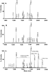
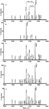
The decoration of the lipid A headgroups of the lipooligosaccharide (LOS) by the LOS phosphoethanolamine (PEA) transferase (LptA) in Neisseria spp. is central for resistance to polymyxin. The structure of the globular domain of LptA shows that the protein has five disulphide bonds, indicating that it is a potential substrate of the protein oxidation pathway in the bacterial periplasm. When neisserial LptA was expressed in Escherichia coli in the presence of the oxidoreductase, EcDsbA, polymyxin resistance increased 30-fold. LptA decorated one position of the E. coli lipid A headgroups with PEA. In the absence of the EcDsbA, LptA was degraded in E. coli. Neisseria spp. express three oxidoreductases, DsbA1, DsbA2 and DsbA3, each of which appear to donate disulphide bonds to different targets. Inactivation of each oxidoreductase in N. meningitidis enhanced sensitivity to polymyxin with combinatorial mutants displaying an additive increase in sensitivity to polymyxin, indicating that the oxidoreductases were required for multiple pathways leading to polymyxin resistance. Correlates were sought between polymyxin sensitivity, LptA stability or activity and the presence of each of the neisserial oxidoreductases. Only meningococcal mutants lacking DsbA3 had a measurable decrease in the amount of PEA decoration on lipid A headgroups implying that LptA stability was supported by the presence of DsbA3 but did not require DsbA1/2 even though these oxidoreductases could oxidise the protein. This is the first indication that DsbA3 acts as an oxidoreductase in vivo and that multiple oxidoreductases may be involved in oxidising the one target in N. meningitidis. In conclusion, LptA is stabilised by disulphide bonds within the protein. This effect was more pronounced when neisserial LptA was expressed in E. coli than in N. meningitidis and may reflect that other factors in the neisserial periplasm have a role in LptA stability.
Conflict of interest statement
Figures


Similar articles
-
The structure of the neisserial lipooligosaccharide phosphoethanolamine transferase A (LptA) required for resistance to polymyxin.J Mol Biol. 2013 Sep 23;425(18):3389-402. doi: 10.1016/j.jmb.2013.06.029. Epub 2013 Jun 28. J Mol Biol. 2013. PMID: 23810904
-
Modification of lipooligosaccharide with phosphoethanolamine by LptA in Neisseria meningitidis enhances meningococcal adhesion to human endothelial and epithelial cells.Infect Immun. 2008 Dec;76(12):5777-89. doi: 10.1128/IAI.00676-08. Epub 2008 Sep 29. Infect Immun. 2008. PMID: 18824535 Free PMC article.
-
Cationic antimicrobial peptide resistance in Neisseria meningitidis.J Bacteriol. 2005 Aug;187(15):5387-96. doi: 10.1128/JB.187.15.5387-5396.2005. J Bacteriol. 2005. PMID: 16030233 Free PMC article.
-
Lipid A Phosphoethanolamine Transferase: Regulation, Structure and Immune Response.J Mol Biol. 2020 Aug 21;432(18):5184-5196. doi: 10.1016/j.jmb.2020.04.022. Epub 2020 Apr 27. J Mol Biol. 2020. PMID: 32353363 Review.
-
A comparison of the endotoxin biosynthesis and protein oxidation pathways in the biogenesis of the outer membrane of Escherichia coli and Neisseria meningitidis.Front Cell Infect Microbiol. 2012 Dec 20;2:162. doi: 10.3389/fcimb.2012.00162. eCollection 2012. Front Cell Infect Microbiol. 2012. PMID: 23267440 Free PMC article. Review.
Cited by
-
Knowledge integration and decision support for accelerated discovery of antibiotic resistance genes.Nat Commun. 2022 Apr 29;13(1):2360. doi: 10.1038/s41467-022-29993-z. Nat Commun. 2022. PMID: 35487919 Free PMC article.
-
Breaking antimicrobial resistance by disrupting extracytoplasmic protein folding.Elife. 2022 Jan 13;11:e57974. doi: 10.7554/eLife.57974. Elife. 2022. PMID: 35025730 Free PMC article.
-
Insights into the Mechanistic Basis of Plasmid-Mediated Colistin Resistance from Crystal Structures of the Catalytic Domain of MCR-1.Sci Rep. 2017 Jan 6;7:39392. doi: 10.1038/srep39392. Sci Rep. 2017. PMID: 28059088 Free PMC article.
-
Molecular Mechanisms of Bacterial Resistance to Antimicrobial Peptides in the Modern Era: An Updated Review.Microorganisms. 2024 Jun 21;12(7):1259. doi: 10.3390/microorganisms12071259. Microorganisms. 2024. PMID: 39065030 Free PMC article. Review.
-
Down-Regulation of Flagellar, Fimbriae, and Pili Proteins in Carbapenem-Resistant Klebsiella pneumoniae (NDM-4) Clinical Isolates: A Novel Linkage to Drug Resistance.Front Microbiol. 2019 Dec 17;10:2865. doi: 10.3389/fmicb.2019.02865. eCollection 2019. Front Microbiol. 2019. PMID: 31921045 Free PMC article.
References
-
- Snyder LA, Davies JK, Ryan CS, Saunders NJ (2005) Comparative overview of the genomic and genetic differences between the pathogenic Neisseria strains and species. Plasmid 54: 191–218. - PubMed
-
- Abeysuriya SD, Speers DJ, Gardiner J, Murray RJ (2010) Penicillin-resistant Neisseria meningitidis bacteraemia, Kimberley region, March 2010. Commun Dis Intell 34: 342–344. - PubMed
-
- du Plessis M, de Gouveia L, Skosana H, Thomas J, Blumberg L, et al. (2010) Invasive Neisseria meningitidis with decreased susceptibility to fluoroquinolones in South Africa, 2009. J Antimicrob Chemother 65: 2258–2260. - PubMed
Publication types
MeSH terms
Substances
LinkOut - more resources
Full Text Sources
Other Literature Sources
Medical
Molecular Biology Databases
Miscellaneous

