Review
. 2015 Jan;34(1):6-18.
doi: 10.1089/dna.2014.2552.

Involvement of the IRE1α-XBP1 pathway and XBP1s-dependent transcriptional reprogramming in metabolic diseases

Affiliations
Review

Involvement of the IRE1α-XBP1 pathway and XBP1s-dependent transcriptional reprogramming in metabolic diseases

Rong Wu et al. DNA Cell Biol. 2015 Jan.

Abstract

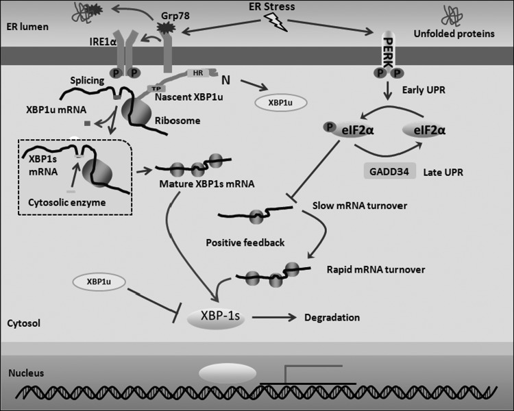
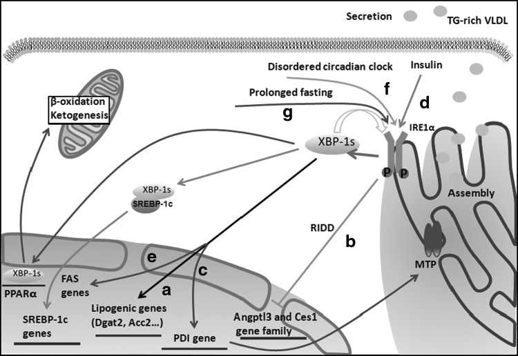
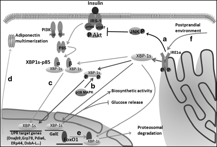
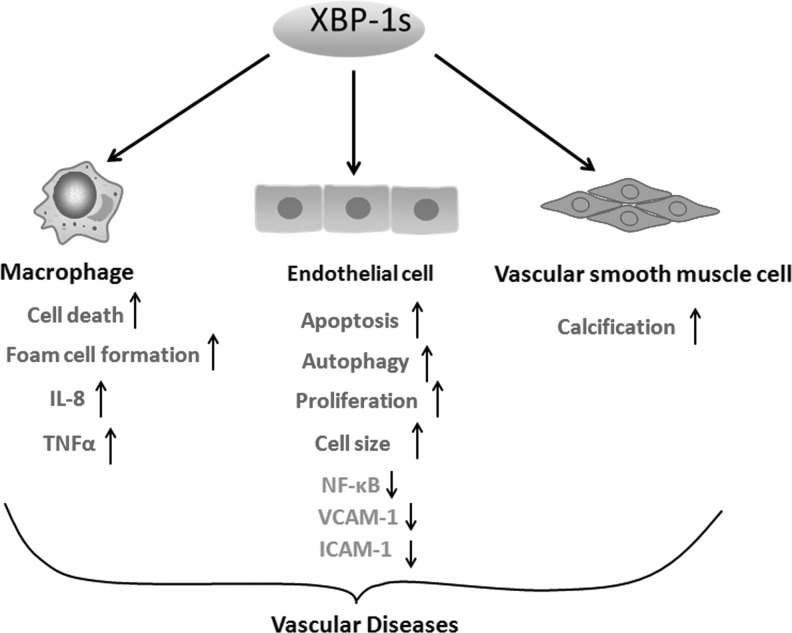
The X-box binding protein 1 (XBP1) is not only an important component of the unfolded protein response (UPR), but also an important nuclear transcription factor. Upon endoplasmic reticulum stress, XBP1 is spliced by inositol-requiring enzyme 1 (IRE1), thereby generating functional spliced XBP1 (XBP1s). XBP1s functions by translocating into the nucleus to initiate transcriptional programs that regulate a subset of UPR- and non-UPR-associated genes involved in the pathophysiological processes of various diseases. Recent reports have implicated XBP1 in metabolic diseases. This review summarizes the effects of XBP1-mediated regulation on lipid metabolism, glucose metabolism, obesity, and atherosclerosis. Additionally, for the first time, we present XBP1s-dependent transcriptional reprogramming in metabolic diseases under different conditions, including pathology and physiology. Understanding the function of XBP1 in metabolic diseases may provide a basic knowledge for the development of novel therapeutic targets for ameliorating these diseases.


Figures

<b>FIG. 1.</b>
FIG. 1.
The schematic diagram of XBP1s production. Under normal circumstances, IRE1α is located on the ER membrane and binds to Grp78. Upon ER stress, IRE1α separates from Grp78 and oligomerizes, resulting in its autophosphorylation. Nascent XBP1u polypeptide recruits its own mRNA to the ER membrane through a hydrophobic region (HR) within XBP1u, facilitating the activated IRE1α to splice 26 nucleotides from the XBP1u mRNA. In addition, the C-terminus of XBP1u results in translational pausing (TP), which increases the efficiency of this process. After splicing, the generated 2′,-3′-cyclic phosphate structure at the cleavage site is enzymatically joined to form the mature XBP1s mRNA. During the early UPR, eIF2α phosphorylation by PERK inhibits XBP1s mRNA translation. Then, both the number of XBP1s mRNAs generated by splicing and translation suppression result in the stabilization and slow turnover of XBP1s mRNA. During the progression of the UPR, eIF2α is dephosphorylated by GADD34, an ER-associated phosphatase regulatory subunit, and translation is then reinitiated, resulting in rapid XBP1s mRNA turnover. Finally, synthesized XBP1s protein stimulates XBP1 transcription and its nuclear translocation to mediate the transcription of additional factors through a positive feedback mechanism. In addition, XBP1u may interact with XBP1s to mediate its proteasomal degradation in the cytosol. eIF2α, eukaryotic initiation factor-2α; ER, endoplasmic reticulum; IRE1, inositol-requiring enzyme 1; PERK, PKR-like ER kinase; UPR, unfolded protein response; XBP1, X-box binding protein 1; XBP1s, spliced XBP1; XBP1u, unspliced XBP1.
<b>FIG. 2.</b>
FIG. 2.
The IRE1α-XBP1 pathway in hepatic lipogenesis. (a) XBP1s directly regulates the expression of a subset of lipogenic genes. (b) XBP1 ablation activates IRE1α because of feedback regulation of IRE1α activity caused by the abundance of its substrate XBP1s. Hyperactivated IRE1α then downregulates the expression of a group of lipid metabolism genes, including Angptl3 and the carboxylesterase1 (Ces1) gene family, by regulated IRE1-dependent decay (RIDD). (c) XBP1s regulates TG-rich VLDL assembly and secretion in hepatocyte-specific IRE1α deletion mice under physiological conditions, which affects MTP activity by regulating PDI at the transcriptional level. (d) XBP1s can interact with the promoter of the SREBP-1c gene following insulin treatment, thereby mediating lipogenesis. (e) XBP1s overexpression stimulates transcription by the FAS promoter. (f) A disordered circadian clock results in the constitutive activation of the IRE1α-XBP1 pathway, thus influencing hepatic lipid metabolism. (g) Hepatic IRE1α-XBP1 signaling is activated by prolonged fasting. Then, XBP1s can directly bind to the endogenous PPARα promoter and upregulate PPARα expression, thereby modulating mitochondrial β-oxidation and ketogenesis. MTP, microsomal triglyceride transfer protein; PPARα, peroxisome proliferator activator receptor α; PDI, protein disulfide isomerase; TG, triglyceride; VLDL, very-low-density lipoprotein.
<b>FIG. 3.</b>
FIG. 3.
The IRE1α-XBP1 pathway in glucose homeostasis. (a) IRE1α-JNK signaling is activated by ER stress, which reduces IRS1 (pY896) tyrosine phosphorylation and Akt phosphorylation whereas enhancing IRS1 (pS307) serine phosphorylation, thereby increasing insulin resistance. (b) p38 MAPK phosphorylates the spliced form of XBP1 on residues Thr48 and Ser61 and greatly enhances its nuclear migration. Then, nuclear XBP1s induces the expression of UPR target genes. (c) Insulin promotes p85α and p85β (subunits of PI3K) association with XBP1s by disrupting their heterodimerization, which subsequently facilitates XBP1s nuclear translocation independent of PI3K catalytic activity. Nuclear XBP1s then induces the expression of UPR target genes. (d) During the UPR, nuclear XBP1s can improve insulin sensitivity by regulating the expression of UPR target genes that are involved in adiponectin multimerization in adipocytes. (e) In the nucleus, XBP1s also directly interacts with FoxO1, a transcription factor involved in gluconeogenesis resulting in its proteasomal degradation and thereby promoting glucose tolerance in the liver. (f) IRE1α-XBP1 signaling is activated by the postprandial environment. XBP1s regulates UDP-galactose-4-epimerase (GalE) expression at the transcriptional level, thereby increasing its biosynthetic activity and reducing hepatic glucose release. FoxO1, Forkhead box O1; JNK, c-Jun N-terminal kinase; p38 MAPK, p38 mitogen-activated protein kinase.
<b>FIG. 4.</b>
FIG. 4.
XBP1s in vascular diseases. XBP1s mediates macrophage cell death, foam cell formation, and IL-8 and TNFα up-expression. XBP1s attenuates NF-κB, VCAM-1, and ICAM-1 expression in retinal vascular endothelial cells. XBP1s induces endothelial cell apoptosis, autophagy and proliferation, and increases cell size. XBP1s results in smooth muscle cell calcification. These different cellular events are involved in vascular diseases, including atherosclerotic initiation and progression and tissue and organ ischemia. ICAM-1, intercellular adhesion molecule-1; IL-8, interleukin-8; TNFα, tumor necrosis factor alpha; VCAM-1, vascular cell adhesion molecule-1.

References

    1. Acosta-Alvear D., Zhou Y., Blais A., Tsikitis M., Lents N.H., Arias C., Lennon C.J., Kluger Y., and Dynlacht B.D. (2007). XBP1 controls diverse cell type- and condition-specific transcriptional regulatory networks. Mol Cell 27,53–66 - PubMed
    1. Akiyama M., Liew C.W., Lu S., Hu J., Martinez R., Hambro B., Kennedy R.T., and Kulkarni R.N. (2013). X-box binding protein 1 is essential for insulin regulation of pancreatic alpha-cell function. Diabetes 62,2439–2449 - PMC - PubMed
    1. Aragon I.V., Barrington R.A., Jackowski S., Mori K., and Brewer J.W. (2012). The specialized unfolded protein response of B lymphocytes: ATF6alpha-independent development of antibody-secreting B cells. Mol Immunol 51,347–355 - PMC - PubMed
    1. Basseri S., Lhotak S., Sharma A.M., and Austin R.C. (2009). The chemical chaperone 4-phenylbutyrate inhibits adipogenesis by modulating the unfolded protein response. J Lipid Res 50,2486–2501 - PMC - PubMed
    1. Benhamron S., Hadar R., Iwawaky T., So J.S., Lee A.H., and Tirosh B. (2014). Regulated IRE1-dependent decay participates in curtailing immunoglobulin secretion from plasma cells. Eur J Immunol 44,867–876 - PubMed

Publication types

MeSH terms