. 2014 Dec;113(3):337-44.
doi: 10.1016/j.radonc.2014.08.026. Epub 2014 Sep 9.

Human papillomavirus type 16 E7 oncoprotein causes a delay in repair of DNA damage

Affiliations

Human papillomavirus type 16 E7 oncoprotein causes a delay in repair of DNA damage

Jung Wook Park et al. Radiother Oncol. 2014 Dec.

Abstract

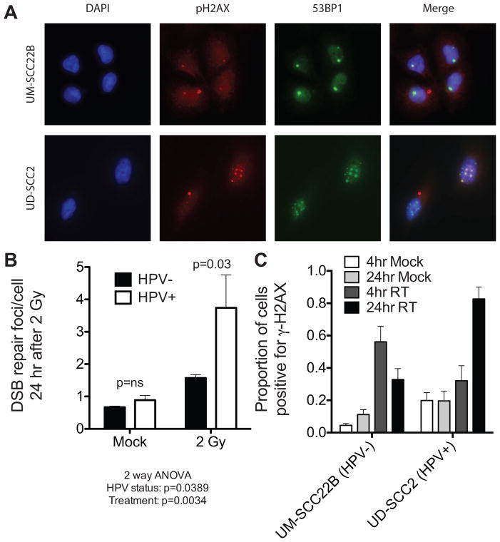
Background and purpose: Patients with human papillomavirus related (HPV+) head and neck cancers (HNCs) demonstrate improved clinical outcomes compared to traditional HPV negative (HPV-) HNC patients. We have recently shown that HPV+ HNC cells are more sensitive to radiation than HPV- HNC cells. However, roles of HPV oncogenes in regulating the response of DNA damage repair remain unknown.

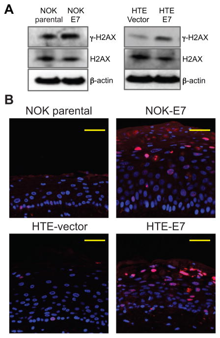
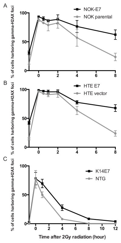
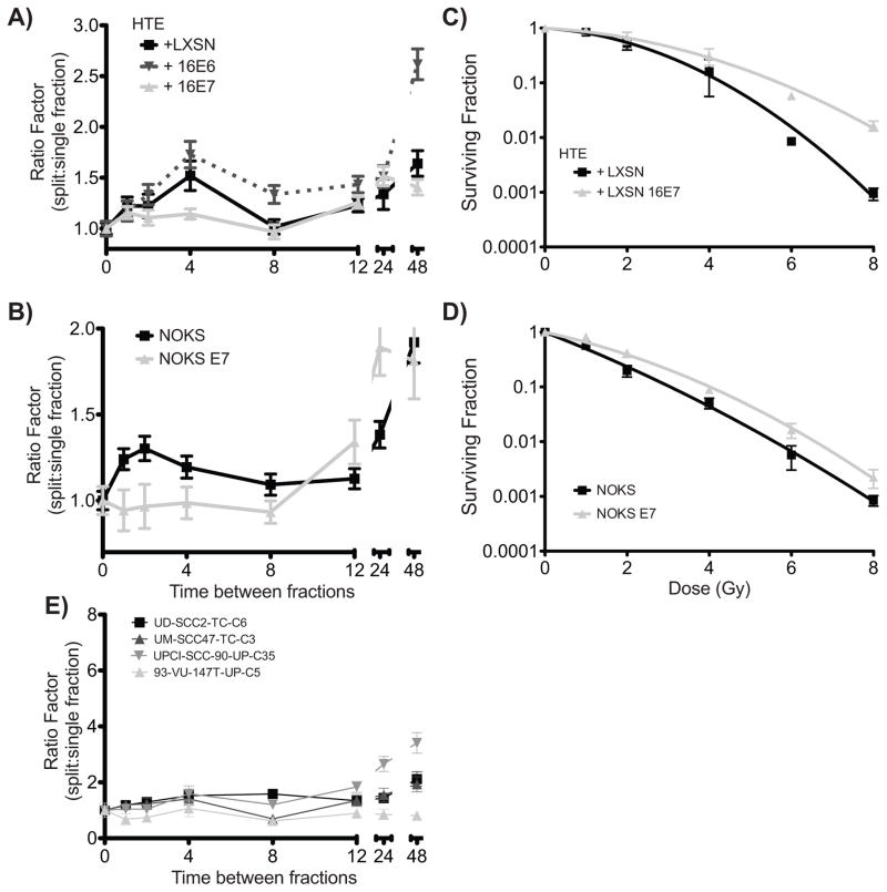
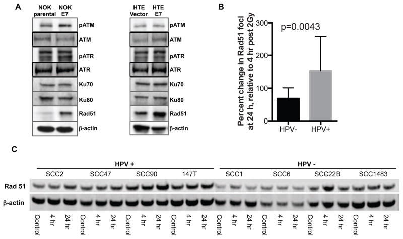
Material and methods: Using immortalized normal oral epithelial cell lines, HPV+ HNC derived cell lines, and HPV16 E7-transgenic mice we assessed the repair of DNA damage using γ-H2AX foci, single and split dose clonogenic survival assays, and immunoblot. The ability of E7 to modulate expression of proteins associated with DNA repair pathways was assessed by immunoblot.

Results: HPV16 E7 increased retention of γ-H2AX nuclear foci and significantly decreased sublethal DNA damage repair. While phospho-ATM, phospho-ATR, Ku70, and Ku80 expressions were not altered by E7, Rad51 was induced by E7. Correspondingly, HPV+ HNC cell lines showed retention of Rad51 after γ-radiation.

Conclusions: Our findings provide further understanding as to how HPV16 E7 manipulates cellular DNA damage responses that may underlie its oncogenic potential and influence the altered sensitivity to radiation seen in HPV+ HNC as compared to HPV- HNC.

Keywords: DNA damage repair; HPV-positive head and neck cancer; HPV16 E7.


Conflict of interest statement

Conflict of Interest Statement: The authors have no conflicts of interest to disclose.

Figures

Figure 1
Figure 1. Prolonged activation of DNA damage repair signals in HPV+ HNC cell lines
A, immunofluorescence staining was performed for anti-γ-H2AX (red) and anti-53BP1 (green) in HPV+ and HPV− HNC cells 24 hours after a 2 Gy dose of radiation. Nuclei counterstained with DAPI (blue). B, Assessment of dual γ-H2AX/53BP1 nuclear foci 24 hours following radiation (2Gy) shows significant radiation induced DNA damage foci in aggregate within HPV+ HNC cells (P=0.03), but not HPV− HNC cells (P>0.05). C, Xenografts of UD-SCC2 (HPV+) and UM-SCC22B (HPV−) HNC cells confirm a failure to resolve γ-H2AX nuclear foci in vivo after a single 2Gy dose of radiation.
Figure 2
Figure 2. HPV16 E7 expression led to an induced γ-H2AX expression and an increased γ-H2AX foci formation
A, E7-expressing NOK and HTE cells showed more γ-H2AX level compared with their no E7 expressing cells in total cellular lysate. The expression of total H2AX was not altered by E7. β-actin was used as a loading control (30μg). B, The amount of γ-H2AX nuclear foci (red) positive cells was increased by E7 in our three-dimensional organotypic raft culture of NOK and HTE cells. DAPI staining (blue) was utilized to detect nuclei.
Figure 3
Figure 3. Retention of γ-H2AX nuclear foci positive cells in E7-expressing NOK/HTE cell lines and animal models
A and B, Immortalized human oral epithelial cells were treated with a single 2Gy dose of radiation and scored for γ-H2AX nuclear foci at indicated time points. Both NOK (A) and HTE (B) cells expressing HPV16 E7 demonstrated a significant delay in radiation-induced DNA-damage repair (P<0.05). ‘−1 hour’ indicates no radiation treatment and the basal level of γ-H2AX nuclear foci positive cells. C, γ-H2AX nuclear foci positive cells were measured in the animal dorsal skin of non-transgenic (NTG) and HPV16 E7-transgenic (K14E7) mice. K14E7 mice show a significantly higher number of γ-H2AX foci positive cells compared with unirradiated K14E7 mice (−1 hour). All assays were performed three independent times.
Figure 4
Figure 4. Delayed repair of radiation-induced DNA damage in E7-expressing NOK/HTE cells via sublethal DNA damage repair assay (SLDR)
A and B, SLDR was assessed by delivering two identical radiation doses separated by indicated timepoints. The plating efficiency with no interval between doses (i.e. single dose) was set as 1 and the ratio between this plating efficiency and that of each indicated time point (ratio factor) determined by colony formation assay. No initial repair of sublethal damage was seen in E7 expressing cells while vector alone and E6 expressing cells demonstrated the expected increase in plating efficiency between 1 and 4 hours after radiation. C and D, Clonogenic survival of HTE and NOK cells expressing vector alone of HPV-16 E7 demonstrate increased colony formation across a range of radiation doses of HPV-16 E7 expressing cells. E, SLDR assay of HPV+ cell lines demonstrates minimal repair of sublethal damage.
Figure 5
Figure 5. Rad51 expression is modulated by E7 expression
A, western blot analysis was performed for several cellular proteins associated with DNA damage repair systems using total cell lysate. Among the selected proteins, Rad51 is up-regulated by E7 in NOK as well as HTE cells. B, The proportion of cells with 5 or more Rad51 foci were counted at baseline, 4 and 24 hours after a 2 Gy dose of radiation. Increased Rad51 foci was seen in all cells 4 hours after radiation. In HPV− cells the number of foci-positive cells returned towards baseline by 24 hours while in HPV+ cells, the proportion increased. C, To monitor Rad51 expression following single 2Gy dose radiation, western blot analysis was performed in the above time points in HPV+ and HPV− HNC cell lines. HPV+ HNC cells showed higher levels of Rad51 at baseline and following radiation. Rad51 expression stayed elevated from 4 to 24 hours after radiation in HPV+ cell lines, but returned towards baseline in HPV− cell lines.

References

    1. Gillison ML, Koch WM, Capone RB, Spafford M, Westra WH, Wu L, et al. Evidence for a causal association between human papillomavirus and a subset of head and neck cancers. J Natl Cancer Inst. 2000;92:709–20. - PubMed
    1. McDougall JK. Immortalization and transformation of human cells by human papillomavirus. Curr Top Microbiol Immunol. 1994;186:101–19. - PubMed
    1. Duensing S, Munger K. Centrosome abnormalities and genomic instability induced by human papillomavirus oncoproteins. Prog Cell Cycle Res. 2003;5:383–91. - PubMed
    1. Munger K, Howley PM. Human papillomavirus immortalization and transformation functions. Virus Res. 2002;89:213–28. - PubMed
    1. Gillison ML. Human papillomavirus-associated head and neck cancer is a distinct epidemiologic, clinical, and molecular entity. Semin Oncol. 2004;31:744–54. - PubMed

Publication types

Substances