. 2014 Oct;20(10):1187-92.
doi: 10.1038/nm.3611. Epub 2014 Sep 14.

The intracellular Ca²⁺ channel MCOLN1 is required for sarcolemma repair to prevent muscular dystrophy

Affiliations

The intracellular Ca²⁺ channel MCOLN1 is required for sarcolemma repair to prevent muscular dystrophy

Xiping Cheng et al. Nat Med. 2014 Oct.

Abstract

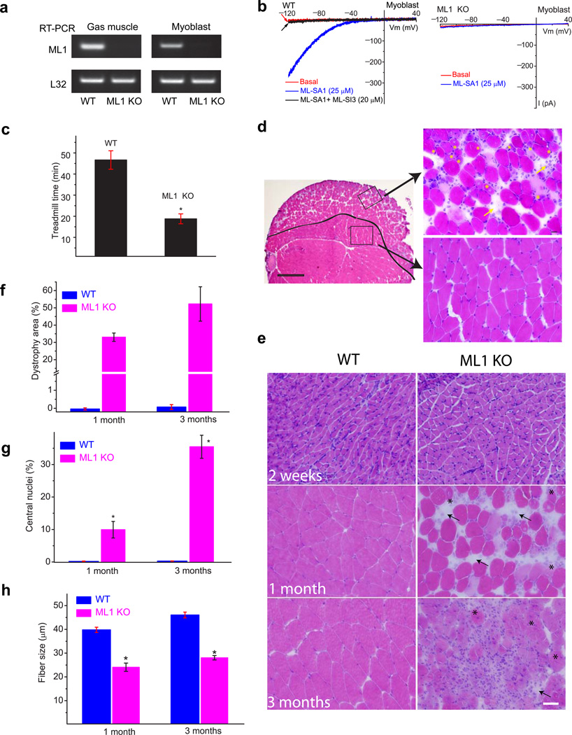
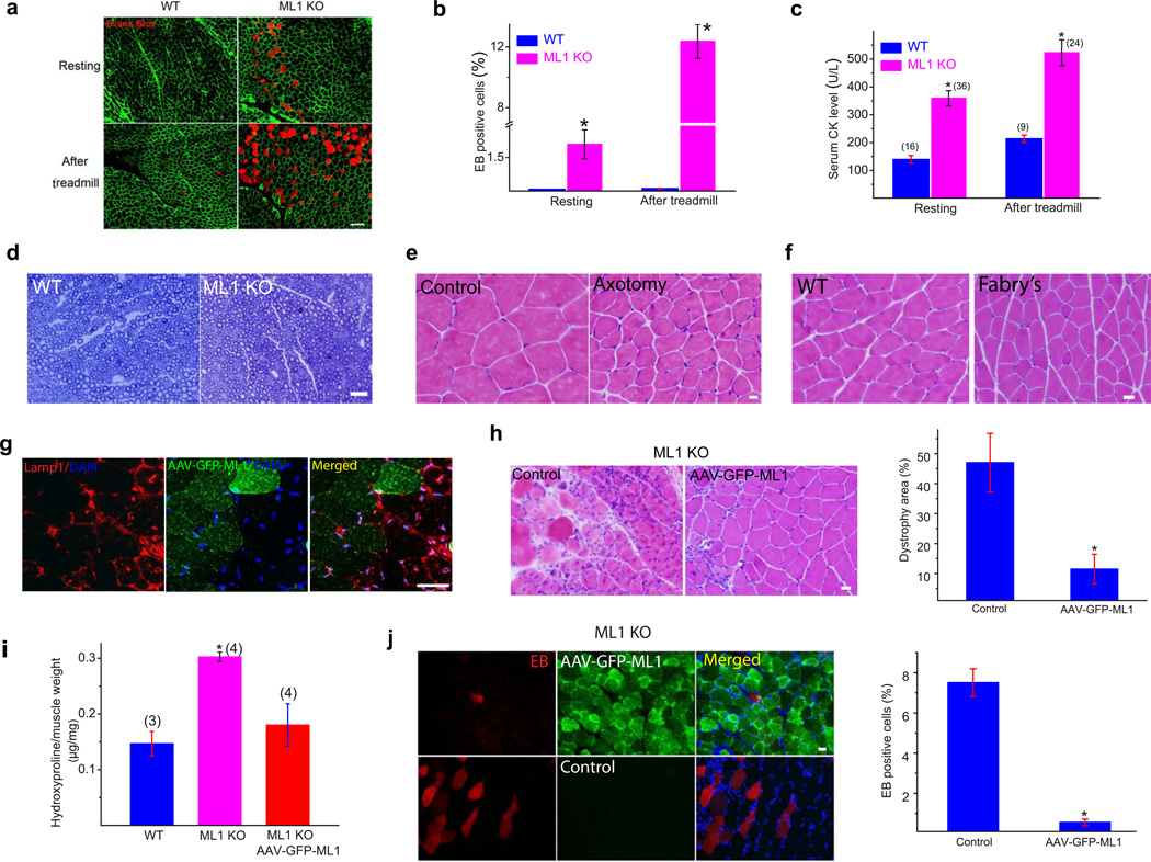
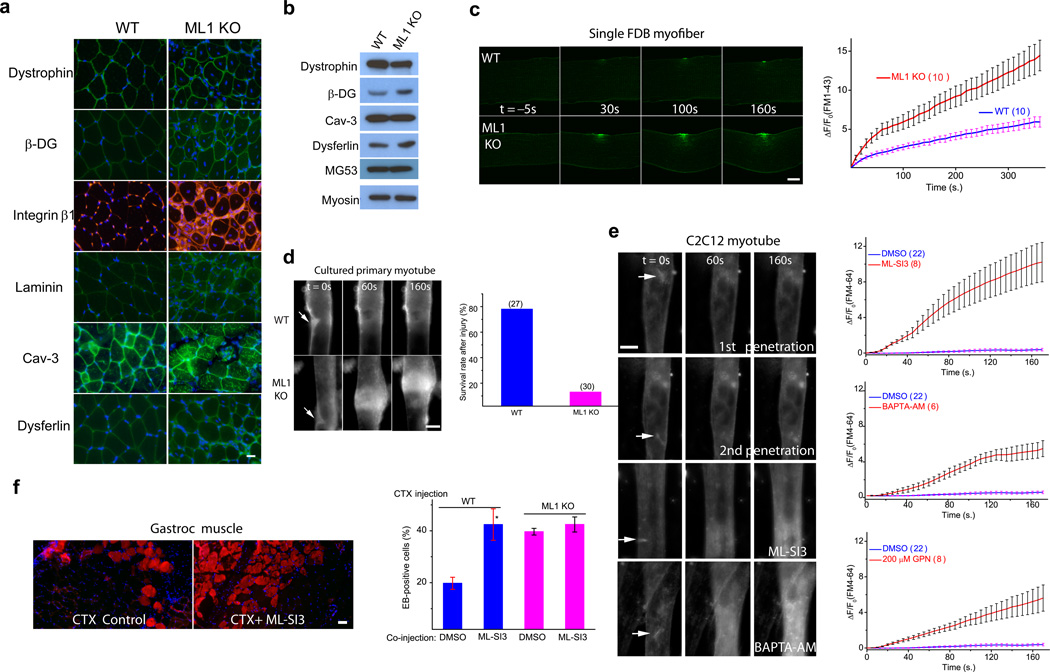
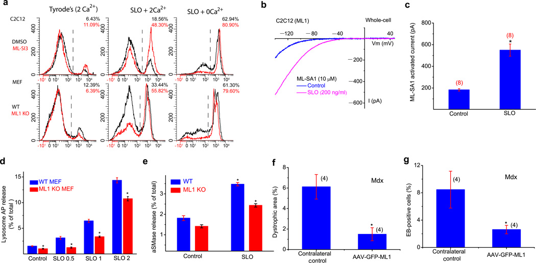
The integrity of the plasma membrane is maintained through an active repair process, especially in skeletal and cardiac muscle cells, in which contraction-induced mechanical damage frequently occurs in vivo. Muscular dystrophies (MDs) are a group of muscle diseases characterized by skeletal muscle wasting and weakness. An important cause of these group of diseases is defective repair of sarcolemmal injuries, which normally requires Ca(2+) sensor proteins and Ca(2+)-dependent delivery of intracellular vesicles to the sites of injury. MCOLN1 (also known as TRPML1, ML1) is an endosomal and lysosomal Ca(2+) channel whose human mutations cause mucolipidosis IV (ML4), a neurodegenerative disease with motor disabilities. Here we report that ML1-null mice develop a primary, early-onset MD independent of neural degeneration. Although the dystrophin-glycoprotein complex and the known membrane repair proteins are expressed normally, membrane resealing was defective in ML1-null muscle fibers and also upon acute and pharmacological inhibition of ML1 channel activity or vesicular Ca(2+) release. Injury facilitated the trafficking and exocytosis of vesicles by upmodulating ML1 channel activity. In the dystrophic mdx mouse model, overexpression of ML1 decreased muscle pathology. Collectively, our data have identified an intracellular Ca(2+) channel that regulates membrane repair in skeletal muscle via Ca(2+)-dependent vesicle exocytosis.


Figures

Figure 1
Figure 1. ML1-null mice develop early-onset progressive MD
(a) Reverse transcription-PCR analysis of WT and TRPML1 (ML1) KO Gastroc muscle and primary cultured myoblasts with a primer pair targeting the deleted region (exons 3, 4, and 5) of the TRPML1 gene. The housekeeping gene L32 served as a loading control. (b) ML-SA1 (25 µM) activated endogenous whole-endolysosome ML1-like currents (IML1) in WT but not ML1 KO myoblasts. ML-SA1-activated currents were inhibited by ML-SI3 (20 µM). Currents were stimulated by using a ramp voltage protocol from −120 to +40 mV. (c) One-month-old ML1-null mice exhibited a shorter running time in a treadmill test compared with WT controls. Animals were trained to run on a 15° downhill treadmill at a speed of 20 m/min. n = 5 animals each. (d) H&E staining of a Gastroc muscle section from a 1-month-old ML1 KO mouse. Scale bar = 400 µm. The black line indicates the boundary between the “dystrophic” region at the periphery and the “normal” region in the center. The representative images on the right are from the boxed areas (left) and show the centrally nucleated fibers (indicated with *) and fibrosis (arrows). Scale bar = 20 µm. (e) H&E staining of Gastroc muscle sections from ML1 KO mice at 2 weeks, and 1 and 3 months of age. Scale bar = 20 µm. (f) The percentage of the dystrophic area in ML1 KO (pink) and WT (blue) muscle. (g) The percentage of centrally nucleated fibers increased with age in ML1 KO. (h) The diameters of muscle fibers were substantially smaller in ML1 KO. For panels f, g, h, n = 3 animals for each condition. Data are presented as the mean ± s.e.m.
Figure 2
Figure 2. MD and muscle membrane damage of ML1 KO mice are not caused by neural degeneration and can be rescued by muscle expression of ML1
(a) Evans blue (EB) dye staining in WT and ML1 KO Gastroc muscles either at rest or after a 30-min treadmill test at the speed of 12 meter/min. The immuostaining of α-DG was used to label the sarcolemma. Scale bar = 40 µm. (b) The percentage of EB-positive muscle fibers in WT and ML1 KO mice. n = 3 animals for each condition. (c) Serum CK levels in WT and ML1 KO mice, either before or after treadmill exercise. The number of animals for each condition is indicated in the parentheses. (d) Lack of obvious myelination defects in the sciatic nerve of 1-month-old ML1 KO mice. (e) Sciatic axotomy induced atrophy of WT Gastroc muscle. (f) Atrophic Gastroc muscle of 4-month-old Fabry's mice. Scale bar = 20 µm. (g) AAV-GFP-ML1-infection reduced Lamp1 elevation in ML1-null muscle cells compared with neighboring noninfected controls. Scale bar = 40 µm. (h) The effect of AAV-GFP-ML1 infection on the dystrophic phenotype in ML1 KO muscles; H&E staining revealed deceased levels of necrosis and fibrosis. The right panel shows a decrease in the percentage of the dystrophic area in ML1-null muscle. n = 4 animals for each condition. (i) AAV-GFPML1 infection on collagen content in ML1 KO muscle. The content of hydroxyproline, a marker for collagen content, was measured in Gastroc skeletal muscle from 3-month-old WT and ML1 KO mice. (g) The effect of AAV-GFP-ML1 infection on EB uptake in ML1-null muscle. n = 3 animals for each condition. Data are presented as the mean ± s.e.m. Scale bars = 10 µm for panels d, e, h, and i.
Figure 3
Figure 3. Defective membrane repair capacity in ML1 KO muscle
(a) Immunofluorescence of dystrophin, β-dystroglycan (β-DG), integrin β1, laminin, caveolin-3 (Cav-3), and dysferlin in ML1-null Gastroc muscle. Scale bar = 10 µm. (b) Western blotting analysis of the DGC components, Cav-3, dysferlin, and MG53 in ML1 KO mice. Myosin served as a loading control. (c) A membrane repair assay performed on single FDB muscle fibers isolated from WT and ML1 KO mice. Membrane damage was induced with a two-photon laser at t = 0 s. Scale bar = 10 µm. The right panel shows the time-dependent changes (ΔF) in FM1–43 fluorescence intensity normalized to the basal fluorescence (F0) for WT (blue) and ML1 KO (red) fibers. (d) Representative images of in vitro differentiated myotubes in response to mechanical damage elicited by a microelectrode (arrows). The lower panel shows the percentage of “surviving” myotubes in response to microelectrode penetration. The experiments were performed in the presence of extracellular Ca2+, and defective membrane resealing led to excessive Ca2+ influx that triggered prolonged muscle contraction. The “surviving” cells are those without prolonged contraction. Scale bar = 50 µm. (e) Responses of C2C12-derived myotubes to microelectrode penetration in Tyrode's solution (2 mM Ca2+) in the presence of DMSO (0.1%; 1st and 2nd penetration), ML-SI3 (20 µM), BAPTA-AM (20 µM), and GPN (200 µM). The right panels show the time course of FM4–64 accumulation (ΔF/F0) at injury sites following microelectrode penetration. Scale bar = 50 µm. Note that the same set of DMSO control data was compared with all groups of drug treatment. (f) The effect of ML-SI3, a TRPML-specific synthetic inhibitor, on EB dye uptake in WT Gastroc muscles injected with the cardiotoxin VII4 (CTX), a cytolytic toxin that disrupts cell membrane in living animals. The lower panel shows that intramuscular coinjection of ML-SI3 with CTX on EB dye uptake in WT and ML1 KO muscle. Scale bar = 10 µm. Data are presented as the mean ± s.e.m, n = 3 animals for each condition.
Figure 4
Figure 4. An essential role of ML1 in lysosomal exocytosis, membrane repair, and protection of muscle damage in vivo
(a) C2C12 and MEF cells were treated with pore-forming toxin streptolysin O (SLO; 2–5 µg/ml), and stained with PI, a marker for membrane damage. FACS quantification of PI staining was performed in cells with or without SLO or external Ca2+ (2 mM). (b, c) SLO (0.2 µg/ml) treatment increased whole-cell IML1 in C2C12 cells transfected with GFP-ML1. d, SLO (0.5, 1 and 2 µg/ml for 15 min) treatment on the release of lysosomal acid phosphatase (AP; determined using an AP activity colorimetric assay kit) into the culture medium in WT and ML1 KO MEFs. The data are presented as the percentage of the activity of the released vs. total cell-associated enzymes. e, SLO (0.5 µg/mL for 15 min) treatment on the release of lysosomal acid SMase (aSMase; determined using an aSMase activity assay kit) in WT and ML1 KO MEFs. (f, g) The effect of AAV-GFP-ML1 infection on EB uptake and dystrophic area in mdx Gastroc muscle. Data are presented as the mean ± s.e.m.

References

    1. Clarke MS, Khakee R, McNeil PL. Loss of cytoplasmic basic fibroblast growth factor from physiologically wounded myofibers of normal and dystrophic muscle. J Cell Sci. 1993;106(Pt 1):121–133. - PubMed
    1. McNeil PL, Khakee R. Disruptions of muscle fiber plasma membranes. Role in exercise-induced damage. Am J Pathol. 1992;140:1097–1109. - PMC - PubMed
    1. Davies KE, Nowak KJ. Molecular mechanisms of muscular dystrophies: old and new players. Nat Rev Mol Cell Biol. 2006;7:762–773. - PubMed
    1. Rahimov F, Kunkel LM. The cell biology of disease: cellular and molecular mechanisms underlying muscular dystrophy. J Cell Biol. 2013;201:499–510. - PMC - PubMed
    1. Bansal D, et al. Defective membrane repair in dysferlin-deficient muscular dystrophy. Nature. 2003;423:168–172. - PubMed

Method References

    1. Bolsover FE, Murphy E, Cipolotti L, Werring DJ, Lachmann RH. Cognitive dysfunction and depression in Fabry disease: a systematic review. J Inherit Metab Dis. 2013 - PubMed
    1. Springer ML, Rando TA, Blau HM. Gene delivery to muscle. Curr Protoc Hum Genet. 2002;Chapter 13(Unit13):14. - PubMed
    1. Dong XP, et al. PI(3,5)P(2) Controls Membrane Traffic by Direct Activation of Mucolipin Ca Release Channels in the Endolysosome. Nat Commun. 2010;1 - PMC - PubMed
    1. Wang X, et al. TPC proteins are phosphoinositide-activated sodium-selective ion channels in endosomes and lysosomes. Cell. 2012;151:372–383. - PMC - PubMed
    1. Brooke MH, Kaiser KK. Muscle fiber types: how many and what kind? Arch Neurol. 1970;23:369–379. - PubMed

Publication types

MeSH terms

Substances