. 2014 Nov 3;33(21):2473-91.
doi: 10.15252/embj.201489729. Epub 2014 Sep 12.

USP8 regulates mitophagy by removing K6-linked ubiquitin conjugates from parkin

Affiliations

USP8 regulates mitophagy by removing K6-linked ubiquitin conjugates from parkin

Thomas M Durcan et al. EMBO J. .

Abstract

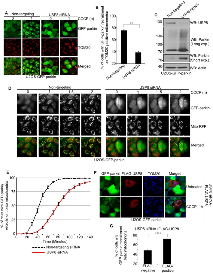
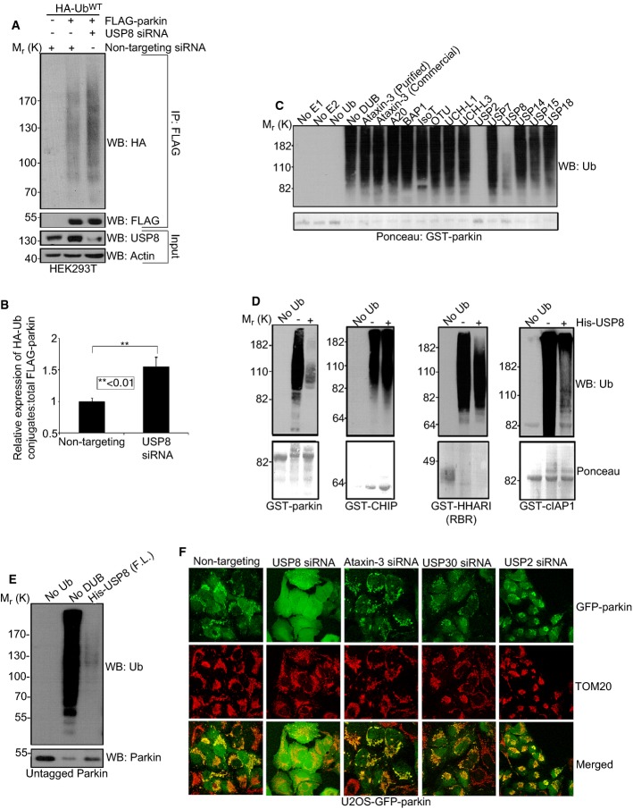
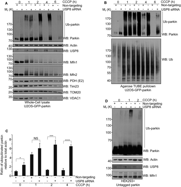
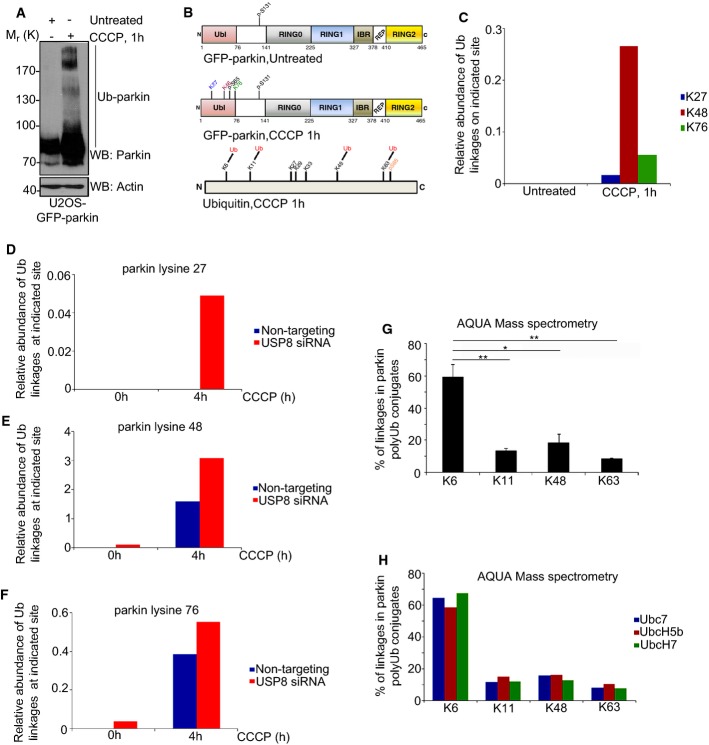
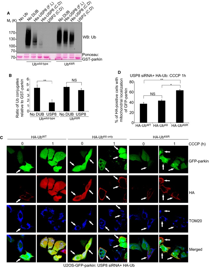
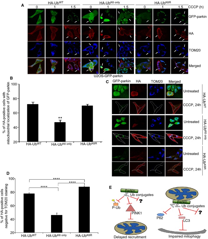
Mutations in the Park2 gene, encoding the E3 ubiquitin-ligase parkin, are responsible for a familial form of Parkinson's disease (PD). Parkin-mediated ubiquitination is critical for the efficient elimination of depolarized dysfunctional mitochondria by autophagy (mitophagy). As damaged mitochondria are a major source of toxic reactive oxygen species within the cell, this pathway is believed to be highly relevant to the pathogenesis of PD. Little is known about how parkin-mediated ubiquitination is regulated during mitophagy or about the nature of the ubiquitin conjugates involved. We report here that USP8/UBPY, a deubiquitinating enzyme not previously implicated in mitochondrial quality control, is critical for parkin-mediated mitophagy. USP8 preferentially removes non-canonical K6-linked ubiquitin chains from parkin, a process required for the efficient recruitment of parkin to depolarized mitochondria and for their subsequent elimination by mitophagy. This work uncovers a novel role for USP8-mediated deubiquitination of K6-linked ubiquitin conjugates from parkin in mitochondrial quality control.

Keywords: USP8; deubiquitination; mitophagy; parkin; ubiquitin.


Figures

Figure 1
Figure 1. USP8 siRNA delays parkin recruitment onto mitochondria
A, B USP8 siRNA impedes parkin recruitment onto mitochondria following CCCP treatment. U2OS-GFP-parkin cells were transfected with non-targeting or USP8 siRNA (10 nM) for 60 h (A). Untreated cells or cells treated with CCCP for 1 h were fixed, and images were acquired after staining for the mitochondrial protein TOM20. After 1-h CCCP treatment, cells were analyzed for GFP-parkin co-localization onto TOM20-positive mitochondria (B). Experiments were blinded and performed in triplicate with 100 cells analyzed for each condition. The vertical bars represent SEM for three independent experiments. For statistical analysis, a two-way ANOVA with Tukey post-test was performed, **P < 0.01. C Validation of USP8 siRNA knockdown. U2OS-GFP-parkin cells were transfected with non-targeting or USP8 siRNA oligos (10 nM) for 60 h. Cells were lysed and analyzed by immunoblotting for USP8, parkin (long and short exposure), and actin. D, E A delay in parkin recruitment onto mitochondria is observed in cells transfected with USP8 siRNA by live-cell microscopy. U2OS-GFP-parkin cells were transfected with non-targeting or USP8 siRNA (10 nM) for 60 h (D). 16 h prior to imaging, cells were infected with CellLight® mitochondria-RFP (Mito-RFP) to visualize mitochondria. Live-cell imaging was initiated 5 min after CCCP treatment, and images were acquired every 5 min over a 140-min period. Parkin recruitment upon membrane depolarization is visualized by the appearance of punctate GFP fluorescence superposed onto mitochondrial RFP fluorescence. Quantification of GFP-parkin recruitment to mitochondria (E) is facilitated by calculating the percentage of cells showing recruitment of GFP-parkin onto mitochondria at 5-min intervals over a period of 145 min. Experiments were performed in triplicate with 350 cells analyzed in each condition. The vertical bars represent the mean ± SEM for three independent experiments (see also Supplementary Video S1). F, G Expression of FLAG-HA-USP8 rescues the effects of USP8 siRNA. U2OS-GFP-parkin cells were co-transfected with USP8 RNAi (5 nM) and FLAG-USP8 (0.5 μg) for 60 h (F). Cells treated with CCCP for 1 h were fixed, and images were acquired after staining for the mitochondrial protein, TOM20. After 1-h CCCP treatment, cells were analyzed for GFP-parkin co-localization onto TOM20-positive mitochondria in cells either negative or positive for FLAG-USP8 (G). Experiments were blinded and performed in triplicate with 100 cells analyzed for each condition. For statistical analysis, a two-way ANOVA with Tukey post-test was performed, ****P < 0.0001. The vertical bars represent the mean ± SEM for three independent experiments (see also Supplementary Fig S3D and E). Source data are available online for this figure.
Figure 2
Figure 2. USP8 siRNA impairs parkin-mediated mitophagy
A–E USP8 siRNA impairs parkin-mediated mitophagy. U2OS-GFP-parkin cells were transfected with non-targeting or USP8 siRNA (10 nM) for 48 h (A). Transfected cells were either left untreated or were treated with CCCP for 24 or 48 h before fixation. Immunofluorescence images of cells were acquired after staining for TOM20. Cells were outlined in white as a result of low GFP-parkin levels. The percentage of all U2OS-GFP-parkin cells after 24 h (B, C) or 48 h (D, E) CCCP treatment lacking TOM20-positive mitochondria or containing GFP-parkin puncta co-localizing with TOM20-positive mitochondria was quantified in cells transfected with non-targeting or USP8 siRNA. Experiments were blinded and performed in triplicate with 100 cells analyzed for each condition. The vertical bars represent SEM. For statistical analysis, a two-way ANOVA with Tukey post-test was performed, *P < 0.05, **P < 0.01; NS, not significant. F USP8 siRNA has no effect on CCCP-induced mitochondrial depolarization. U2OS-GFP-parkin cells transfected with non-targeting or USP8 siRNA (10 nM) for 60 h were first incubated with the potentiometric dye TMRM (600 nM) 20 min prior to imaging. Prior to CCCP treatment, cells were imaged for 10 min to confirm TMRM staining. Following CCCP treatment, images of cells were acquired every minute for 10 min (see also Supplementary Video S2). The membrane potential in untreated cells was tested by imaging untreated cells for 1 h (see also Supplementary Video S3). G USP8 siRNA has no discernible effect on CCCP-induced PINK1 accumulation. U2OS-GFP-parkin cells transfected with non-targeting, PINK1 siRNA, or USP8 siRNA (10 nM) for 60 h were either left untreated or were treated with CCCP for 1 or 3 h. Cells were lysed and immunoblotted for parkin, actin, and PINK1. Source data are available online for this figure.
Figure 3
Figure 3. USP8 deubiquitinates parkin
A, B USP8 knockdown causes Ub conjugates to accumulate on FLAG-parkin in HEK293T cells. HEK293T cells were co-transfected with FLAG-parkin (0.5 μg), HA-UbWT (0.5 μg) and non-targeting or USP8 siRNA (5 nM). Lysates were immunoprecipitated with FLAG resin and analyzed by immunoblotting for HA and FLAG. Input lysates (5% of total input) were analyzed by immunoblotting for USP8 and actin. The optical densities of the Ub-parkin conjugates were quantified using NIH ImageJ, and the data represent the mean ± SEM for three independent experiments. For statistical analysis, a two-way ANOVA with Tukey post-test was performed, **P < 0.01. C The activity of USP8 toward parkin relative to other DUBs. GST-parkin bound to glutathione beads was left to ubiquitinate for 2 h alone at 37°C. After 2 h, the beads were washed to remove reaction components and ubiquitinated GST-parkin was then incubated in the presence or absence of the indicated DUB for 1 h at 37°C. Reactions were immunoblotted for Ub and Ponceau stained for GST-parkin. D USP8 preferentially hydrolyzes preassembled parkin Ub conjugates. For these reactions, GST-parkin, GST-CHIP, GST-HHARI (RBR domain), or GST-cIAP1 bound to glutathione beads were left to ubiquitinate for 2 h alone at 37°C. After 2 h, the beads were washed to remove reaction components. The ubiquitinated E3 was then incubated in the presence or absence of the full-length His-USP8 for 1 h at 37°C. Reactions were analyzed by Ponceau S staining and immunoblotting for Ub. E USP8 can hydrolyze preassembled Ub conjugates on untagged parkin. For these reactions, untagged parkin was left to ubiquitinate for 2 h alone at 37°C with UbcH7 as the E2. After 2 h, apyrase was added for 20 min to terminate the reaction. Ubiquitinated parkin was now incubated for 1 h at 37°C in the presence or absence of His-tagged full-length USP8. Reactions were immunoblotted for Ub and parkin. F Knockdown of other DUBs compared to USP8 had no effect on the recruitment of parkin onto mitochondria following CCCP treatment. U2OS-GFP-parkin cells were transfected with either non-targeting, USP8, ataxin-3, USP30, or USP2 siRNA (10 nM) for 60 h. Cells treated with CCCP for 1 h were fixed, and images were acquired after staining for the mitochondrial protein, TOM20 (see also Supplementary Fig S6). Source data are available online for this figure.
Figure 4
Figure 4. Parkin auto-ubiquitination is enhanced following USP8 siRNA
A–C USP8 siRNA prolongs parkin auto-ubiquitination following treatment with CCCP over 4 h. U2OS-GFP-parkin cells were transfected with non-targeting or USP8 siRNA (10 nM) for 60 h (A). Cells were either left untreated or were treated with CCCP for the indicated time intervals. Lysates were analyzed by immunoblotting for parkin, actin, USP8, Mfn1, Mfn2, PDH (E2), Tim23, TOM20, and VDAC1. From these lysates, ubiquitinated proteins were purified with Agarose-TUBEs (B). Bound proteins were analyzed by immunoblotting for parkin and Ub. The optical densities of ubiquitinated parkin (parkin smear excluding unmodified parkin) and total actin were quantified using NIH ImageJ (C), and the data represent the mean ± SEM for three independent experiments. For statistical analysis, a two-way ANOVA with Tukey post-test was performed, *P < 0.05, ***P < 0.001, ****P < 0.0001; NS, not significant. D USP8 siRNA enhances the auto-ubiquitination of untagged parkin following treatment with CCCP. HEK293T cells were co-transfected with untagged parkin and either non-targeting or USP8 siRNA (10 nM) for 60 h. Cells were either left untreated or were treated with CCCP for the indicated time intervals. Lysates were analyzed by immunoblotting for parkin, actin, USP8, and Mfn1. Source data are available online for this figure.
Figure 5
Figure 5. Knockdown of USP8 leads to an increase in Ub conjugates on parkin
A–C Mass spectrometry analysis of GFP-parkin in untreated and CCCP-treated U2OS-GFP-parkin cells. U2OS-GFP-parkin cells were left untreated or treated with CCCP for 1 h (A). Lysates (400 μg) were immunoprecipitated with GFP-nAb Agarose. Lysates and beads (5% of total bead volume) were analyzed by immunoblotting for parkin and actin. Following on-bead digestion, GFP-parkin samples were analyzed by LC-MS/MS mass spectrometry (B). Identified sites of ubiquitination and phosphorylation on parkin and Ub are indicated. Peptides for each of the 3 sites of ubiquitination on GFP-parkin (K27, K48 and K76) and an unmodified control parkin peptide were selected for time-scheduled parallel reaction monitoring (PRM) (C). The relative abundance of each peptide between samples was calculated, and the relative amount of Ub conjugates present at each individual lysine in parkin was normalized relative to total parkin levels. The data represent the mean value of two technical replicates. D–F PRM mass spectrometry analysis of GFP-parkin following USP8 knockdown U2OS-GFP-parkin cells were transfected with either non-targeting or USP8 siRNA (10 nM). After 60 h, cells were either left untreated or were treated with CCCP for 4 h. The abundance of Ub conjugates at the indicated sites in GFP-parkin was analyzed by parallel reaction monitoring. The relative abundance of each peptide between samples was calculated, and the relative amount of Ub conjugates present at each individual lysine in parkin was normalized relative to total parkin levels. The data represent the mean value of two technical replicates. G, H AQUA analysis and quantification of Ub linkages in parkin poly-Ub conjugates. In vitro ubiquitination reactions were carried out with GST-parkin bound to glutathione beads for 2 h at 37°C in the presence or absence of Ub, E1, UbcH7, and GST alone (G). Ubiquitinated GST-parkin was eluted, trypsinized, and analyzed on an LC-ESI-TOF. Internal peptides for Ub and K6, K11, K33, K48, and K63 Ub linkages were used in the analysis. Ub conjugates were not detected on GST alone. K27, K29, or K33 linkages were not detected in parkin poly-Ub conjugates. Poly-Ub conjugates did not form on parkin when Ub, E1, or E2 were absent (data not shown). The data represent the mean ± SEM for two independent experiments. For statistical analysis, a one-way ANOVA with Tukey post-test was performed, **P < 0.01, *P < 0.05. AQUA analysis of parkin self-ubiquitination in the presence of different E2 Ub-conjugating enzymes (H). UbcH7, UbcH5b, and Ube7 were the E2s tested in the analysis. Source data are available online for this figure.
Figure 6
Figure 6. USP8 hydrolyzes K6 linkages in parkin Ub conjugates
A, B USP8 is unable to hydrolyze preassembled parkin Ub conjugates when K6 linkages are absent. GST-parkin bound to glutathione beads was left to ubiquitinate for 2 h alone at 37°C in the presence of wild-type Ub or UbK6R (A). After 2 h, the beads were washed to remove reaction components. Ubiquitinated GST-parkin was then incubated in the presence or absence of His-tagged full-length USP8, the USP8 catalytic domain, or the untagged USP2 catalytic domain for 1 h at 37°C. Reactions were immunoblotted for Ub and GST-parkin was stained with Ponceau S. The optical densities of the Ub conjugates relative to total GST-parkin were quantified using NIH ImageJ (B), and the data represent the mean ± SEM for three independent experiments. For statistical analysis, a two-way ANOVA with Tukey post-test was performed, **P < 0.01; NS, not significant. C, D Expression of HA-UbK6R rescues the delay in mitochondrial recruitment of parkin following USP8 siRNA. U2OS-GFP-parkin cells were co-transfected with USP8 siRNA (5 nM) and either HA-Ubwild-type, HA-UbK6 only, or HA-UbK6R (1 μg) for 60 h (C). Cells were treated with CCCP for the indicated time periods and fixed. Immunofluorescence images of cells were acquired after staining for HA and TOM20. After 1-h treatment with CCCP, cells were analyzed for the recruitment of GFP-parkin onto TOM20-positive mitochondria in HA-positive cells (D). Experiments were blinded and performed in triplicate with 100 cells analyzed for each condition. The vertical bars represent SEM. For statistical analysis, a two-way ANOVA with Tukey post-test was performed, *P < 0.05, **P < 0.01; NS, not significant. Source data are available online for this figure.
Figure 7
Figure 7. Increasing K6-linked Ub conjugates impairs parkin-mediated mitophagy
A, B Expression of HA-UbK6 only delays the mitochondrial recruitment of parkin following treatment with CCCP. U2OS-GFP-parkin cells were transfected with either HA-Ubwild-type, HA-UbK6 only, or HA-UbK6R (2 μg) for 60 h (A). Cells were treated with CCCP for indicated time periods and fixed. Immunofluorescence images of cells were acquired after staining for HA and TOM20. After 1-h treatment with CCCP, cells overexpressing either HA-Ubwild-type, HA-UbK6 only, or HA-UbK6R (2 μg) were analyzed for the recruitment of GFP-parkin onto TOM20-positive mitochondria in HA-positive cells (B). Experiments were blinded and performed in triplicate with 100 cells analyzed for each condition. The vertical bars represent SEM. For statistical analysis, a two-way ANOVA with Tukey post-test was performed, **P < 0.01. C, D Expression of HA-UbK6 only impairs parkin-mediated mitophagy. U2OS-GFP-parkin cells were transfected with either HA-Ubwild-type, HA-UbK6 only, or HA-UbK6R (2 μg) for 60 h (C). Cells were treated with CCCP for 24 h and fixed. Immunofluorescence images of cells were acquired after staining for HA and TOM20. After 24 h treatment with CCCP, the percentage of HA-positive cells negative for TOM20 staining was quantified (D). Experiments were blinded and performed in triplicate with 100 cells analyzed for each condition. For statistical analysis, a two-way ANOVA with Tukey post-test was performed, **P < 0.01. E Schematic for how USP8 knockdown delays parkin-mediated mitophagy. At steady state, parkin is auto-inhibited with minimal levels of auto-ubiquitination observed. Ub chains that do form can be removed by USP8 acting upon the K6 linkages within these conjugates. However, when USP8 is absent, Ub conjugates persist on parkin, whose presence appears to delay parkin recruitment onto depolarized mitochondria by impeding the interaction of parkin with either PINK1/phosphorylated Ub or another parkin activating protein. During mitophagy, a robust increase in parkin auto-ubiquitination occurs, followed by removal of Ub conjugates and subsequent clearance of damaged mitochondria. When USP8 is knocked down, these chains are now left intact. Thus, their presence delays parkin-mediated clearance by potentially impeding parkin from interacting with p62, LC3, or another autophagic adaptor protein required for the successful completion of mitophagy.

Comment in

References

    1. Ali N, Zhang L, Taylor S, Mironov A, Urbe S, Woodman P. Recruitment of UBPY and ESCRT exchange drive HD-PTP-dependent sorting of EGFR to the MVB. Curr Biol. 2013;23:453–461. - PubMed
    1. Ben-Saadon R, Zaaroor D, Ziv T, Ciechanover A. The polycomb protein Ring1B generates self atypical mixed ubiquitin chains required for its in vitro histone H2A ligase activity. Mol Cell. 2006;24:701–711. - PubMed
    1. Berlin I, Schwartz H, Nash PD. Regulation of epidermal growth factor receptor ubiquitination and trafficking by the USP8.STAM complex. J Biol Chem. 2010;285:34909–34921. - PMC - PubMed
    1. de Bie P, Zaaroor-Regev D, Ciechanover A. Regulation of the Polycomb protein RING1B ubiquitination by USP7. Biochem Biophys Res Commun. 2010;400:389–395. - PubMed
    1. de Bie P, Ciechanover A. Ubiquitination of E3 ligases: self-regulation of the ubiquitin system via proteolytic and non-proteolytic mechanisms. Cell Death Differ. 2011;18:1393–1402. - PMC - PubMed

Publication types

MeSH terms