. 2014 Nov;137(Pt 11):2922-37.
doi: 10.1093/brain/awu257. Epub 2014 Sep 12.

c-Jun activation in Schwann cells protects against loss of sensory axons in inherited neuropathy

Affiliations

c-Jun activation in Schwann cells protects against loss of sensory axons in inherited neuropathy

Janina Hantke et al. Brain. 2014 Nov.

Abstract

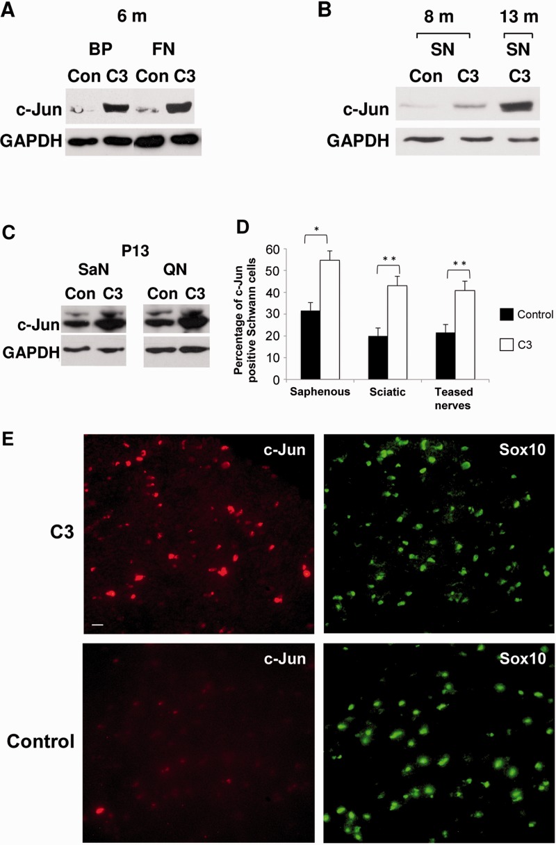
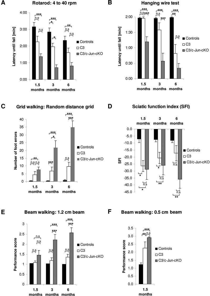
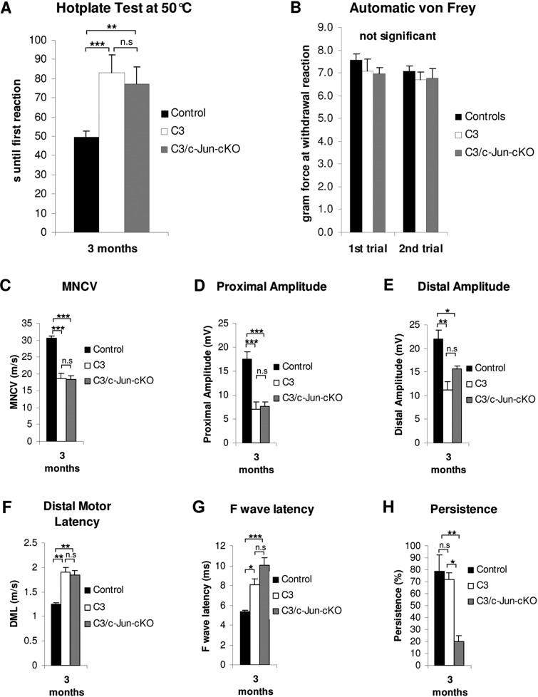
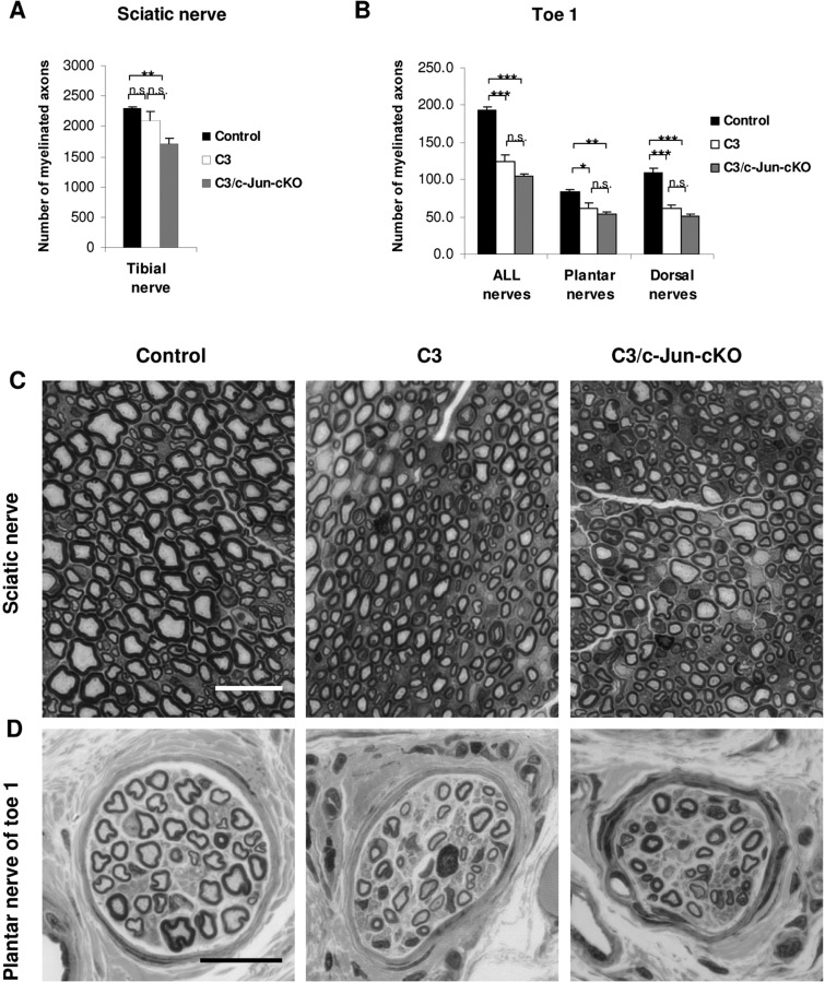
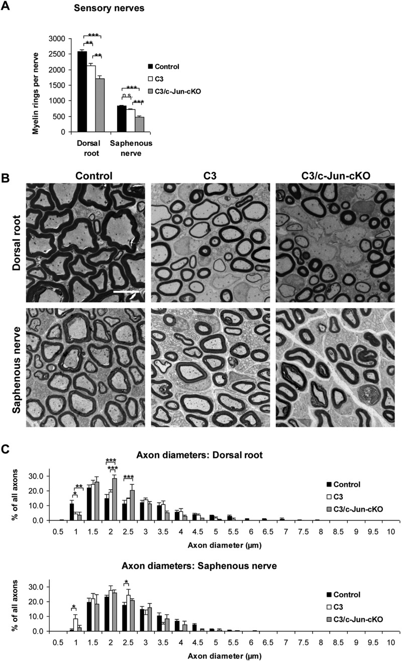
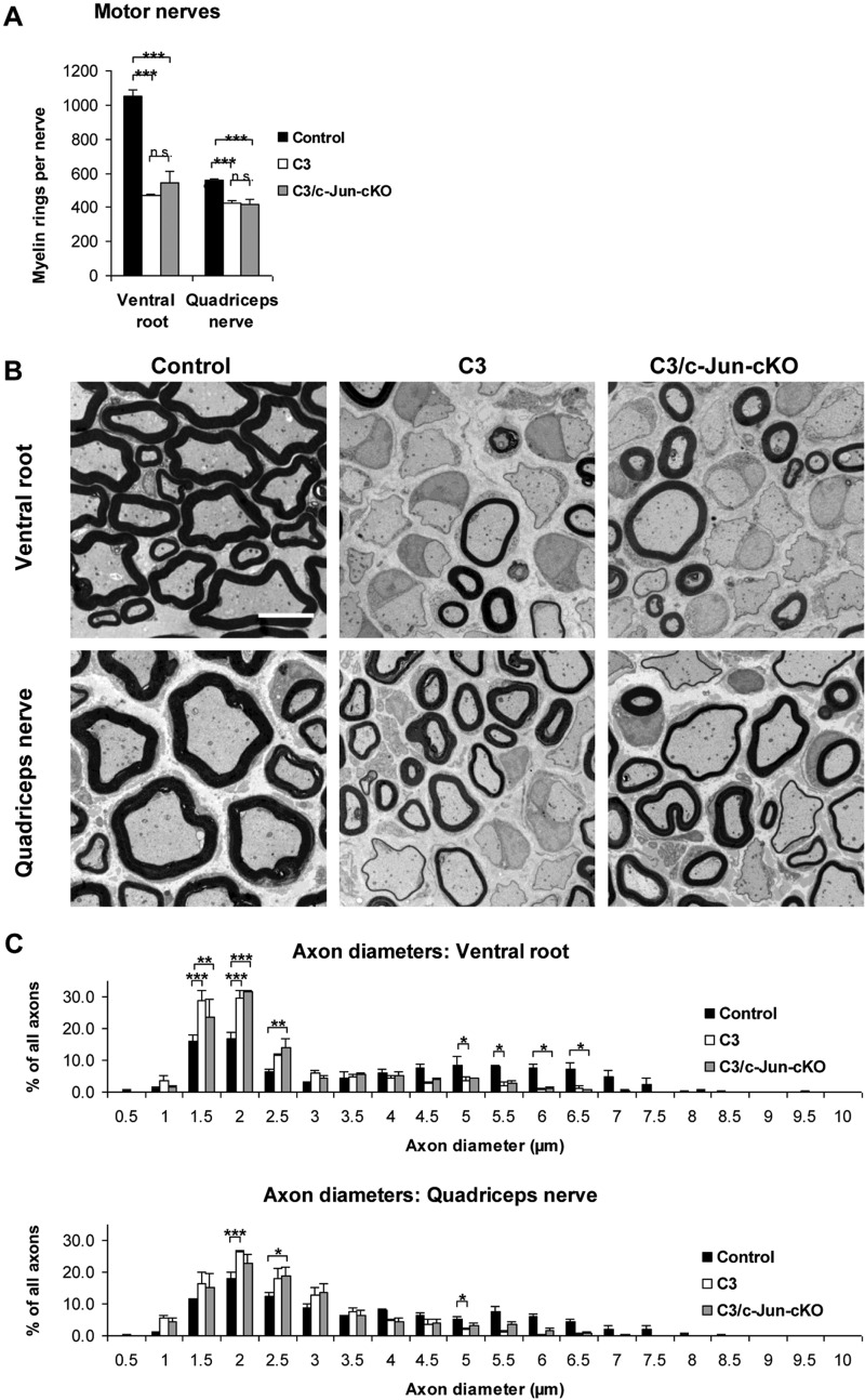
Charcot-Marie-Tooth disease type 1A is the most frequent inherited peripheral neuropathy. It is generally due to heterozygous inheritance of a partial chromosomal duplication resulting in over-expression of PMP22. A key feature of Charcot-Marie-Tooth disease type 1A is secondary death of axons. Prevention of axonal loss is therefore an important target of clinical intervention. We have previously identified a signalling mechanism that promotes axon survival and prevents neuron death in mechanically injured peripheral nerves. This work suggested that Schwann cells respond to injury by activating/enhancing trophic support for axons through a mechanism that depends on upregulation of the transcription factor c-Jun in Schwann cells, resulting in the sparing of axons that would otherwise die. As c-Jun orchestrates Schwann cell support for distressed neurons after mechanical injury, we have now asked: do Schwann cells also activate a c-Jun dependent neuron-supportive programme in inherited demyelinating disease? We tested this by using the C3 mouse model of Charcot-Marie-Tooth disease type 1A. In line with our previous findings in humans with Charcot-Marie-Tooth disease type 1A, we found that Schwann cell c-Jun was elevated in (uninjured) nerves of C3 mice. We determined the impact of this c-Jun activation by comparing C3 mice with double mutant mice, namely C3 mice in which c-Jun had been conditionally inactivated in Schwann cells (C3/Schwann cell-c-Jun(-/-) mice), using sensory-motor tests and electrophysiological measurements, and by counting axons in proximal and distal nerves. The results indicate that c-Jun elevation in the Schwann cells of C3 nerves serves to prevent loss of myelinated sensory axons, particularly in distal nerves, improve behavioural symptoms, and preserve F-wave persistence. This suggests that Schwann cells have two contrasting functions in Charcot-Marie-Tooth disease type 1A: on the one hand they are the genetic source of the disease, on the other, they respond to it by mounting a c-Jun-dependent response that significantly reduces its impact. Because axonal death is a central feature of much nerve pathology it will be important to establish whether an axon-supportive Schwann cell response also takes place in other conditions. Amplification of this axon-supportive mechanism constitutes a novel target for clinical intervention that might be useful in Charcot-Marie-Tooth disease type 1A and other neuropathies that involve axon loss.

Keywords: axonal degeneration; demyelinating disease; neural repair; neuron-glial interaction; neuropathy.


Figures

Figure 1
Figure 1
c-Jun levels are elevated in Schwann cells of the C3 mouse. (A–C) Western blots of (uninjured) peripheral nerves from control and C3 mice showing elevated c-Jun protein levels at 6, 8 and 13 months, and post-natal Day 13. (A) Strongly elevated c-Jun levels are seen in the brachial plexus (BP) and femoral nerve (FN) of C3 mice compared to controls (6-month-old mice). (B) In the sciatic nerve (SN) of C3 mice, c-Jun protein levels increase with age (m = months). (C) In C3 mice, elevated c-Jun is already seen in saphenous (SaN) and quadriceps (QN) nerves at post-natal Day 13. (D) Counts showing that in C3 nerves, the number of c-Jun+ Schwann cell nuclei is about double that in control nerves. The numbers for saphenous and sciatic nerves represent the percentage of SOX10+ Schwann cell nuclei that were also c-Jun+ in double immunolabelled transverse sections. The numbers for teased nerves represent the percentage of DAPI labelled nuclei that were also c-Jun+ in myelin Schwann cells identified by phase contrast in teased sciatic and saphenous nerves immunolabelled with L1 antibodies to identify non-myelin (Remak) cells. (E) Transverse sciatic nerve sections double labelled with SOX10 antibodies to identify Schwann cells, and c-Jun antibodies. The images for c-Jun were captured using identical parameters and illustrate the difference in intensity of c-Jun labelling typically seen between C3 and control nerves. In controls, c-Jun is hard to detect in this comparison, although the signal is unambiguous using other imaging settings. Scale bar = 10 µm.
Figure 2
Figure 2
C3 mice show sensory-motor deficits that are amplified in the absence of Schwann cell c-Jun in C3/c-Jun-cKO mice. (A) Rotarod: Using the accelerating rotarod all mouse lines show some decline in performance with age. C3 mice show a trend towards worse performance than controls which reaches significance in 3-month-old mice. Similarly, C3/c-Jun-cKO mice tend to perform worse than C3 mice, reaching significance in 3-month-old mice. (B) Hanging wire test: C3 mice perform significantly worse than controls at 6 months. C3/c-Jun-cKO mice perform significantly worse than C3 mice at 1.5 and 3 months. (C) Grid walking: C3 mice show a trend towards worse performance than controls, which is significant at 3 months. C3/c-Jun-cKO mice perform significantly worse that C3 mice at 3 and 6 months and show a strong trend towards deteriorating performance with age. (D) Sciatic function index: C3 mice tend to perform worse than controls at all ages and this is significant at 1.5 and 3 months. In 6-month-old mice there is a clear trend towards worse performance by C3/c-Jun-cKO mice compared to C3 but this does not reach significance. (E) Beam walking 1.2 cm beam: There is little difference between C3 and control mice. But C3/c-Jun-cKO mice show a strong decline in performance compared to C3 that is significant at 3 and 6 months. (F) Beam walking 0.5 cm beam: C3 mice perform significantly worse than controls at 1.5 months but the deterioration in C3/c-Jun-cKO mice compared to C3 mice is not significant. *P < 0.05, **P < 0.01, ***P < 0.001.
Figure 3
Figure 3
Sensory tests and electrophysiological measurement. (A) Hotplate test: both C3 and C3/c-Jun-cKO mice show a prominent sensory deficit in perception of thermal stimuli, which is significantly different from control animals (B) Automatic von Frey: Applying the von Frey test in an automatic version detected no significant differences between the three mouse lines. (C–H) Electrophysiology. The figures obtained in the C3 mouse reveal several deficits compared to control animals that are typical for CMT1A. One parameter, F wave persistence, is normal in C3 mice but significantly reduced in C3/c-Jun-cKO mice. Nerve conduction studies were performed on 3-month-old control, C3 and C3/c-Jun-cKO animals. (C–F) C3 and C3/c-Jun-cKO mice have significantly decreased motor neuron conduction velocity (MNCV), and proximal and distal amplitude, whereas distal motor latency is increased in both mouse lines in comparison to the control. (G) F wave latency was increased in the C3 and C3/c-Jun-cKO mice. (H) C3/c-Jun-cKO mice showed significantly reduced F wave persistence compared to both C3 and control mice. *P < 0.05, **P < 0.01, ***P < 0.001.
Figure 4
Figure 4
Light microscopy of mixed nerves. (A) There is a trend towards lower numbers of myelinated axons from control to C3 to C3/c-Jun-cKO mice. (B) In the distally located nerves in toe 1, the number of myelinated axons in nerves from C3 and C3/c-Jun-cKO mice is significantly lower than in controls. Removal of Schwann cell c-Jun (the C3/c-Jun-cKO compared to the C3 mouse) does not increase the number of myelin sheaths. (C and D) General appearance of the tibial and toe nerves from control, C3 and C3/c-Jun-cKO mice, as shown in semithin sections. Scale bar = 20 µm. *P < 0.05, **P < 0.01, ***P < 0.001.
Figure 5
Figure 5
Light microscopy of sensory nerves. (A) Counts of myelinated fibres in light microscopy photographs show that C3 mice have small but significant loss of myelin sheaths in dorsal roots but not in saphenous nerves when compared to control nerves. Both nerves showed significant loss of myelin sheaths in C3/c-Jun-cKO compared to C3 mice. (B) Electron micrographs illustrating amyelinated and thickly and thinly myelinated axons. These myelin abnormalities are less pronounced in the saphenous nerve than in the dorsal root. Scale bar = 5 µm. (C) Analysis of myelinated axon diameters in dorsal root and saphenous nerve in control, C3 and C3/c-Jun-cKO mice. *P < 0.05, **P < 0.01, ***P < 0.001.
Figure 6
Figure 6
Light microscopy of motor nerves. (A) Counts of myelinated fibres in light microscopy photographs show that C3 mice have about 50% fewer myelin sheaths in ventral roots and ∼20% fewer sheaths in quadriceps nerves compared to controls. No significant increase in the number of myelin sheaths was seen in C3/c-Jun-cKO nerves compared to C3 nerves. (B) Electron micrographs show the marked dysmyelination in motor nerves of C3 and C3/c-Jun-cKO mice. Scale bar = 5 µm. (C) Analysis of myelinated axon diameters in ventral root and quadriceps nerve in control, C3 and C3/c-Jun-cKO mice. *P < 0.05, **P < 0.01, ***P < 0.001.
Figure 7
Figure 7
Analysis of muscle fibres. (A) Light micrographs of muscles in control, C3 and C3/c-Jun-cKO mice. Both C3 and C3/c-Jun-cKO have immature (2c), angulated (black arrowhead), hypertrophic (black arrow) and atrophied fibres (empty arrow head). Scale bar = 50 µm. (B) Fibre type distribution in control, C3 and C3/c-Jun-cKO animals. (C–E) Area distribution of type 1 and type 2 fibres. *P < 0.05, **P < 0.01, ***P < 0.001.

References

    1. Arthur-Farraj PJ, Latouche M, Wilton DK, Quintes S, Chabrol E, Banerjee A, et al. c-Jun Reprograms Schwann cells of injured nerves to generate a repair cell essential for regeneration. Neuron. 2012;75:633–47. - PMC - PubMed
    1. Behrens A, Sibilia M, David JP, Mohle-Steinlein U, Tronche F, Schutz G, et al. Impaired postnatal hepatocyte proliferation and liver regeneration in mice lacking c-jun in the liver. EMBO J. 2002;21:1782–90. - PMC - PubMed
    1. Birouk N, Gouider R, Le Guern E, Gugenheim M, Tardieu S, Maisonobe T, et al. Charcot-Marie-Tooth disease type 1A with 17p11.2 duplication. Clinical and electrophysiological phenotype study and factors influencing disease severity in 119 cases. Brain. 1997;120:813–23. - PubMed
    1. Brooke MH, Kaiser KK. Muscle fiber types: how many and what kind? Arch Neurol. 1970;23:369–79. - PubMed
    1. D'Antonio M, Musner N, Scapin C, Ungaro D, Del Carro U, Ron D, et al. Resetting translational homeostasis restores myelination in Charcot-Marie-Tooth disease type 1B mice. J Exp Med. 2013;210:821–38. - PMC - PubMed

Publication types

Substances