Carotid magnetic resonance imaging for monitoring atherosclerotic plaque progression: a multicenter reproducibility study
- PMID: 25216871
- PMCID: PMC4297722
- DOI: 10.1007/s10554-014-0532-7
Carotid magnetic resonance imaging for monitoring atherosclerotic plaque progression: a multicenter reproducibility study
Abstract
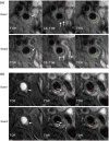
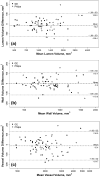
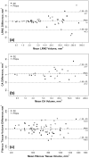
This study sought to determine the multicenter reproducibility of magnetic resonance imaging (MRI) and the compatibility of different scanner platforms in assessing carotid plaque morphology and composition. A standardized multi-contrast MRI protocol was implemented at 16 imaging sites (GE: 8; Philips: 8). Sixty-eight subjects (61 ± 8 years; 52 males) were dispersedly recruited and scanned twice within 2 weeks on the same magnet. Images were reviewed centrally using a streamlined semiautomatic approach. Quantitative volumetric measurements on plaque morphology (lumen, wall, and outer wall) and plaque tissue composition [lipid-rich necrotic core (LRNC), calcification, and fibrous tissue] were obtained. Inter-scan reproducibility was summarized using the within-subject standard deviation, coefficient of variation (CV) and intraclass correlation coefficient (ICC). Good to excellent reproducibility was observed for both morphological (ICC range 0.98-0.99) and compositional (ICC range 0.88-0.96) measurements. Measurement precision was related to the size of structures (CV range 2.5-4.9 % for morphology, 36-44 % for LRNC and calcification). Comparable measurement variability was found between the two platforms on both plaque morphology and tissue composition. In conclusion, good to excellent inter-scan reproducibility of carotid MRI can be achieved in multicenter settings with comparable measurement precision between platforms, which may facilitate future multicenter endeavors that use serial MRI to monitor atherosclerotic plaque progression.
Trial registration: ClinicalTrials.gov NCT00880178 NCT01178320.
Conflict of interest statement
Figures


Similar articles
-
In vivo semi-automatic segmentation of multicontrast cardiovascular magnetic resonance for prospective cohort studies on plaque tissue composition: initial experience.Int J Cardiovasc Imaging. 2016 Jan;32(1):73-81. doi: 10.1007/s10554-015-0704-0. Epub 2015 Jul 14. Int J Cardiovasc Imaging. 2016. PMID: 26169389 Free PMC article.
-
In-vivo quantitative T2 mapping of carotid arteries in atherosclerotic patients: segmentation and T2 measurement of plaque components.J Cardiovasc Magn Reson. 2013 Aug 16;15(1):69. doi: 10.1186/1532-429X-15-69. J Cardiovasc Magn Reson. 2013. PMID: 23953780 Free PMC article.
-
Bilateral symmetry of human carotid artery atherosclerosis: a multi-contrast weighted MR study.Int J Cardiovasc Imaging. 2016 Aug;32(8):1219-26. doi: 10.1007/s10554-016-0890-4. Epub 2016 May 2. Int J Cardiovasc Imaging. 2016. PMID: 27139458
-
Imaging of the high-risk carotid plaque: magnetic resonance imaging.Semin Vasc Surg. 2017 Mar;30(1):54-61. doi: 10.1053/j.semvascsurg.2017.04.009. Epub 2017 Apr 27. Semin Vasc Surg. 2017. PMID: 28818259 Review.
-
Advanced MRI for carotid plaque imaging.Int J Cardiovasc Imaging. 2016 Jan;32(1):83-9. doi: 10.1007/s10554-015-0743-6. Epub 2015 Aug 21. Int J Cardiovasc Imaging. 2016. PMID: 26293362 Free PMC article. Review.
Cited by
-
Assessment of Therapeutic Response to Statin Therapy in Patients With Intracranial or Extracranial Carotid Atherosclerosis by Vessel Wall MRI: A Systematic Review and Updated Meta-Analysis.Front Cardiovasc Med. 2021 Oct 27;8:742935. doi: 10.3389/fcvm.2021.742935. eCollection 2021. Front Cardiovasc Med. 2021. PMID: 34778404 Free PMC article.
-
Carotid Plaque Lipid Content and Fibrous Cap Status Predict Systemic CV Outcomes: The MRI Substudy in AIM-HIGH.JACC Cardiovasc Imaging. 2017 Mar;10(3):241-249. doi: 10.1016/j.jcmg.2016.06.017. JACC Cardiovasc Imaging. 2017. PMID: 28279371 Free PMC article. Clinical Trial.
-
Cardiovascular imaging 2015 in the International Journal of Cardiovascular Imaging.Int J Cardiovasc Imaging. 2016 May;32(5):697-709. doi: 10.1007/s10554-016-0877-1. Int J Cardiovasc Imaging. 2016. PMID: 27086358 Review. No abstract available.
-
In vivo semi-automatic segmentation of multicontrast cardiovascular magnetic resonance for prospective cohort studies on plaque tissue composition: initial experience.Int J Cardiovasc Imaging. 2016 Jan;32(1):73-81. doi: 10.1007/s10554-015-0704-0. Epub 2015 Jul 14. Int J Cardiovasc Imaging. 2016. PMID: 26169389 Free PMC article.
-
Vessel wall characterization using quantitative MRI: what's in a number?MAGMA. 2018 Feb;31(1):201-222. doi: 10.1007/s10334-017-0644-x. Epub 2017 Aug 14. MAGMA. 2018. PMID: 28808823 Free PMC article. Review.
References
-
- Corti R, Fayad ZA, Fuster V, Worthley SG, Helft G, Chesebro J, Mercuri M, Badimon JJ. Effects of lipid-lowering by simvastatin on human atherosclerotic lesions: a longitudinal study by high-resolution, noninvasive magnetic resonance imaging. Circulation. 2001;104:249–252. - PubMed
-
- Lima JA, Desai MY, Steen H, Warren WP, Gautam S, Lai S. Statin-induced cholesterol lowering and plaque regression after 6 months of magnetic resonance imaging-monitored therapy. Circulation. 2004;110:2336–2341. - PubMed
-
- Yonemura A, Momiyama Y, Fayad ZA, Ayaori M, Ohmori R, Higashi K, Kihara T, Sawada S, Iwamoto N, Ogura M, Taniguchi H, Kusuhara M, Nagata M, Nakamura H, Tamai S, Ohsuzu F. Effect of lipid-lowering therapy with atorvastatin on atherosclerotic aortic plaques detected by noninvasive magnetic resonance imaging. J Am Coll Cardiol. 2005;45:733–742. - PubMed
-
- Lee JM, Wiesmann F, Shirodaria C, Leeson P, Petersen SE, Francis JM, Jackson CE, Robson MD, Neubauer S, Channon KM, Choudhury RP. Early changes in arterial structure and function following statin initiation: quantification by magnetic resonance imaging. Atherosclerosis. 2008;197:951–958. - PMC - PubMed
-
- Underhill HR, Yuan C, Zhao XQ, Kraiss LW, Parker DL, Saam T, Chu B, Takaya N, Liu F, Polissar NL, Neradilek B, Raichlen JS, Cain VA, Waterton JC, Hamar W, Hatsukami TS. Effect of rosuvastatin therapy on carotid plaque morphology and composition in moderately hypercholesterolemic patients: a high-resolution magnetic resonance imaging trial. Am Heart J. 2008;155:581–584. - PubMed
Publication types
MeSH terms
Substances
Associated data
Grants and funding
LinkOut - more resources
Full Text Sources
Other Literature Sources
Medical

