

Endothelial progenitor cells: from senescence to rejuvenation
- PMID: 25217265
- PMCID: PMC4163195
- DOI: 10.1016/j.semnephrol.2014.06.003
Endothelial progenitor cells: from senescence to rejuvenation
Abstract
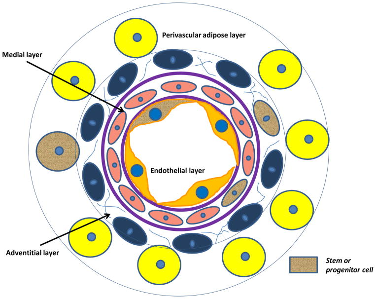
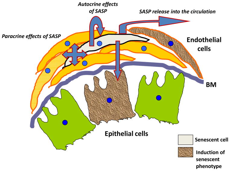
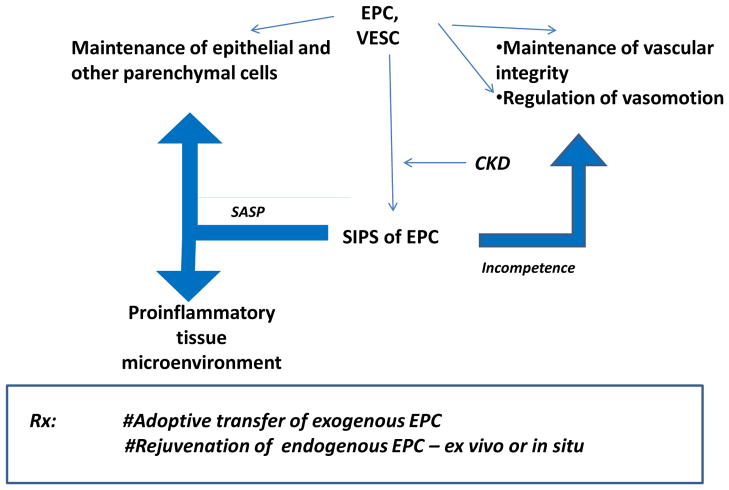
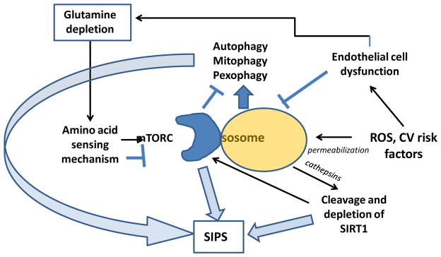
Discovered more than 15 years ago, endothelial progenitor cells attract both basic and translational researchers. It has become clear that they represent a heterogeneous population of endothelial colony-forming cells, early or late outgrowth endothelial cells, or blood outgrowth endothelial cells, each characterized by differing proliferative and regenerative capacity. Scattered within the vascular wall, these cells participate in angiogenesis and vasculogenesis and support regeneration of epithelial cells. There is growing evidence that this cell population is impaired during the course of chronic cardiovascular and kidney disease when it undergoes premature senescence and loss of specialized functions. Senescence-associated secretory products released by such cells can affect the neighboring cells and further exacerbate their regenerative capacity. For these reasons, adoptive transfer of endothelial progenitor cells is being used in more than 150 ongoing clinical trials of diverse cardiovascular diseases. Attempts to rejuvenate this cell population either ex vivo or in situ are emerging. The progress in this field is paramount to regenerate the injured kidney.
Keywords: Stress-induced premature senescence (SIPS); cell therapy; regeneration; rejuvenation therapy; senescence-associated secretory products (SASP).
Copyright © 2014 Elsevier Inc. All rights reserved.
Conflict of interest statement
Author states no conflict of interest
Figures
Similar articles
-
Stress-Induced Premature Senescence of Endothelial and Endothelial Progenitor Cells.Adv Pharmacol. 2016;77:281-306. doi: 10.1016/bs.apha.2016.04.007. Epub 2016 Jun 6. Adv Pharmacol. 2016. PMID: 27451101 Free PMC article. Review.
-
Endothelial and cardiac progenitor cells for cardiovascular repair: A controversial paradigm in cell therapy.Pharmacol Ther. 2018 Jan;181:156-168. doi: 10.1016/j.pharmthera.2017.08.004. Epub 2017 Aug 19. Pharmacol Ther. 2018. PMID: 28827151 Review.
-
Ex vivo expansion of human outgrowth endothelial cells leads to IL-8-mediated replicative senescence and impaired vasoreparative function.Stem Cells. 2013 Aug;31(8):1657-68. doi: 10.1002/stem.1414. Stem Cells. 2013. PMID: 23629812
-
Rejuvenation of human cardiac progenitor cells with Pim-1 kinase.Circ Res. 2013 Oct 25;113(10):1169-79. doi: 10.1161/CIRCRESAHA.113.302302. Epub 2013 Sep 17. Circ Res. 2013. PMID: 24044948 Free PMC article.
-
Restoring the function of a diseased kidney via its microvasculature.Nephron Exp Nephrol. 2014;126(2):82. doi: 10.1159/000360672. Epub 2014 May 19. Nephron Exp Nephrol. 2014. PMID: 24854646 Review.
Cited by
-
Pravastatin Promotes Endothelial Colony-Forming Cell Function, Angiogenic Signaling and Protein Expression In Vitro.J Clin Med. 2021 Jan 6;10(2):183. doi: 10.3390/jcm10020183. J Clin Med. 2021. PMID: 33419165 Free PMC article.
-
Silica-coated magnetic nanoparticles labeled endothelial progenitor cells alleviate ischemic myocardial injury and improve long-term cardiac function with magnetic field guidance in rats with myocardial infarction.J Cell Physiol. 2019 Aug;234(10):18544-18559. doi: 10.1002/jcp.28492. Epub 2019 Apr 14. J Cell Physiol. 2019. PMID: 30982985 Free PMC article.
-
Endothelial Progenitor Cells Physiology and Metabolic Plasticity in Brain Angiogenesis and Blood-Brain Barrier Modeling.Front Physiol. 2016 Dec 1;7:599. doi: 10.3389/fphys.2016.00599. eCollection 2016. Front Physiol. 2016. PMID: 27990124 Free PMC article. Review.
-
Genetically modified endothelial progenitor cells with hNotch1.ICN overexpression display facilitated angiogenesis.Ann Transl Med. 2020 Oct;8(20):1316. doi: 10.21037/atm-20-6362. Ann Transl Med. 2020. PMID: 33209896 Free PMC article.
-
Recent Advances in the Anti-Inflammatory Activity of Plant-Derived Alkaloid Rhynchophylline in Neurological and Cardiovascular Diseases.Pharmaceutics. 2021 Jul 29;13(8):1170. doi: 10.3390/pharmaceutics13081170. Pharmaceutics. 2021. PMID: 34452133 Free PMC article. Review.
References
-
- Sen S, McDonald SP, Coates PT, Bonder CS. Endothelial progenitor cells: novel biomarker and promising therapy for cardiovascular disease. Clin Sci. 2011;120:263–283. - PubMed
-
- Asahara T, Murohara T, Sullivan A, et al. Isolation of putative progenitor endothelial cells for angiogenesis. Science. 1997;275:964–967. - PubMed
-
- Khakoo A, Finkel T. Endothelial progenitor cells. Annu Rev Med. 2005;56:79–101. - PubMed
-
- Ingram D, Mead L, Moore D, et al. Vessel wall-derived endothelial cells rapidly proliferate because they contain a complete hierarchy of endothelial progenitor cells. Blood. 2005;105:2783–2786. - PubMed
Publication types
MeSH terms
Grants and funding
LinkOut - more resources
Full Text Sources
Other Literature Sources
Medical
