Review
. 2014 Jul;34(4):374-83.
doi: 10.1016/j.semnephrol.2014.06.004. Epub 2014 Jun 13.

Kidney pericytes: roles in regeneration and fibrosis

Affiliations
Review

Kidney pericytes: roles in regeneration and fibrosis

Rafael Kramann et al. Semin Nephrol. 2014 Jul.

Abstract

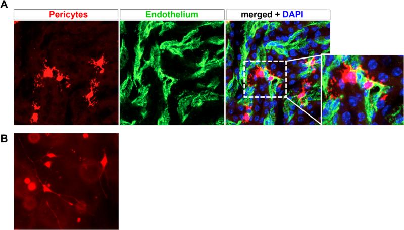
Renal pericytes have been neglected for many years, but recently they have become an intensively studied cell population in renal biology and pathophysiology. Pericytes are stromal cells that support vasculature, and a subset of pericytes are mesenchymal stem cells. In kidney, pericytes have been reported to play critical roles in angiogenesis, regulation of renal medullary and cortical blood flow, and serve as progenitors of interstitial myofibroblasts in renal fibrogenesis. They interact with endothelial cells through distinct signaling pathways and their activation and detachment from capillaries after acute or chronic kidney injury may be critical for driving chronic kidney disease progression. By contrast, during kidney homeostasis it is likely that pericytes serve as a local stem cell population that replenishes differentiated interstitial and vascular cells lost during aging. This review describes both the regenerative properties of pericytes as well as involvement in pathophysiologic conditions such as fibrogenesis.

Keywords: Pericytes; capillary rarefaction; kidney fibrosis; mesenchymal stem cells.


Figures

Figure 1
Figure 1
Renal pericytes associate closely with vasculature. (A) Col1α1-CreERt2 mice were crossed against R26RtdTomato reporter mice, and pulsed with tamoxifen to induce recombination in medullary pericytes. A high-power view of tdTomato+ pericytes is shown, note the extensive branching processes emanating from the cell body. Endothelial cells were co-stained green using fluorescein isothiocyanate FITC-CD31 antibodies, and the merged image shows how tightly the pericytes associate with and wraparound peritubular capillaries. Genetically tagged renal pericytes (left panel) surrounding peritubular capillaries (middle panel, CD31). (B) Genetically labeled pericytes were dissociated from kidney by enzymatic dissociation and purified by fluorescence activated cell sorting. Subsequently, they were plated in a three-dimensional collagen gel, where long delicate processes extend from the cell body, similar to the in vivo situation. DAPI, 4',6-diamidino-2-phenylindole.
Figure 2
Figure 2
Fate of FoxD1+ cells in kidney development. FoxD1-Cre mice were crossed against the R26RtdTomato reporter line to genetically label FoxD1+ cells and all of their descendants. (A) cells include thin branched stromal cells located in the interstitium (arrows). These cells represent pericytes and perivascular fibro-blasts. In addition, the glomerular staining indicates that both mesangial cells and podocytes express FoxD1 (arrowheads). However, podocytes acquire FoxD1 expression later in development, so only mesangial cells have a lineage relationship with FoxD1 precursors in development. (B) FoxD1+ lineage cells also generate vascular smooth cells that line arteries and kidney arterioles (asterisks).
Figure 3
Figure 3
Capillary rarefaction after kidney injury. Sham kidneys were co-stained for peritubular capillaries with CD31 antibodies (left panels). Before death, mice were perfused with FITC-conjugated microbeads in agarose to generate a fluorescence microangiogram (FMA, middle panels). The merged image illustrates luminal filling of CD31-positive capillaries with green microbeads, providing a readout of vascular perfusion. In the lower panels, mice were subject to severe AKI and allowed to recover for 8 weeks. They were subject to the same protocol as the sham mice. The results show substantially reduced perfusion of peritubular capillaries, as well as endothelial cell density. This capillary rarefaction strongly correlated with the degree of kidney injury at day 1 after AKI, suggesting an inability of damaged capillaries to repair. DAPI, 4',6-diamidino-2-phenylindole.

References

    1. Diaz-Flores L, Gutierrez R, Madrid JF, et al. Pericytes. Morphofunction, interactions and pathology in a quiescent and activated mesenchymal cell niche. Histol Histopathol. 2009;24:909–69. - PubMed
    1. Armulik A, Genove G, Betsholtz C. Pericytes: developmental, physiological, and pathological perspectives, problems, and promises. Dev Cell. 2011;21:193–215. - PubMed
    1. Mugford JW, Sipila P, McMahon JA, McMahon AP. Osr1 expression demarcates a multi-potent population of intermediate mesoderm that undergoes progressive restriction to an Osr1-dependent nephron progenitor compartment within the mammalian kidney. Dev Biol. 2008;324:88–98. - PMC - PubMed
    1. Humphreys BD, Lin SL, Kobayashi A, et al. Fate tracing reveals the pericyte and not epithelial origin of myofibroblasts in kidney fibrosis. Am J Pathol. 2010;176:85–97. - PMC - PubMed
    1. Humphreys BD. Targeting pericyte differentiation as a strategy to modulate kidney fibrosis in diabetic nephropathy. Semin Nephrol. 2012;32:463–70. - PMC - PubMed

Publication types