Review
. 2014 Jul;34(4):418-28.
doi: 10.1016/j.semnephrol.2014.06.008. Epub 2014 Jun 13.

Progenitor cells and podocyte regeneration

Affiliations
Review

Progenitor cells and podocyte regeneration

Stuart J Shankland et al. Semin Nephrol. 2014 Jul.

Abstract

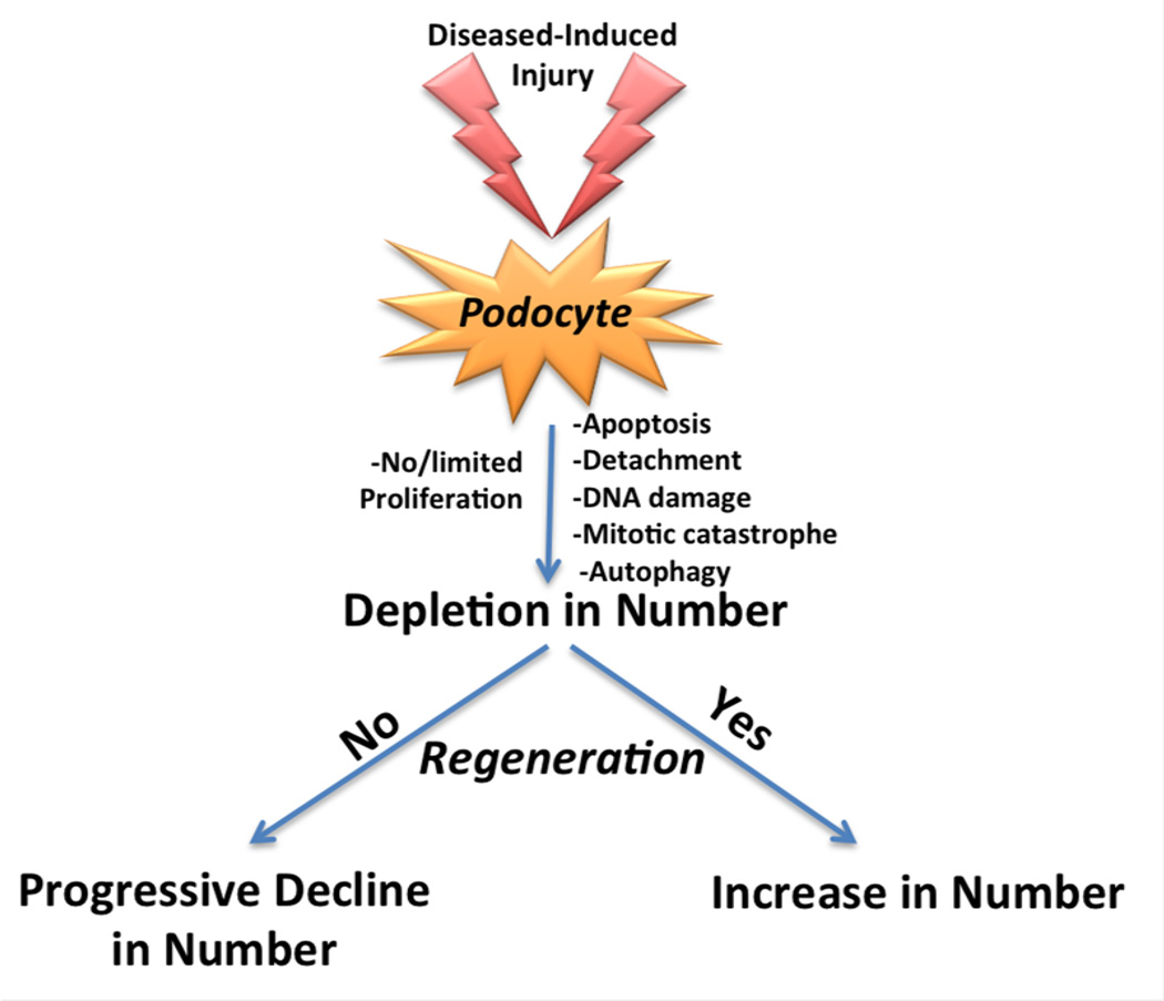
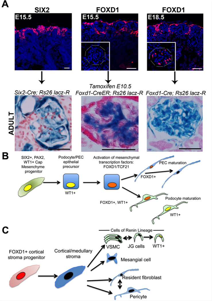
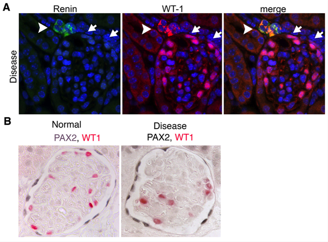
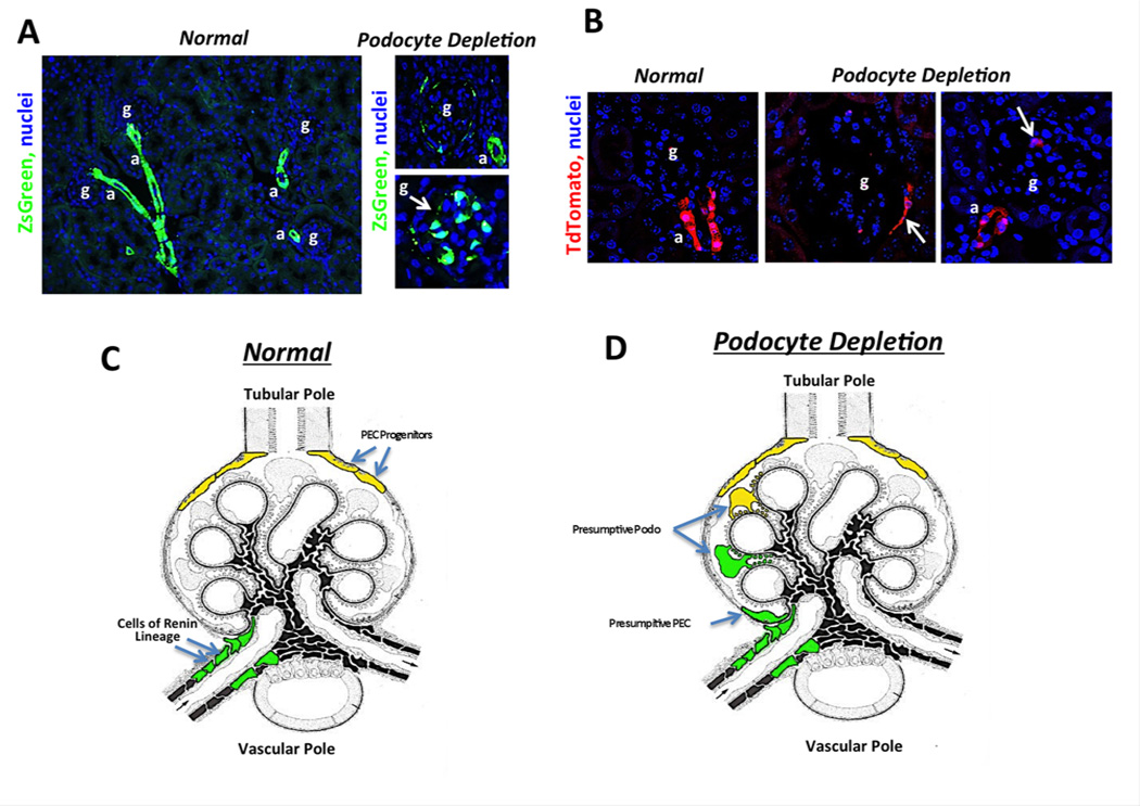
The very limited ability of adult podocytes to proliferate in vivo is clinically significant because podocytes form a vascular barrier that is functionally critical to the nephron, podocyte hypoplasia is a characteristic of disease, and inadequate regeneration of podocytes is a major cause of persistent podocyte hypoplasia. Excessive podocyte loss or inadequate replacement leads to glomerulosclerosis in many progressive kidney diseases. Thus, restoration of podocyte cell density almost certainly is reliant on regeneration by podocyte progenitors. However, such putative progenitors have remained elusive until recently. In this review, we describe the developmental processes leading to podocyte and parietal epithelial cell (PEC) formation during glomerulogenesis. We compare evidence that in normal human kidneys PECs expressing progenitor markers CD133 and CD24 can differentiate into podocytes in vitro and in vivo, with evidence from animal models suggesting a more limited role of the PEC's capacity to serve as a podocyte progenitor in adults. We highlight tantalizing new evidence that specialized vascular wall cells of afferent arterioles, including those that produce renin in healthy kidney, provide a novel local progenitor source of new PECs and podocytes in response to podocyte hypoplasia in the adult, and draw comparisons with glomerulogenesis.

Keywords: Glomerulus; WT-1; cells of renin lineage; glomerulosclerosis; parietal epithelial cells; proteinuria.


Conflict of interest statement

Conflict of Interest: JSD is employed by Biogen Idec.; SJS and JWP have no conflicts of interest

Figures

Figure 1
Figure 1. Schema showing the fate of podocytes in response to injury
Figure 2
Figure 2. Podocytes and PECs derive from SIX2+ epithelial progenitor cells in cap mesenchyme but late in nephrogenesis activate FOXD1, a transcription factor critical in formation of mural cells in the kidney
(A) Images (upper) showing two discrete progenitor populations in the nephrogenic outer zone of the developing kidney at E15.5 defined by expression of SIX2 and FOXD1 respectively. Below shows results of fate mapping of these progenitors using Cre/LoxP transgenic reporter mice, indicating in adult mice they given rise to podocytes/PECs and mesangial cells respectively. At E18.5 FOXD1 is now active additionally in podocytes of primitive glomeruli (inset), and mapping of FOXD1 expression shows that all podocytes and PECs have expressed FOXD1 at this late time in nephrogenesis. (B) Schema showing the results of fate mapping of SIX2+ epithelial progenitors which co-express WT1 and PAX2 in development. These progenitors give rise to precursor cells for podocytes and PECs which have a cuboidal appearance. Late in development they activate FOXD1 and TCF21 which appear to regulate the final differentiation into mature cells, and they persistently express WT1. (C) Schema showing the results of fate mapping of FOXD1+ stromal progenitors which give rise to mesangial cells, fibroblasts, pericytes and vascular smooth muscle including JG cells. In glomerular disease, JG cells activate WT1.
Figure 3
Figure 3. Renin producing vascular smooth muscle cells activate WT1 and PECs upregulate PAX2 in adult glomerular disease with podocyte loss
(A) Split panel images of a glomerulus following administration of anti-podocyte antibodies showing de novo expression of WT1 in the renin producing cells of the JGA (arrowhead). In addition to expression of WT1 in podocytes attached to the tuft, WT1 can also be seen in some PECs (arrows). (B) Images of normal and diseased glomeruli following anti-podocyte antibody administration. Note increased expression intensity of PAX2 in PECs
Figure 4
Figure 4. Images showing the fate of Cells of Renin Lineage in glomerular regeneration and Schema showing proposed dual role for PECs and CoRL as adult podocyte progenitors in glomerular disease
(A) Images from normal adult kidney and following podocytes loss during kidney disease in RenCre;ZsGreen reporter mice. Note all of the vascular arteriolar wall is labeled due to Renin expression during development. Note that the adjacent glomerulus is devoid of cells of renin lineage but following podocyte loss cells can be seen along Bowman’s capsule and in the glomerular tuft. (B) Images from RenCreER;tdTomato reporter mice that received tamoxifen to induce recombination to activate tdTomato expression in renin producing cells at 6 weeks of age. Only cells in the JGA at the tip of the afferent arteriole are permanently labeled. Note that following podocytes loss during kidney disease, there is migration of renin labeled cells from the JGA to Bowman’s capsule and the glomerular tuft. (C-D) Schema showing the fate of juxtaglomerular cells of renin lineage and tubular pole PECs in the setting of kidney disease. Both of these cells from fate mapping experiments have the capacity to acquire podocytes like qualities.

References

    1. Susztak K, Raff AC, Schiffer M, Bottinger EP. Glucose-induced reactive oxygen species cause apoptosis of podocytes and podocyte depletion at the onset of diabetic nephropathy. Diabetes. 2006;55(1):225–233. - PubMed
    1. Brinkkoetter PT, Olivier P, Wu JS, Henderson S, Krofft RD, Pippin JW, et al. Cyclin I activates Cdk5 and regulates expression of Bcl-2 and Bcl-XL in postmitotic mouse cells. The Journal of clinical investigation. 2009 - PMC - PubMed
    1. Cybulsky AV, Takano T, Papillon J, Kitzler TM, Bijian K. Endoplasmic reticulum stress in glomerular epithelial cell injury. Am J Physiol Renal Physiol. 2011;301(3):F496–F508. - PubMed
    1. Hartleben B, Wanner N, Huber TB. Autophagy in glomerular health and disease. Semin Nephrol. 2014;34(1):42–52. - PubMed
    1. Petermann AT, Pippin J, Krofft R, Blonski M, Griffin S, Durvasula R, et al. Viable podocytes detach in experimental diabetic nephropathy: potential mechanism underlying glomerulosclerosis. Nephron Experimental nephrology. 2004;98(4):e114–e123. - PubMed

Publication types