. 2014 Nov 15;592(22):5079-92.
doi: 10.1113/jphysiol.2014.282079. Epub 2014 Sep 12.

Suprachiasmatic nucleus function and circadian entrainment are modulated by G protein-coupled inwardly rectifying (GIRK) channels

Affiliations

Suprachiasmatic nucleus function and circadian entrainment are modulated by G protein-coupled inwardly rectifying (GIRK) channels

L M Hablitz et al. J Physiol. .

Abstract

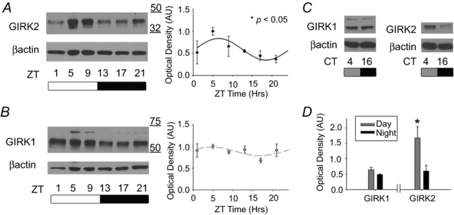
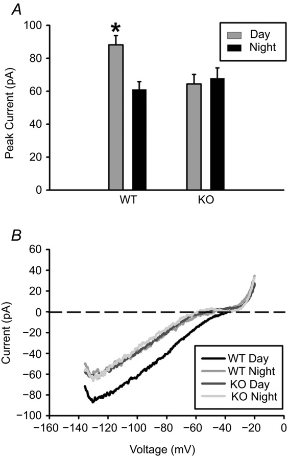
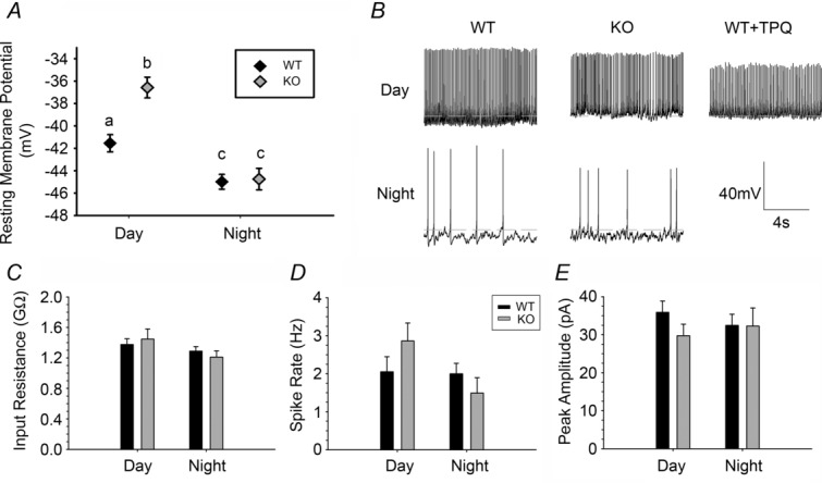
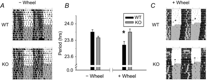
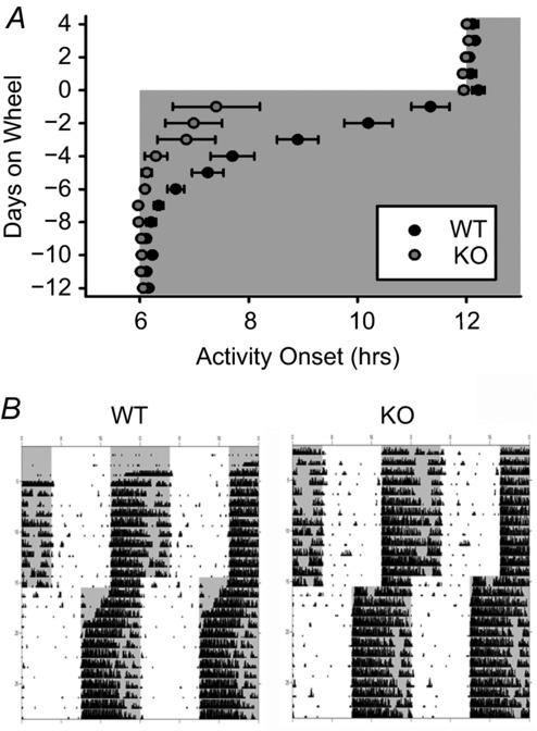
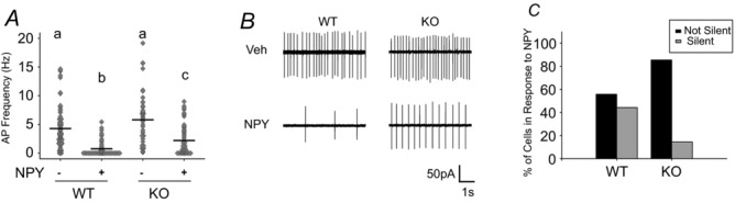
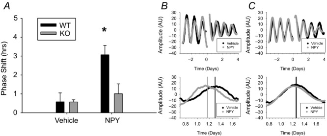
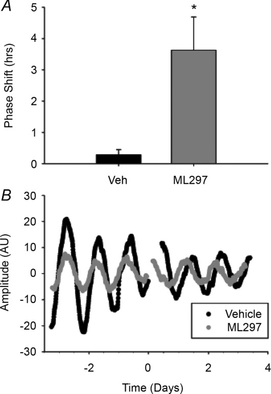
G protein signalling within the central circadian oscillator, the suprachiasmatic nucleus (SCN), is essential for conveying time-of-day information. We sought to determine whether G protein-coupled inwardly rectifying potassium channels (GIRKs) modulate SCN physiology and circadian behaviour. We show that GIRK current and GIRK2 protein expression are greater during the day. Pharmacological inhibition of GIRKs and genetic loss of GIRK2 depolarized the day-time resting membrane potential of SCN neurons compared to controls. Behaviourally, GIRK2 knockout (KO) mice failed to shorten free running period in response to wheel access in constant darkness and entrained more rapidly to a 6 h advance of a 12 h:12 h light-dark (LD) cycle than wild-type (WT) littermate controls. We next examined whether these effects were due to disrupted signalling of neuropeptide Y (NPY), which is known to mediate non-photic phase shifts, attenuate photic phase shifts and activate GIRKs. Indeed, GIRK2 KO SCN slices had significantly fewer silent cells in response to NPY, likely contributing to the absence of NPY-induced phase advances of PER2::LUC rhythms in organotypic SCN cultures from GIRK2 KO mice. Finally, GIRK channel activation is sufficient to cause a non-photic-like phase advance of PER2::LUC rhythms on a Per2(Luc+/-) background. These results suggest that rhythmic regulation of GIRK2 protein and channel function in the SCN contributes to day-time resting membrane potential, providing a mechanism for the fine tuning responses to non-photic and photic stimuli. Further investigation could provide insight into disorders with circadian disruption comorbidities such as epilepsy and addiction, in which GIRK channels have been implicated.


Figures

Figure 1
Figure 1. GIRK2, but not GIRK1, protein levels are regulated in a circadian manner
A, representative immunoblots for GIRK2 from mouse SCN out of an LD cycle (left). Relative optical density (mean ± SEM) for GIRK2 throughout the day with lines indicating the predicted cosinor curve (*< 0.05, right). n = 5–6 per time point. B, representative immunoblot for GIRK1 in mouse SCN across an LD cycle (left). Relative optical density (mean ± SEM) for GIRK1 at each time point throughout the day with predicted cosinor curve (right). n = 3–5 per time point. C, representative GIRK1 (left) and GIRK2 (right) blots from mouse SCN out of DD at CT 4 (subjective day) and CT 16 (subjective night). D, relative optical density (mean ± SEM) between subjective day and night for GIRK1 and GIRK2. *< 0.05, n = 3–4 per time point.
Figure 2
Figure 2. GIRK currents are increased during the day in SCN neurons
A, peak inward current (mean ± SEM) during the day vs. night (*< 0.05) for GIRK2 knockout (KO) and wild-type (WT) mice. B, representative voltage-clamp ramp traces. Recordings were done in the presence of TTX (1 μm), bicuculline (30 μm), d-AP5 (50 μm), CNQX (10 μm), and CdCl2 (200 μm) in order to block synaptic transmission. All recordings were done in 30 mm KCl to increase potassium conductance. n = at least 3 animals and 20 cells per group.
Figure 3
Figure 3. GIRK2 knockout SCN neurons exhibit more depolarized resting membrane potential compared to wild-type controls
A, resting membrane potential of WT and KO SCN neurons during the day and night. Lowercase letters (a, b, c) indicate groups that are significantly different (*P < 0.05). B, representative gap-free current clamp of KO and WT SCN neurons during the day and at night. A representative trace from WT slices treated with the GIRK2 antagonist Tertiapin-Q (0.2 μm) during the day is also included. Dotted line indicates −40 mV. C–E, input resistance, action potential frequency from gap-free current clamp recordings, and action potential amplitudes from gap-free current clamp recordings, respectively, from WT and KO neurons during the day and night. n = 3–5 animals and at least 25 cells per group.
Figure 4
Figure 4. GIRK2 knockout mice fail to shorten free-running period in response to wheel-running activity
A, representative, double-plotted actograms of WT (top) and KO (bottom) mice without wheel access (activity measured by infrared motion sensors), n = 14–15 per group. B, wheel-locked infrared free-running period (mean ± SEM), and wheel-running activity-based free-running period (mean ± SEM) for KO and WT mice. *< 0.05. C, representative actograms for WT and KO mice with wheel access (right, activity measured by wheel revolutions); n = 7–8 animals per each group. Time of lights off is indicated by dark grey; black tick marks indicate activity counts.
Figure 5
Figure 5. GIRK2 knockout mice entrain more rapidly to a 6 h light cycle advance
A, activity onset (mean ± SEM) in days prior and following a 6 h advance of an LD cycle (day 0) for WT and KO mice. Lights off represented by grey shading. B, representative actograms of WT (left) and KO (right) mice housed on wheels in a 12 h:12 h LD cycle. Lights off represented by grey shading; black tick marks indicate activity counts. n = 6 animals per group.
Figure 6
Figure 6. Loss of GIRK2 reduces effect of NPY on SCN neuron spontaneous firing rate
A, frequency plot of individual WT and KO SCN neurons treated with 2.35 μm NPY or vehicle from ZT 4 to 6. Mean value indicated by black continuous line. Lowercase letters (a, b, c) indicate groups that are significantly different (*P < 0.05). B, representative loose patch traces (5 s) of neurons in A. C, percentage of silent cells vs. non-silent cells with NPY treatment. n = 3 animals and ≥45 cells per group.
Figure 7
Figure 7. GIRK2 is necessary for NPY-induced phase shifts in the molecular clock
A, phase shifts of PER2::LUC rhythms in response to a 1 h treatment of 2.35 μm NPY or vehicle at CT 4 in WT and KO mice. *P < 0.05. B and C, representative bioluminescence traces from two WT (B) or two KO (C) SCN cultures treated with vehicle (black) or NPY (grey) for three cycles before and after treatment (top) or day 1 after treatment (bottom). Time 0: onset of NPY application. Predicted peak time, determined from 3 cycles post treatment, is indicated by vertical bars for each group. n = 6–8 cultures per group.
Figure 8
Figure 8. Activation of GIRK channels induces non-photic-like phase advances
A, phase shifts of PER2::LUC rhythms in response to a 1 h treatment of 10 μm ML297 or vehicle starting between CT 3–4.5. B, representative bioluminescence traces from SCN cultures treated with vehicle (black) or ML297 (grey) for three cycles before and after treatment. n = 6 cultures per group.

References

    1. Acuna-Goycolea C, Tamamaki N, Yanagawa Y, Obata K. van den Pol AN. Mechanisms of neuropeptide Y, peptide YY, and pancreatic polypeptide inhibition of identified green fluorescent protein-expressing GABA neurons in the hypothalamic neuroendocrine arcuate nucleus. J Neurosci. 2005;25:7406–7419. - PMC - PubMed
    1. Aguado C, Colon J, Ciruela F, Schlaudraff F, Cabanero MJ, Perry C, Watanabe M, Liss B, Wickman K. Lujan R. Cell type-specific subunit composition of G protein-gated potassium channels in. J Neurochem. 2008;105:497–511. - PubMed
    1. Albrecht U. Timing to perfection: the biology of central and peripheral circadian clocks. Neuron. 2012;74:246–260. - PubMed
    1. Arora D, Hearing M, Haluk DM, Mirkovic K, Fajardo-Serrano A, Wessendorf MW, Watanabe M, Lujan R. Wickman K. Acute cocaine exposure weakens GABAB receptor-dependent G-protein-gated inwardly rectifying K+ signaling in dopamine neurons of the ventral tegmental area. J Neurosci. 2011;31:12251–12257. - PMC - PubMed
    1. Aton SJ, Huettner JE, Straume M. Herzog ED. GABA and Gi/o differentially control circadian rhythms and synchrony in clock neurons. Proc Natl Acad Sci U S A. 2006;103:19188–19193. - PMC - PubMed

Publication types

MeSH terms

Substances

LinkOut - more resources