. 2014 Sep;26(9):3809-22.
doi: 10.1105/tpc.114.128736. Epub 2014 Sep 12.

Adjustment of host cells for accommodation of symbiotic bacteria: vacuole defunctionalization, HOPS suppression, and TIP1g retargeting in Medicago

Affiliations

Adjustment of host cells for accommodation of symbiotic bacteria: vacuole defunctionalization, HOPS suppression, and TIP1g retargeting in Medicago

Aleksandr Gavrin et al. Plant Cell. 2014 Sep.

Abstract

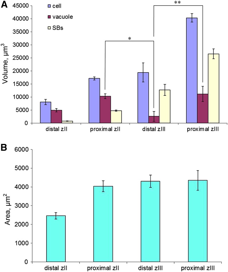
In legume-rhizobia symbioses, the bacteria in infected cells are enclosed in a plant membrane, forming organelle-like compartments called symbiosomes. Symbiosomes remain as individual units and avoid fusion with lytic vacuoles of host cells. We observed changes in the vacuole volume of infected cells and thus hypothesized that microsymbionts may cause modifications in vacuole formation or function. To examine this, we quantified the volumes and surface areas of plant cells, vacuoles, and symbiosomes in root nodules of Medicago truncatula and analyzed the expression and localization of VPS11 and VPS39, members of the HOPS vacuole-tethering complex. During the maturation of symbiosomes to become N2-fixing organelles, a developmental switch occurs and changes in vacuole features are induced. For example, we found that expression of VPS11 and VPS39 in infected cells is suppressed and host cell vacuoles contract, permitting the expansion of symbiosomes. Trafficking of tonoplast-targeted proteins in infected symbiotic cells is also altered, as shown by retargeting of the aquaporin TIP1g from the tonoplast membrane to the symbiosome membrane. This retargeting appears to be essential for the maturation of symbiosomes. We propose that these alterations in the function of the vacuole are key events in the adaptation of the plant cell to host intracellular symbiotic bacteria.


Figures

Figure 1.
Figure 1.
Nodule Zonation, Stages of Symbiosome Development, and 3D Reconstructed Cells of M. truncatula Nodules. (A) Scheme of the nodule zones: M, meristem; zII, infection zone; zIII, zone of nitrogen fixation. The stages of symbiotic cells in development are shown next to each other (1 to 4). Zones described here correspond to the light microscopy image of the nodule (B) The arrow points to the zone of transition between the infection and fixation zones. The vacuoles of infected cells in this zone are highlighted by red. Here, the arrows point to the cells in consequent stages of development given in high-magnification images ([C] to [E]) and as examples of 3D reconstruction ([F] to [H]). (C) and (F) Cells from the zone of bacteria release. (D) and (G) The transition zone (interzone 2/3). It is recognized by the appearance of starch grains. (E) and (H) Mature infected cells. Flaccid vacuoles of a mature symbiotic cell have a “scalloped” surface, whereas vacuoles of an uninfected cell are roundish and turgid. (I) An example of Imaris rendering based on a confocal Z-stack of a GFP-SYP22–expressing nodule. Frames show the reconstructed images of vacuoles from the neighboring cells. Samples were contrasted by PI (red). IC, infected cell; It, infection thread; s, symbiosomes; SG, starch grains; UC, uninfected cell; Uv, vacuole of uninfected cell; v, vacuole. Bar in (B) = 100 µm; bar in (C) = 25 µm; bars in (D) and (E) = 75 µm; bar in (H) = 12.5 µm; bar in (F) = 5 µm; bar in (G) = 10 µm; bar in (I) = 50 µm.
Figure 2.
Figure 2.
Quantification of Cell, Vacuole, and Symbiosome Volume and Vacuole Surface Area from Imaris 3D Reconstructed Z-Stacks of Infected Cells. (A) Dynamics of vacuole, symbiosome (SBs), and cell volume. *P < 0.001 and **P < 0.05. (B) Dynamics of vacuole surface area. Error bars indicate sd calculated from the data sets [See online article for color version of this figure.]
Figure 3.
Figure 3.
NR Staining to Determine Vacuolar pH in Nodule Cells. (A) Infection zone. (B) Fixation zone. Confocal images display the red color of the acidotrophic dye showing acid compartments–vacuoles. Symbiosomes were counterstained by Sytox Green. inf, infected cells; R, bacteria release; UC uninfected cells. Asterisks indicate dead bacteria stained by NR inside the vacuole lumen. Bar in (A) = 20 µm; bar in (B) = 50 µm.
Figure 4.
Figure 4.
Expression of ProVPS11:GUS and ProVPS39:GUS in Young (14-DAI) Nodules and the Localization of GFP-VPS11 and GFP-VPS39 in Mature and Senescent Infected Cells Undergoing Lysis. (A) and (B) GUS staining shows the expression of VPS11 in all cells of the apical part of nodules and only in uninfected cells in the zone of nitrogen fixation. (C) and (G) GFP-VPS labels young infected cells during bacteria release and only noninfected cells in the fixation zone. (D) and (H) VPS11 appears on symbiosomes in senescent infected cells where symbiosomes are scattered in a disintegrated cytoplasm, a bacteria in a vacuole-like structure formed by the fusion of senescent symbiosomes. VPS signal is marked by 15-nm gold particles in (H). (E) and (F) GUS staining shows the expression of VPS39 in all cells of the apical part of nodules and only in uninfected cells in the zone of nitrogen fixation. Membranes are contrasted by staining with FM4-64 in (C), (D), and (G). B, bacteroid; IC, infected cell; It, infection thread; UC, uninfected cell; zII, infection zone; zIII, fixation zone. Arrowheads indicate starch grains of infected cells in the first cell layer of the fixation zone. Bars in (A) and (D) = 75 µm; bars in (B) and (E) = 25 µm; bars in (C), (D), and (G) = 5 µm; bar in (H) = 200 nm.
Figure 5.
Figure 5.
Silencing of VPS39 Disturbs the Formation of the Central Vacuole in the Infected Cell. (A) In the zone of infection, infected cells have numerous small vacuoles. (B) Magnification of (A). (C) and (D) Empty vector control. v, vacuole. Bars in (A) and (C) = 25 µm; bars in (B) and (D) = 5 µm. [See online article for color version of this figure.]
Figure 6.
Figure 6.
Localization of TIP1g. (A) Confocal image of ProUBQ3:GFP-TIP1g–expressing nodules. GFP-TIP1g labels the tonoplast of the apical nodule cells. (B) Confocal image of ProLB:GFP-TIP1g–expressing nodules. GFP-TIP1g labels the tonoplast and symbiosome membrane in infected cells of the fixation zone. (C) TIP1g promoter activity. Histochemical GUS staining is observed throughout the developing and fixing nodule zones on young 14-DAI nodules. (D) Confocal image of the apical part of a ProTIP1g:GFP-TIP1g–expressing nodule. GFP-TIP1g labels the symbiosome membrane from the first cell layer of the fixation zone. (E) Magnification of the first cell layer of the fixation zone. (F) EM immunogold labeling with anti-GFP antibody. Signal (arrowhead) is present over the symbiosome membrane. The immunogold labeling has been quantified from five different frames, each containing 6 to 8 symbiosomes (37 symbiosomes in total), and 85% of the symbiosomes showed the labeling. Inf, infected cell; It, infection thread; zII, infection zone; zIII, fixation zone. Bars in (A), (B), and (D) = 20 µm; bar in (C) = 75 µm; bar in (E) = 5 µm; bar in (F) = 200 nm.
Figure 7.
Figure 7.
nifH Expression Is Not Induced in ProLB:RNAi-TIP1g Transgenic Nodules. (A) and (B) nifH expression is switched on in the first cell layer of the fixation zone in control nodules. (B) is a magnification of (A); the rectangle highlights starch grains in the infected cell of the first cell layer of the fixation zone. (C) In ProLB:RNAi-TIP1g–expressing nodules, the zone of nitrogen fixation is not developed, such that the zone of transition, which contains small undeveloped symbiosomes, is expanded to four to six cell layers. Hand sections of M. truncatula nodules formed by S. meliloti (nifH:GFP) were counterstained by FM4-64. zII, infection zone; zIII, fixation zone. Bar in (A) = 100 µm; bars in (B) and (C) = 20 µm.
Figure 8.
Figure 8.
TIP1g Is Required for Symbiosome Development and Infected Cell Turgidity. (A), (B), (E), and (F) Light microscopy of ProLB:RNAi-TIP1g 14-DAI nodules. Symbiosome development in transgenic nodules did not proceed further than stage 3. Detachment of the plasma membrane from the cell wall in infected cells is indicated by arrows. Secondary release of bacteria in infected cells is indicated by arrowheads. (C), (D), (G), and (H) Electron microscopy of ProLB:RNAi-TIP1g 14-DAI nodules. Symbiosomes reached only developmental stage 2 or 3, and cells showed extreme ER proliferation (C). Detachment of plasma membrane formed a gap between the plasma membrane and the cell wall (D). The asterisk indicates autophagic bodies containing small parts of the cytoplasm (G). Secondary release of rhizobia into already populated cells is indicated in (H). (I) and (J) Light microscopy of empty vector control 14-DAI nodules. (K) and (L) EM of empty vector control 14-DAI nodules. A young infected cell populated by symbiosomes in stage 2 to 3 is shown in (K), and mature radially aligned symbiosomes are shown in (L). The asterisk indicates starch grains. Bars in (A), (E), and (I) = 75 µm; bars in (B), (F), and (J) = 25 µm; bar in (C) = 1 µm; bars in (D) and (G) = 0.5 µm; bars in (H), (K), and (L) = 2 µm. [See online article for color version of this figure.]

References

    1. Avila-Ospina, L., Moison, M., Yoshimoto, K., Masclaux-Daubresse, C. (2014). Autophagy, plant senescence, and nutrient recycling. J. Exp. Bot. 65: 3799–3811 - PubMed
    1. Bak, G., Lee, E.-J., Lee, Y., Kato, M., Segami, S., Sze, H., Maeshima, M., Hwang, J.-U., Lee, Y. (2013). Rapid structural changes and acidification of guard cell vacuoles during stomatal closure require phosphatidylinositol 3,5-bisphosphate. Plant Cell 25: 2202–2216 - PMC - PubMed
    1. Balderhaar, H.J., Ungermann, C. (2013). CORVET and HOPS tethering complexes—Coordinators of endosome and lysosome fusion. J. Cell Sci. 126: 1307–1316 - PubMed
    1. Bassham, D.C., Laporte, M., Marty, F., Moriyasu, Y., Ohsumi, Y., Olsen, L.J., Yoshimoto, K. (2006). Autophagy in development and stress responses of plants. Autophagy 2: 2–11 - PubMed
    1. Behnia, R., Munro, S. (2005). Organelle identity and the signposts for membrane traffic. Nature 438: 597–604 - PubMed

Publication types

MeSH terms

LinkOut - more resources