. 2015 Feb 1;24(3):609-24.
doi: 10.1093/hmg/ddu474. Epub 2014 Sep 12.

ILDR1 null mice, a model of human deafness DFNB42, show structural aberrations of tricellular tight junctions and degeneration of auditory hair cells

Affiliations

ILDR1 null mice, a model of human deafness DFNB42, show structural aberrations of tricellular tight junctions and degeneration of auditory hair cells

Eva L Morozko et al. Hum Mol Genet. .

Abstract

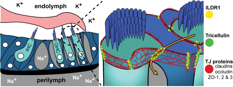
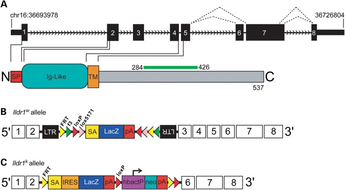
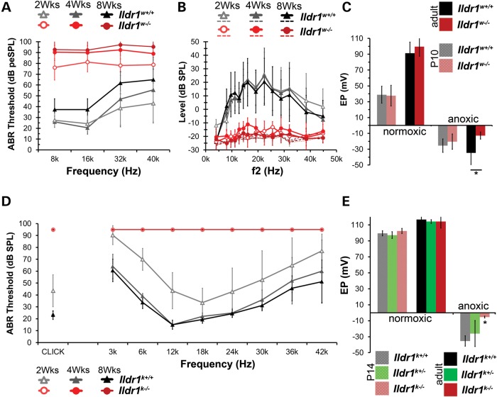
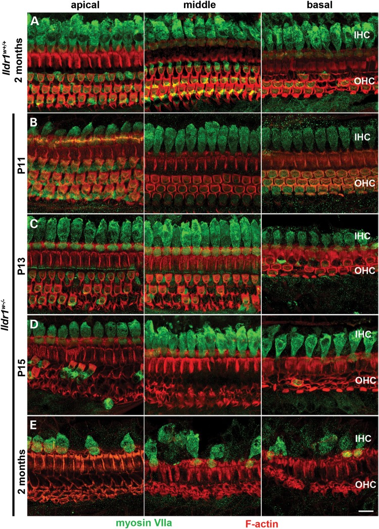
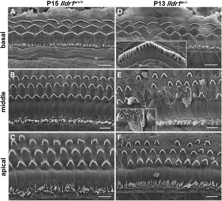
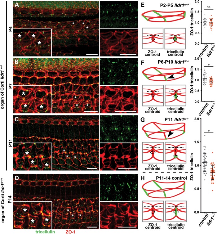
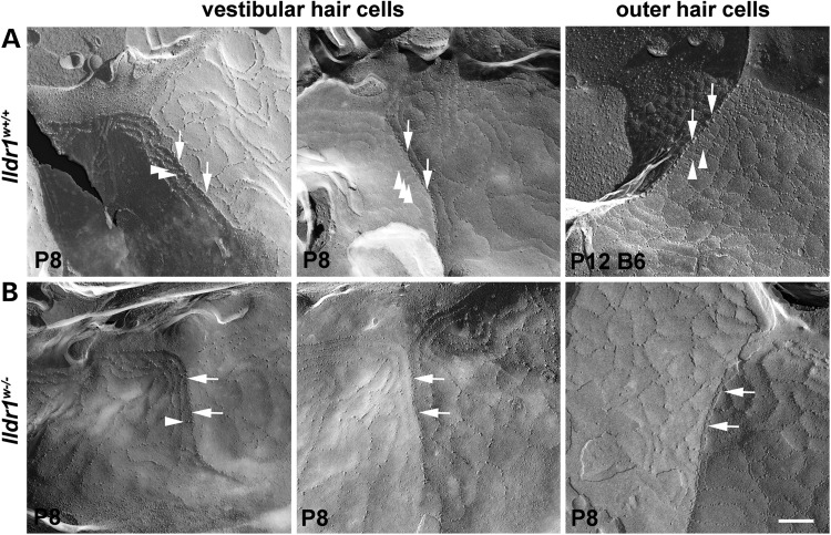
In the mammalian inner ear, bicellular and tricellular tight junctions (tTJs) seal the paracellular space between epithelial cells. Tricellulin and immunoglobulin-like (Ig-like) domain containing receptor 1 (ILDR1, also referred to as angulin-2) localize to tTJs of the sensory and non-sensory epithelia in the organ of Corti and vestibular end organs. Recessive mutations of TRIC (DFNB49) encoding tricellulin and ILDR1 (DFNB42) cause human nonsyndromic deafness. However, the pathophysiology of DFNB42 deafness remains unknown. ILDR1 was recently reported to be a lipoprotein receptor mediating the secretion of the fat-stimulated cholecystokinin (CCK) hormone in the small intestine, while ILDR1 in EpH4 mouse mammary epithelial cells in vitro was shown to recruit tricellulin to tTJs. Here we show that two different mouse Ildr1 mutant alleles have early-onset severe deafness associated with a rapid degeneration of cochlear hair cells (HCs) but have a normal endocochlear potential. ILDR1 is not required for recruitment of tricellulin to tTJs in the cochlea in vivo; however, tricellulin becomes mislocalized in the inner ear sensory epithelia of ILDR1 null mice after the first postnatal week. As revealed by freeze-fracture electron microscopy, ILDR1 contributes to the ultrastructure of inner ear tTJs. Taken together, our data provide insight into the pathophysiology of human DFNB42 deafness and demonstrate that ILDR1 is crucial for normal hearing by maintaining the structural and functional integrity of tTJs, which are critical for the survival of auditory neurosensory HCs.


Figures

Figure 1.
Figure 1.
Tight junction composition and function in the mammalian inner ear. The bTJs and tTJs are composed of integral membrane and membrane-associated proteins that form paired, parallel horizontal and vertical strands with those in adjacent cells (red). In the inner ear, bTJs and tTJs help separate K+ rich endolymph from Na+ rich perilymph. Tricellulin (green) and ILDR1 (yellow) localize to tricellular contacts of both sensory and non-sensory epithelia. In the area where three cells meet, tTJ components form a central tube, which is responsible for most macromolecule paracellular flux (orange arrow).
Figure 2.
Figure 2.
Schematic representation of ILDR1 and the two mutant alleles of mouse Ildr1 used in this study. (A) ILDR1 is a 537 amino acid protein encoded by 8 exons and has an N-terminal signal peptide (red), an Ig-like domain (teal) and a transmembrane domain (orange). To date, three isoforms of ILDR1 have been reported. Exon 6 is alternatively spliced and exon 7 has two donor splice sites (black dotted lines).The epitope for the commercial ILDR1 antibody (Sigma-Aldrich) used in this study, encoded by a portion of exon 7, is shown as green bar above the protein. (B) Gene trap allele of Ildr1Gt(D178D03)Wrst (Ildr1w−/−) was produced from insertion of targeting vector (rFlipROSAβgeo (Cre)) into intron 2 of Ildr1 (197 bp internal to the exon 2/intron 2 boundary). (C) Ildr1tm1(KOMP)Wtsi allele (Ildr1k−/−) was generated by targeted replacement of exons 3–5 with a Lac-Z cassette.
Figure 3.
Figure 3.
ILDR1 localizes to tTJs between epithelial cells of the inner ear and is absent in Ildr1w−/− mice. In wild-type mice, (A) 3D reconstruction image from a confocal z-stack shows ILDR1 (green) localization along the entire depth of the tTJ between HCs and supporting cells, connecting the circumferential rings of ZO-1 (red) at the top and bottom of these junctions (a’–a’’’). Localization of ILDR1 at tTJs between (B) HCs and supporting cells of vestibular sensory epithelia and (C) between stria vascularis marginal cells of Ildr1w+/+ mice. No ILDR1 was detected in Ildr1w−/− mice (D–F). Asterisks in (A) and (D) indicate magnified HCs. Arrowheads in (B) and (C) point to ILDR1 localization at tricellular contacts. Scale bars: 10 µm.
Figure 4.
Figure 4.
Ildr1w−/ mice are severely hearing impaired but have a normal EP. (A) Mean ABR thresholds of Ildr1w−/− and Ildr1w+/+ littermates at 2 weeks (n = 8 each), 4 weeks (n = 11 each) and 8 weeks of age (n = 11 each). Ildr1w−/− mice have higher ABR thresholds than those of Ildr1w+/+ littermates starting at 2 weeks, which increased into adulthood. Ildr1w+/− mice have a hearing phenotype indistinguishable from Ildr1w+/+ littermates (Supplementary Material, Fig. S1). (B) Mean DPOAE levels for the same Ildr1w−/− and Ildr1w+/+ littermates tested with ABRs. Ildr1w−/− mice have no detectable DPOAE levels at 2, 4 and 8 weeks of age (n = 8, 11, and 11, respectively, per genotype). Dotted lines represent noise floor corresponding to each age and genotype. DPOAEs of Ildr1w+/− mice were indistinguishable from Ildr1w+/+ littermates at all three ages tested (Supplementary Material, Fig. S1). (C) EP measurements from adult (P56-85) and P10 Ildr1w−/− and Ildr1w+/+ littermates. P10 Ildr1w−/− mice establish normal EPs under both normoxic and anoxic conditions (n = 6 and 7 for Ildr1w+/+ and Ildr1w−/−, respectively). However, the negative EP under anoxic conditions was smaller in adult Ildr1w−/− mice compared with Ildr1w+/+ mice (Student's t-test, P < 0.05, n = 5, Ildr1w+/+, n = 4, Ildr1w−/−) while the positive EP in adult Ildr1w−/− mice developed a normal magnitude (n = 7, Ildr1w+/+and Ildr1w−/−). (D) Mean ABR thresholds of Ildr1k−/− and Ildr1k+/+ littermates at 2 weeks (n = 7 each), 4 weeks (n = 5 Ildr1k+/+, 6 Ildr1k−/−) and 8 weeks of age (n = 8 Ildr1k+/+, 12 Ildr1k−/−). Ildr1k−/− mice showed no responses at 95 dB SPL, the maximum sound level used. Difference in ABR threshold elevation between Ildr1w−/− and Ildr1k−/− mice by the second postnatal week may due to variation in the genetic background. Ildr1w−/− and Ildr1k−/− are on Swiss Webster and C57BL/6N genetic backgrounds, respectively. Mutant alleles affecting hearing on a C57BL/6 background have a more severe deafness phenotype (32). (E) Positive EP and negative EP measurements made in P14 Ildr1k+/+, Ildr1k+/− and Ildr1k−/− mice. No significant difference was found for positive EP measurements (ANOVA, P > 0.05). However, the anoxic EP showed a significantly lower magnitude in the Ildr1k−/− compared with the Ildr1k+/+ and Ildr1k+/− controls (ANOVA, P < 0.01). Positive EP measurements made in 10 week old Ildr1k+/+, Ildr1k+/− and Ildr1k−/− mice showed no significant difference (ANOVA, P > 0.05). Error bars (A–E) are shown as ±SD.
Figure 5.
Figure 5.
OHCs of Ildr1w−/− mice rapidly degenerate around hearing onset followed by a slow IHC loss in adult mice. Maximum intensity projections show degeneration of auditory HCs in Ildr1w−/− mice. Hair cell bodies were probed with antibody against myosin VIIa (green) and F-actin was labeled with phalloidin (red). (A) Apical, middle and basal sensory epithelium in adult, 2 month old Ildr1w+/+ control mice. (B–E) Apical, middle and basal sensory epithelium from P11 Ildr1w−/−, P13 Ildr1w−/−, P15 Ildr1w−/−, and adult Ildr1w−/− mice indicate that HCs develop normally but OHCs begin to degenerate around hearing onset. (E) In adult, 2 month old Ildr1w−/− mice, gradual loss of IHCs is observed. Scale bar: 10 µm.
Figure 6.
Figure 6.
SEM images show OHC degeneration in the organ of Corti from P13 Ildr1w−/− mice in comparison to P15 Ildr1w+/+ controls. (A, D) basal turn, (B, E) middle turn and (C, F) apical turn. Note degeneration and loss of OHCs in all three turns of Ildr1w−/− mice. Inset in (D) shows a close-up view of a degenerating OHC from another image of the basal turn. Note the loss of shorter-row stereocilia. Inset in (E) shows close-up of degenerating OHC apical membrane fusion. IHC degeneration is not evident at P13. Scale bars: 5 mm.
Figure 7.
Figure 7.
Tricellulin localizes to tTJs in the absence of ILDR1, prior to degeneration of auditory HCs. (A–D) 3D reconstructions of z-stack confocal images from whole-mount organ of Corti samples, probed with antibodies directed against tricellulin (green) and ZO-1 (red). (A) In P4 Ildr1w−/− mice, tricellulin is visualized along the entire tTJ depth. (E) Cartoon schematic of tricellulin localization. Velocity measurement parameters and graph show that tricellulin is localized normally in P2–P5 Ildr1w−/− mice (mean age = 3.81 ± 1.19) compared with control animals, P > 0.05, n = 4 control animals and 5 experimental animals. (B) By P7 (mean age of P6–P10 group = 7.54 ± 0.72 SD), some mislocalization begins to be observed in Ildr1w−/− mice. (F) Arrowhead in cartoon depicts mislocalization at P7, P < 0.005, n = 5 animals per group. (C) In P11 Ildr1w−/− mice, mislocalization is apparent due to lack of tricellulin signal toward the top of the tTJ and its concentration near the basal end of the tTJ. (G) Arrowhead in cartoon shows interpreted mislocalization at P11; and velocity measurements and graph for the P11–P14 control group show a clear statistical significant mislocalization of tricellulin, P < 0.000001, n = 6 animals per group. 3D reconstruction of (D–H) Ildr1w+/+ control mice show tricellulin localization as outlined in the cartoon. Drawings and arrowheads show interpretation of tricellulin mislocalization in Ildr1w−/− mice as the cuticular plate thickens from P4 to P11 and visual description of Velocity measurements. Insets show high magnification of OHCs from the corresponding age and asterisks indicate HCs depicted in magnified panels. Error bars (E–H) are shown as ±SD. Scale bars 10 µm.
Figure 8.
Figure 8.
ILDR1 is required for the normal ultrastructure of the tTJ. Freeze-fracture images of the TJs in the tricellular region (tTJ) where a HC and two supporting cells are in contact. (A) P8 Ildr1w+/+ mouse (left and central panels) and P12 C57Bl6/J wild-type mouse (right panel). (B) P8 Ildr1w−/− mouse. Vestibular sensory epithelium shown in the left and central panels (A and B), and OHC of the organ of Corti in the panels at the right. In the wild type (three panels in (A)), the contact between the three cells is defined as a prominent perpendicular strand of particles, which is the central element of the tTJ, to which several near horizontal strands, which are elements of bTJs, connect to tTJs. In the Ildr1w−/− animals (three panels in B), the central element of tTJs is less prominent with fewer particles (arrows) and the majority of the bTJ strands as they approach the tricellular junction run parallel to the tricellular junctional element rather than running perpendicular to it; only a few run into and join it (arrowheads). Scale bar: 200 nm.

Similar articles

Cited by

References

    1. Bazzoni G., Dejana E. Endothelial cell-to-cell junctions: molecular organization and role in vascular homeostasis. Physiol. Rev. 2004;84:869–901. - PubMed
    1. Anderson J.M., Van Itallie C.M. Physiology and function of the tight junction. Cold Spring Harb. Perspect. Biol. 2009;1:a002584. - PMC - PubMed
    1. Steed E., Balda M.S., Matter K. Dynamics and functions of tight junctions. Trends Cell Biol. 2010;20:142–149. - PubMed
    1. Wade J.B., Karnovsky M.J. The structure of the zonula occludens. A single fibril model based on freeze-fracture. J. Cell Biol. 1974;60:168–180. - PMC - PubMed
    1. Fanning A.S., Van Itallie C.M., Anderson J.M. Zonula occludens-1 and -2 regulate apical cell structure and the zonula adherens cytoskeleton in polarized epithelia. Mol. Biol. Cell. 2012;23:577–590. - PMC - PubMed

Publication types

MeSH terms

Substances

Supplementary concepts