. 2014 Oct;42(18):11763-76.
doi: 10.1093/nar/gku793. Epub 2014 Sep 12.

Ms1, a novel sRNA interacting with the RNA polymerase core in mycobacteria

Affiliations

Ms1, a novel sRNA interacting with the RNA polymerase core in mycobacteria

Jarmila Hnilicová et al. Nucleic Acids Res. 2014 Oct.

Abstract

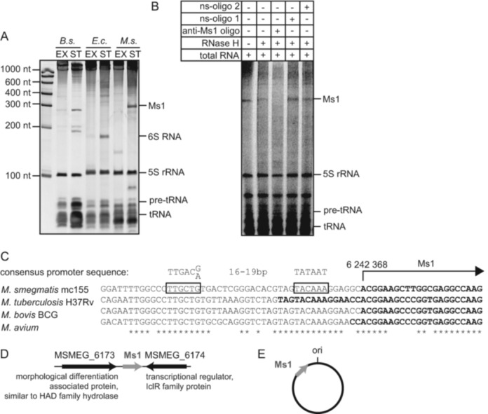
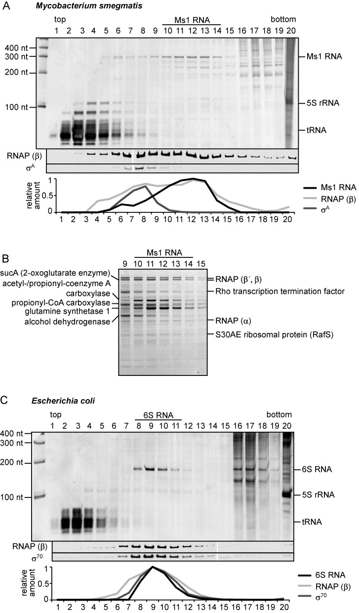
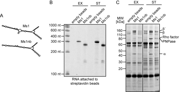
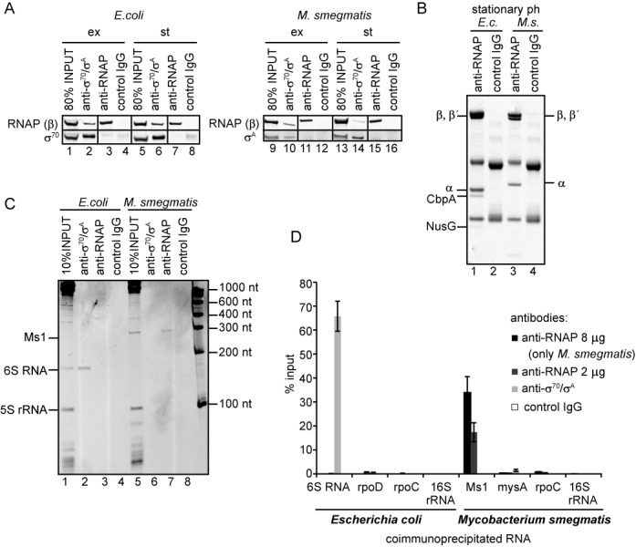
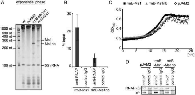
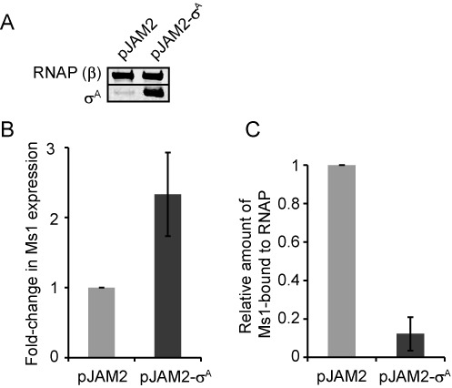
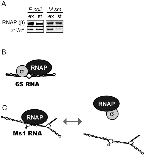
Small RNAs (sRNAs) are molecules essential for a number of regulatory processes in the bacterial cell. Here we characterize Ms1, a sRNA that is highly expressed in Mycobacterium smegmatis during stationary phase of growth. By glycerol gradient ultracentrifugation, RNA binding assay, and RNA co-immunoprecipitation, we show that Ms1 interacts with the RNA polymerase (RNAP) core that is free of the primary sigma factor (σA) or any other σ factor. This contrasts with the situation in most other species where it is 6S RNA that interacts with RNAP and this interaction requires the presence of σA. The difference in the interaction of the two types of sRNAs (Ms1 or 6S RNA) with RNAP possibly reflects the difference in the composition of the transcriptional machinery between mycobacteria and other species. Unlike Escherichia coli, stationary phase M. smegmatis cells contain relatively few RNAP molecules in complex with σA. Thus, Ms1 represents a novel type of small RNAs interacting with RNAP.


Figures

Figure 1.
Figure 1.
Mycobacterial Ms1 sRNA is expressed in amounts comparable to 6S RNAs. (A) Total RNA was isolated from Bacillus subtilis (B.s.), Escherichia coli (E.c.) and Mycobacterium smegmatis (M.s.) in exponential (EX) or stationary (ST) phase. RNAs were resolved on denaturing polyacrylamide gels and stained with GelRed. In M. smegmatis, an ∼300 nt sRNA was present in stationary phase cells in amounts comparable to B. subtilis or E. coli 6S RNAs. (B) Before loading onto the gel, total RNA from M. smegmatis stationary phase was incubated either with a complementary DNA oligonucleotide (anti-Ms1 oligo) or nonspecific control oligonucleotides (ns-oligo 1 and ns-oligo 2) and treated with RNase H. (C) The first nucleotide of Ms1 is adenine transcribed from position 6 242 368 in the genome. The putative −10 and −35 promoter sequences (framed) are perfectly conserved in M. smegmatis, Mycobacterium tuberculosis, Mycobacterium bovis BCG and Mycobacterium avium. The 5′ end sequences of previously identified Ms1 homologs in these species are highlighted in bold. The consensus promoter sequence was adopted from (36). (D) The flanking genes of Ms1 in M. smegmatis are shown. (E) Scheme of Ms1's position in the genome of M. smegmatis with respect to the origin of replication (ori).
Figure 2.
Figure 2.
Ms1 is present in a large protein complex. Lysates from stationary phase Mycobacterium smegmatis (A) or Escherichia coli (C) cells were fractionated by ultracentrifugation in glycerol gradients; individual fractions (1–20; top to bottom) were collected and the RNAs present in each fraction were resolved on denaturing polyacrylamide gels and stained with GelRed. The RNAP β subunit and primary σ factors were visualized by western blotting. Relative amounts of sRNAs (Ms1 and 6S RNA visualized by GelRed) and proteins detected by western blotting are shown below. (B) Proteins from Ms1 fractions were separated by SDS-PAGE and stained with Coomassie. RNAP subunits but no σ factors were among the most abundant proteins in Ms1 fractions. For details on the mass spectrometry analysis, see Supplementary Table S2. This experiment was performed 3× with identical results.
Figure 3.
Figure 3.
Ms1 pulls down RNA polymerase core. (A) Predicted structures of Ms1 and its mutant variant lacking the central bubble, Ms1nb. Both Ms1 and Ms1nb RNAs were prepared in vitro, biotinylated and coupled to streptavidin beads. (B) One tenth of the RNA-coated streptavidin beads were run on a urea-polyacrylamide gel as a control for the efficiency of biotinylation and coupling. (C) The Ms1- and Ms1nb-beads were incubated with Mycobacterium smegmatis exponential or stationary phase lysates. Proteins that associated with Ms1 and Ms1nb were separated by SDS-PAGE, stained with Coomassie and Ms1-interacting proteins analyzed by mass spectrometry. Core subunits of RNAP interacted with Ms1 sRNA, but considerably less with Ms1nb and not with empty beads. This experiment was repeated 3× with identical results.
Figure 4.
Figure 4.
Ms1 interacts with core RNA polymerase. (A) Antibodies against RNAP (anti-RNAP; clone name 8RB13-it recognizes the core form of RNAP) and σA/σ70 (anti-σA70, clone name 2G10-it recognizes also the holoenzyme containing σA/σ70) efficiently immunoprecipitated proteins from both Mycobacterium smegmatis and Escherichia coli. Immunoprecipitated proteins were visualized by western blotting. ex: exponential phase; st: stationary phase. ‘Control IgG’ is a mouse nonspecific IgG used as a negative control. The experiment was repeated 3× with identical results. (B) The RNAP antibody (8RB13) immunoprecipitated the core form of RNA polymerase. Immunoprecipitated proteins from E. coli (E.c.) and M. smegmatis (M.s.) stationary phase lysates were separated by SDS-PAGE and stained with Coomassie. ‘Control IgG’ stands for a mouse nonspecific IgG used as a negative control. Additional E. coli proteins were identified by mass spectrometry (see Supplementary Table S4). No σ factors were detected. No significant bands besides RNAP subunits were found for M. smegmatis. (C) RNAs coimmunoprecipitated with RNAP (2 μg of antibody) and σA/σ70 antibodies were resolved on PAGE and stained with GelRed or (D) quantified by RT-qPCR and normalized to the input. In immunoprecipitations quantified by RT-qPCR, two amounts of the anti-RNAP antibody (8RB13) were used (2 and 8 μg); the amount of coimmunoprecipitated Ms1 increased with the increased amount of the RNAP antibody, suggesting that the concentration of the RNAP antibody was not saturating. E. coli 6S RNA coimmunoprecipitated with σ70 which is in complex with RNA polymerase. None of the control mRNAs-two mRNAs: rpoD (σ70 mRNA), mysA (σA mRNA), rpoC (RNAP β′ subunit mRNA) and 16S rRNA were coimmunoprecipitated with the antibodies used. ‘Control IgG’ is mouse nonspecific IgG used as a negative control. Error bars are SEM (standard error of the mean) from at least three independent experiments.
Figure 5.
Figure 5.
Overexpression of Ms1 in exponential phase. (A) Total RNA was isolated from the wt control strain that contained the empty pJAM2 vector and from strains carrying plasmids that contained either Ms1 or Ms1nb under the rrnB promoter. The RNAs were then resolved on denaturing polyacrylamide gels and stained with GelRed. Both Ms1 and Ms1nb were highly expressed in exponential phase of growth. (B) Core RNAP was immunoprecipitated with 2 μg of the anti-RNAP antibody (8RB13) from strains overexpressing Ms1 or Ms1nb. Coimmunoprecipitated RNA was isolated and the amount of Ms1 or Ms1nb quantified by RT-qPCR. ‘Control IgG’ is a mouse nonspecific IgG used as a negative control. Error bars are SEM from three independent experiments. (C) Growth curves of the control strain (pJAM2) and strains overexpressing Ms1 (rrnB-Ms1) or Ms1nb (rrnB-Ms1nb) were compared. The graph shows one representative experiment; the experiment was repeated 3×. (D) σA was immunoprecipitated from the control strain (pJAM2) and strains overexpressing Ms1 (rrnB-Ms1) or Ms1nb (rrnB-Ms1nb) and the amount of the coimmunoprecipitated RNAP β subunit was determined by western blotting. No difference in the amount of RNAP-bound σA was detected upon Ms1 or Ms1nb overexpression. ‘Control IgG’ is a mouse nonspecific IgG used as a negative control.
Figure 6.
Figure 6.
σA overexpression decreases the amount of Ms1-RNAP. (A) Protein lysates from Mycobacterium smegmatis carrying pJAM2-σA or the empty pJAM2 plasmid were cultured for 6 h in 0.2% acetamide (which induces expression from the pJAM2 acetamidase promoter) and harvested in stationary phase. The amount of σA and RNA polymerase β subunit (RNA polymerase β served as a loading control) was detected by western blotting. Upon induction with acetamide, the σA level increased in M. smegmatis stationary phase cells carrying pJAM2-σA but not in cells carrying the empty vector. (B) Ms1 sRNA expression increases ∼2.5-fold after the overexpression of σA (Ms1 RNA level was measured by RT-qPCR and normalized to 16S rRNA). (C) The amount of Ms1 coimmunoprecipitated with RNAP core decreased more than 8-fold after the overexpression of σA (coimmunoprecipitated Ms1 was first normalized to the input and then to control cells with the empty pJAM2; the graph shows the averages from two independent experiments and the error bars indicate the range).
Figure 7.
Figure 7.
Modes of interaction of sRNAs with bacterial RNAP. (A) The level of σA relative to β dropped in Mycobacterium smegmatis cells harvested 12 h after entry into stationary phase. In Escherichia coli, the relative protein level of σ70 to β remained unchanged even after 16 h in stationary phase of growth. The experiment was repeated 3× with identical results. (B) 6S RNA (e. g. E. coli, Bacillus subtilis) binds to RNAP containing the main σ factor. (C) Ms1 (mycobacteria) binds to the RNAP core in the absence of σ factors and the presence of σA decreases this interaction.

Similar articles

  • Ms1 RNA increases the amount of RNA polymerase in Mycobacterium smegmatis.
    Šiková M, Janoušková M, Ramaniuk O, Páleníková P, Pospíšil J, Bartl P, Suder A, Pajer P, Kubičková P, Pavliš O, Hradilová M, Vítovská D, Šanderová H, Převorovský M, Hnilicová J, Krásný L. Šiková M, et al. Mol Microbiol. 2019 Feb;111(2):354-372. doi: 10.1111/mmi.14159. Epub 2018 Dec 11. Mol Microbiol. 2019. PMID: 30427073
  • RIP-seq reveals RNAs that interact with RNA polymerase and primary sigma factors in bacteria.
    Vaňková Hausnerová V, Shoman M, Kumar D, Schwarz M, Modrák M, Jirát Matějčková J, Mikesková E, Neva S, Herrmannová A, Šiková M, Halada P, Novotná I, Pajer P, Valášek LS, Převorovský M, Krásný L, Hnilicová J. Vaňková Hausnerová V, et al. Nucleic Acids Res. 2024 May 8;52(8):4604-4626. doi: 10.1093/nar/gkae081. Nucleic Acids Res. 2024. PMID: 38348908 Free PMC article.
  • MoaB2, a newly identified transcription factor, binds to σA in Mycobacterium smegmatis.
    Brezovská B, Narasimhan S, Šiková M, Šanderová H, Kovaľ T, Borah N, Shoman M, Pospíšilová D, Vaňková Hausnerová V, Tužinčin D, Černý M, Komárek J, Janoušková M, Kambová M, Halada P, Křenková A, Hubálek M, Trundová M, Dohnálek J, Hnilicová J, Žídek L, Krásný L. Brezovská B, et al. J Bacteriol. 2024 Dec 19;206(12):e0006624. doi: 10.1128/jb.00066-24. Epub 2024 Nov 5. J Bacteriol. 2024. PMID: 39499088 Free PMC article.
  • Small Noncoding 6S RNAs of Bacteria.
    Burenina OY, Elkina DA, Hartmann RK, Oretskaya TS, Kubareva EA. Burenina OY, et al. Biochemistry (Mosc). 2015 Nov;80(11):1429-46. doi: 10.1134/S0006297915110048. Biochemistry (Mosc). 2015. PMID: 26615434 Review.
  • 6S RNA, a Global Regulator of Transcription.
    Wassarman KM. Wassarman KM. Microbiol Spectr. 2018 May;6(3):10.1128/microbiolspec.rwr-0019-2018. doi: 10.1128/microbiolspec.RWR-0019-2018. Microbiol Spectr. 2018. PMID: 29916345 Free PMC article. Review.

Cited by

References

    1. Stewart G.R., Robertson B.D., Young D.B. Tuberculosis: a problem with persistence. Nat. Rev. Microbiol. 2003;1:97–105. - PubMed
    1. Waagmeester A., Thompson J., Reyrat J.M. Identifying sigma factors in Mycobacterium smegmatis by comparative genomic analysis. Trends Microbiol. 2005;13:505–509. - PubMed
    1. Sharma U.K., Chatterji D. Transcriptional switching in Escherichia coli during stress and starvation by modulation of sigma activity. FEMS Microbiol. Rev. 2010;34:646–657. - PubMed
    1. Paget M.S., Helmann J.D. The sigma70 family of sigma factors. Genome Biol. 2003;4:203.1–203.4. - PMC - PubMed
    1. Österberg S., del Peso-Santos T., Shingler V. Regulation of alternative sigma factor use. Annu. Rev. Microbiol. 2011;65:37–55. - PubMed

Publication types

MeSH terms

LinkOut - more resources