. 2014 Sep 12;3(10):904-12.
doi: 10.1242/bio.20149415.

Co-activator independent differences in how the metaphase and anaphase APC/C recognise the same substrate

Affiliations

Co-activator independent differences in how the metaphase and anaphase APC/C recognise the same substrate

Takahiro Matsusaka et al. Biol Open. .

Abstract

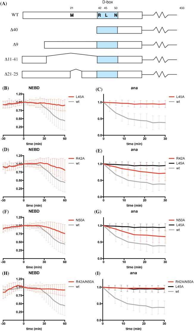
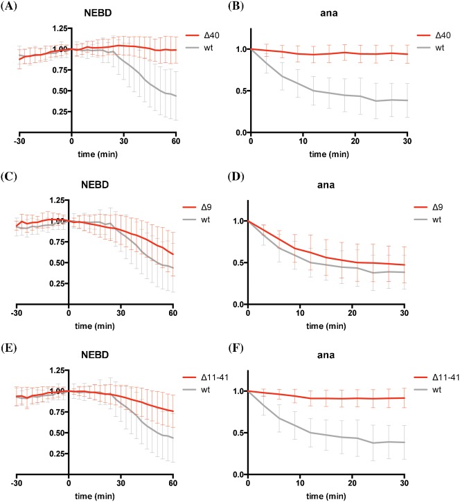
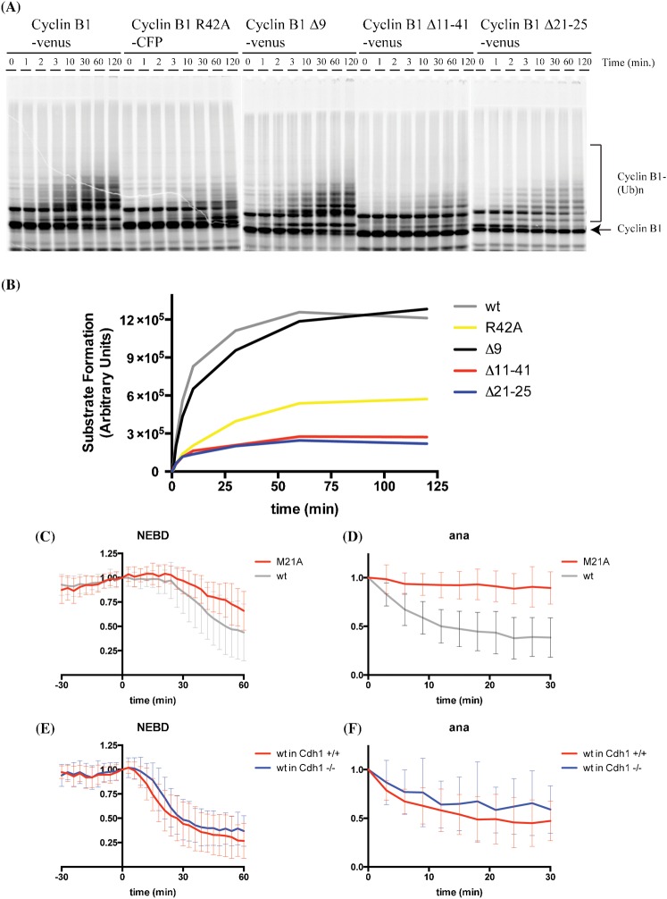
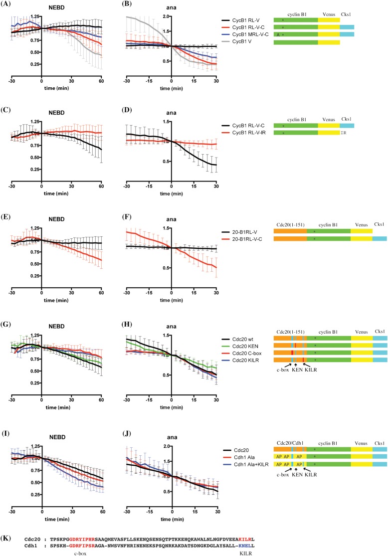
The Anaphase Promoting Complex or Cyclosome (APC/C) is critical to the control of mitosis. The APC/C is an ubiquitin ligase that targets specific mitotic regulators for proteolysis at distinct times in mitosis, but how this is achieved is not well understood. We have addressed this question by determining whether the same substrate, cyclin B1, is recognised in the same way by the APC/C at different times in mitosis. Unexpectedly, we find that distinct but overlapping motifs in cyclin B1 are recognised by the APC/C in metaphase compared with anaphase, and this does not depend on the exchange of Cdc20 for Cdh1. Thus, changes in APC/C substrate specificity in mitosis can potentially be conferred by altering interaction sites in addition to exchanging Cdc20 for Cdh1.

Keywords: Anaphase Promoting Complex/Cyclosome; Cyclin B; Mitosis.


Conflict of interest statement

Competing interests: The authors declare no conflict of interest.

Figures

Fig. 1.
Fig. 1.. L45 of the Cyclin B1 D-box is most important for degradation.
(A) Schematics of cyclin B1 constructs used in this study. The D-box is highlighted in blue. (B,C) HeLa cells were injected with cyclin B1-Venus (grey, n = 36) or cyclin B1 L45A-Venus (red, n = 24) constructs and followed by time-lapse fluorescence and DIC microscopy at 3-min intervals. The total fluorescence minus background was quantified for each cell in successive images of a time series and plotted over time as mean ± SD from 3 independent experiments. Fluorescence of cells at NEBD (B) or anaphase onset (C) was set to 1. Time 0 is NEBD in (B) or anaphase onset in (C). (D,E) HeLa cells were injected with cyclin B1-Venus (grey, n = 36), L45A-Venus (black, n = 24) or cyclin B1 R42A-Venus (red, n = 55) constructs and analysed as in panels B and C. Fluorescence of cells at NEBD (D) or anaphase onset (E) was set to 1. Time 0 is NEBD in (D) or anaphase onset in (E). Data are from 3 independent experiments. (F,G) HeLa cells were injected with cyclin B1-Venus (grey, n = 36), L45A-Venus (black, n = 24) or cyclin B1 N50A-Venus (red, n = 33) constructs and analysed as in panels B and C. Fluorescence of cells at NEBD (F) or anaphase onset (G) was set to 1. Time 0 is NEBD in (F) or anaphase onset in (G). Data are from 3 (wt and L45A) or 2 (N50A) independent experiments. (H,I) HeLa cells were injected with cyclin B1-Venus (grey, n = 36), L45A-Venus (black, n = 24) or cyclin B1 R42A/N50A-Venus (red, n = 38) constructs and analysed as in panels B and C. Data are from 3 independent experiments.
Fig. 2.
Fig. 2.. The first 40 amino acids of cyclin B1 are required for its degradation.
(A,B) HeLa cells were injected with cyclin B1-Venus (grey, n = 36) or cyclin B1 Δ40-Venus (red, n = 45) constructs and analysed as in Fig. 1. Data are from 3 independent experiments. (C,D) HeLa cells were injected with cyclin B1-Venus (grey, n = 36) or cyclin B1 Δ9-Venus (red, n = 43) constructs and analysed as in Fig. 1. Data are from 3 independent experiments. (E,F) HeLa cells were injected with cyclin B1-Venus (grey, n = 36) or cyclin B1 Δ11–41-Venus (red, n = 47) constructs and analysed as in Fig. 1. Data are from 3 independent experiments. Note that the data for wild type cyclin B1 degradation are the same as those in Fig. 1.
Fig. 3.
Fig. 3.. Residues N-terminal to the canonical D-box of cyclin B1 are required for its ubiquitylation and degradation in anaphase.
(A) In vitro ubiquitylation reactions using purified budding yeast APC/C-Cdh1 and the indicated in vitro translated human cyclin B1 mutants. Data are representative of results from two independent experiments. (B) The data from (A) were quantified and the amount of ubiquitylated cyclin B1 was plotted as a function of time. (C,D) HeLa cells were injected with cyclin B1-Venus (grey, n = 36) or cyclin B1 M21A-Venus (red, n = 20) constructs and analysed as in Fig. 1. Data are from 3 independent experiments. (E,F) Cdh1+/+ (red), or Cdh1−/− (blue) mouse embryonic fibroblasts were transfected with cyclin B1-Venus constructs and analysed as in Fig. 1. Error bars indicate mean ± SD of 41 and 50, cells, for panels E and F, respectively.
Fig. 4.
Fig. 4.. The N-terminus of Cdc20 can substitute for a D-box when cyclin B1 is targeted to the APC/C by Cks1.
(A,B) The anaphase APC/C can degrade a D-box mutant of cyclin B1 targeted to it by Cks1. HeLa cells were injected with cyclin B1 R42A/L45A double mutant-venus (RL-V, black, n = 35), cyclin B1 R42A/L45A double mutant-venus-cks1 (RL-V-C, red, n = 24), cyclin B1 M21A/R42A/L45A triple mutant venus-cks1 (MRL-V-C, blue, n = 20), or cyclin B1-venus (grey, n = 36) constructs and analysed as in Fig. 1. Data are from 3 independent experiments. (C,D) Cks1 cannot be substituted by a C-terminal IR motif. HeLa cells were injected with cyclin B1 R42A/L45A double mutant-venus-cks1 (RL-V-C, black, n = 24) or cyclin B1 R42A/L45A double mutant-venus-IR (RL-V-IR, red, n = 29) constructs and analysed as in Fig. 1. Data are from 3 independent experiments. (E,F) The N-terminus of Cdc20 can promote degradation of cyclin B1 lacking its destruction box in metaphase if it is targeted to the APC/C by Cks1. The N-terminal 151 residues of Cdc20 were fused to cyclin B1 lacking its D-box, designated Cdc20 (1–151)-R42A/L45A double mutant cyclin B1, with or without Cks1 at the C-terminus. HeLa cells were injected with Cdc20 (1–151) cyclin B1 R42A/L45A double mutant-venus (20-B1RL-V, black, n = 24) or Cdc20 (1–151) cyclin B1 R42A/L45A double mutant-venus-cks1 (20-B1RL-V-C, red, n = 31) constructs and analysed as in Fig. 1. Data are from 3 independent experiments. (G,H) The N-terminus of Cdc20 requires its C-box and KILR motif to promote degradation. HeLa cells were injected with Cdc20 (1–151) cyclin B1 R42A/L45A double mutant-venus-cks1 (Cdc20 wt, black, n = 31), Cdc20 (1–151) KEN cyclin B1 R42A/L45A double mutant-venus-cks1 (Cdc20 KEN, green, n = 14), Cdc20 (1–151) C-box cyclin B1 R42A/L45A double mutant-venus-cks1 (Cdc20 C-box, red, n = 14), or Cdc20 (1–151) KILR cyclin B1 R42A/L45A double mutant-venus-cks1 (Cdc20 KILR, blue, n = 14) constructs and analysed as in Fig. 1. Data are from 3 independent experiments. (I,J) The non-phosphorylatable mutant N-terminus of Cdh1 can also promote degradation of cyclin B1 lacking its D-box. HeLa cells were injected with plasmids encoding Cdc20 (1–151) cyclin B1 R42A/L45A double mutant-venus-cks1 (Cdc20, black, n = 31), or a construct in which the N-terminus of Cdc20 was replaced with the N-terminus of non-phosphorylatable Cdh1, Cdh1 (1–155) Ala-cyclin B1 R42A/L45A double mutant-venus-cks1 (Cdh1 Ala, red, n = 16), or the N-terminus of non-phosphorylatable Cdh1 with the KILR motif of Cdc20, Cdh1 (1–155) Ala+KILR-cyclin B1 R42A/L45A double mutant-venus-cks1 (Cdh1 Ala+KILR, blue, n = 27). Constructs were analysed as in Fig. 1. Data are from 3 independent experiments. (K) Sequence alignment of Cdc20 and Cdh1 around C-box and KILR motif. The residues marked in blue (KNEL) in Cdh1 were mutated to those found in Cdc20 (KILR).

Similar articles

Cited by

References

    1. Amon A., Irniger S., Nasmyth K. (1994). Closing the cell cycle circle in yeast: G2 cyclin proteolysis initiated at mitosis persists until the activation of G1 cyclins in the next cycle. Cell 77, 1037–1050 10.1016/0092-8674(94)90443-X - DOI - PubMed
    1. Araki M., Wharton R. P., Tang Z., Yu H., Asano M. (2003). Degradation of origin recognition complex large subunit by the anaphase-promoting complex in Drosophila. EMBO J. 22, 6115–6126 10.1093/emboj/cdg573 - DOI - PMC - PubMed
    1. Barford D. (2011). Structural insights into anaphase-promoting complex function and mechanism. Philos. Trans. R. Soc. B 366, 3605–3624 10.1098/rstb.2011.0069 - DOI - PMC - PubMed
    1. Brandeis M., Hunt T. (1996). The proteolysis of mitotic cyclins in mammalian cells persists from the end of mitosis until the onset of S phase. EMBO J. 15, 5280–5289. - PMC - PubMed
    1. Burton J. L., Solomon M. J. (2001). D box and KEN box motifs in budding yeast Hsl1p are required for APC-mediated degradation and direct binding to Cdc20p and Cdh1p. Genes Dev. 15, 2381–2395 10.1101/gad.917901 - DOI - PMC - PubMed