. 2014 Nov 1;127(Pt 21):4634-44.
doi: 10.1242/jcs.151787. Epub 2014 Sep 12.

Notch directly regulates the cell morphogenesis genes Reck, talin and trio in adult muscle progenitors

Affiliations

Notch directly regulates the cell morphogenesis genes Reck, talin and trio in adult muscle progenitors

Guillaume Pézeron et al. J Cell Sci. .

Abstract

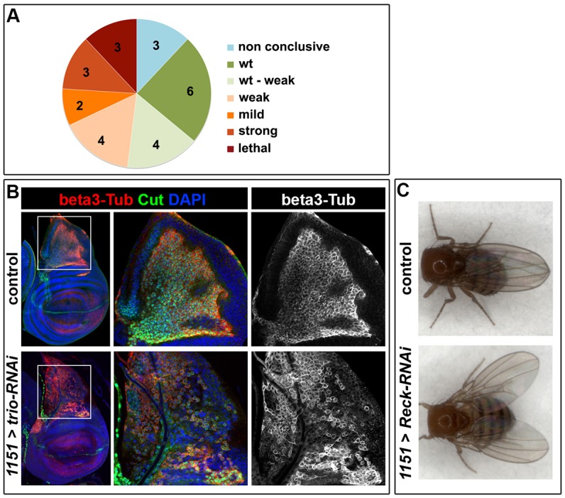
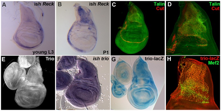
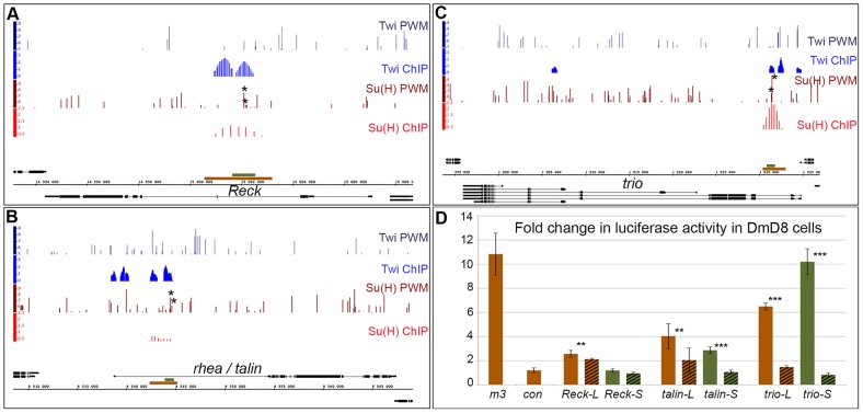
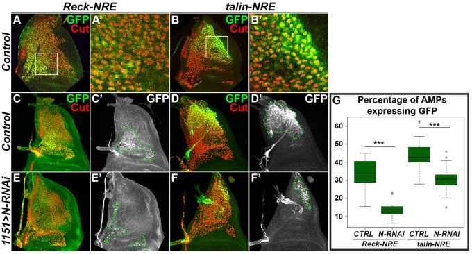
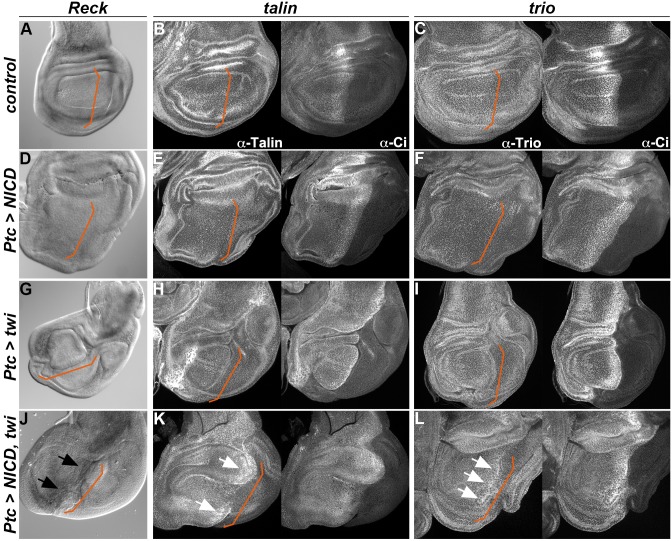
There is growing evidence that activation of the Notch pathway can result in consequences on cell morphogenesis and behaviour, both during embryonic development and cancer progression. In general, Notch is proposed to coordinate these processes by regulating expression of key transcription factors. However, many Notch-regulated genes identified in genome-wide studies are involved in fundamental aspects of cell behaviour, suggesting a more direct influence on cellular properties. By testing the functions of 25 such genes we confirmed that 12 are required in developing adult muscles, consistent with roles downstream of Notch. Focusing on three, Reck, rhea/talin and trio, we verify their expression in adult muscle progenitors and identify Notch-regulated enhancers in each. Full activity of these enhancers requires functional binding sites for Su(H), the DNA-binding transcription factor in the Notch pathway, validating their direct regulation. Thus, besides its well-known roles in regulating the expression of cell-fate-determining transcription factors, Notch signalling also has the potential to directly affect cell morphology and behaviour by modulating expression of genes such as Reck, rhea/talin and trio. This sheds new light on the functional outputs of Notch activation in morphogenetic processes.

Keywords: Drosophila; Gene regulation; Myogenesis; Notch; Reck; Talin; Trio.


Figures

Fig. 1.
Fig. 1.
An RNAi assay identifies genes required for muscle formation. (A) Pie chart showing the proportion of genes whose loss of function resulted in the different phenotypical classes (see Materials and Methods for more details about classes). Note that the 1151-Gal4 driver is expressed in AMPs but not in the larval muscles, to avoid any confounding effects from defects in larval musculature (as might occur with mef2-Gal4). (B) AMP organization in wing imaginal discs from control (1151-Gal4) and trio-RNAi (1151-Gal4 UAS-trio-RNAi) larvae detected with β3-tubulin (beta3-Tub, red). Cut (in green) marks the nuclei of AMPs, and DAPI (blue) labels all nuclei. Altered cell morphology and organization is evident in trio-RNAi-expressing flies compared to control (representative examples of intermediate severity are shown). White squares indicate the regions shown at higher magnification. (C) Dorsal view of control (1151-Gal4) and 1151-Gal4-driven Reck-RNAi adult flies. The defective position of the wings observed, a ‘held out wings’ phenotype, has often been associated with flight muscle defects (Greene et al., 2003; Zaffran et al., 1997).
Fig. 2.
Fig. 2.
Reck, talin and trio are expressed in AMP cells. Expression profiles of Reck, talin and trio in the wing imaginal disc show that all three genes are expressed in AMPs. (A,B) Reck in situ hybridization (ish) in young L3 larvae (A) and P1 pupae (B). (C,D) talin expression profile revealed by immunostaining and co-stained with the AMP marker Cut (maximum projections of z-stacks from confocal acquisitions are presented; D is shown at twice the magnification of C). (E–H) trio expression profile shown by immunostaining (E, Maximum projections), In situ hybridization (F) and an enhancer-trap reporter line (G) also co-stained with the AMP marker mef2 (H) (H is shown at twice the magnification of G).
Fig. 3.
Fig. 3.
Su(H) ChIP identifies NREs in the Reck, talin and trio loci. (A–C) Genomic region surrounding Drosophila Reck (A), talin (B) and trio (C) genes. Black lines and boxes (exons) represent transcribed regions. Graphs show matches to Su(H) PWM (dark red bars; height of bar indicates Patser score 5–9.79), Twist (Twi) PWM (dark blue bars; Patser score 5–9.3), Su(H) [red; enrichment (AvgM log2)] and Twi (blue) ChIP-enriched regions in DmD8 cells (Bernard et al., 2010; Krejci et al., 2009). Brown and green rectangles represent long (brown) and short (green) fragments tested for their Notch signalling activation sensitivity. Asterisks indicate Su(H)-binding sites mutated in subsequent experiments. (D) Response of the indicated long (brown) and short (green) fragment to Notch signalling activation in transient transfection assays in DmD8 cells. Plain bars represent wild-type fragments and striped bars fragments in which Su(H) sites were mutated. E(spl)m3, which is inducible by Notch in all tested conditions (Krejci and Bray, 2007), was used as positive control. Results are mean±s.d. **P<0.05; ***P<0.001.
Fig. 4.
Fig. 4.
Reck and talin NREs drive Notch-dependent GFP expression in AMPs in vivo. (A–B′) Reck (A, a higher magnification is shown in A′) and talin (B, and higher magnification in B′) long NREs (L-NRE) drive GFP expression in a subset of AMPs as shown by GFP colocalizations with the AMP marker Cut. White squares in A and B indicate regions shown at higher magnification in A′ and B′. Single optical sections from confocal acquisitions are presented. (C–F′) Expression from reporters Reck-L-NRE-GFP (C,C′,E,E′) and talin-L-NRE-GFP (D,D′,F,F′) in wild-type (C–D′) and Notch-depleted conditions (E–F′). Cut expression was used to mark the AMPs. Green dotted lines in C′,D′,E′ and F′ outline Cut- and GFP-expressing cells. Maximum projections of z-stacks from confocal acquisitions are presented. (G) Boxplot representing the percentage of AMPs (Cut-expressing cells) expressing GFP from the Reck-L-NRE-GFP and talin-L-NRE-GFP reporters in wild-type (CTRL) and Notch-depleted (N-RNAi) conditions. The percentage of AMPs expressing GFP was estimated by manually measuring areas occupied by cells expressing Cut or Cut and GFP. The box represents the interquartile range, the middle line the median, and the whiskers show ± 1.5× the interquartile range. ***P<0.001.
Fig. 5.
Fig. 5.
Notch can induce ectopic expression of Reck, talin and trio in the presence of Twist. (A–L) ptc-Gal4; Tub-Gal80ts was used to drive expression of NICD and/or Twist (Twi) in the wing pouch. The expression profile of Reck (A,D,G,J, in situ hybridization), talin (B,E,H,K, immunostaining) and trio (C,F,I,L, immunostaining) in a wild-type wing pouch (A–C), in NICD-expressing discs (D–F), Twist-expressing discs (G–I) and NICD- plus Twist-expressing discs (J–L). Note the ectopic expression of Reck (J, black arrows) and the upregulation of talin and trio (K,L, white arrows) induced by Notch in the presence of Twist. Ci staining was used to indicate the limit between anterior and posterior domains, along which ptc-Gal4 is expressed (indicated with orange lines).

References

    1. Anant S., Roy S., VijayRaghavan K. (1998). Twist and Notch negatively regulate adult muscle differentiation in Drosophila. Development 125, 1361–1369 - PubMed
    1. Bate M., Rushton E., Currie D. A. (1991). Cells with persistent twist expression are the embryonic precursors of adult muscles in Drosophila. Development 113, 79–89 - PubMed
    1. Bateman J., Shu H., Van Vactor D. (2000). The guanine nucleotide exchange factor trio mediates axonal development in the Drosophila embryo. Neuron 26, 93–106. 10.1016/S0896-6273(00)81141-1 - DOI - PubMed
    1. Baylies M. K., Bate M. (1996). twist: a myogenic switch in Drosophila. Science 272, 1481–1484. 10.1126/science.272.5267.1481 - DOI - PubMed
    1. Becam I., Milán M. (2008). A permissive role of Notch in maintaining the DV affinity boundary of the Drosophila wing. Dev. Biol. 322, 190–198. 10.1016/j.ydbio.2008.07.028 - DOI - PubMed

Publication types

MeSH terms

LinkOut - more resources