. 2014 Nov 1;127(Pt 21):4549-60.
doi: 10.1242/jcs.153791. Epub 2014 Sep 12.

Novel roles for actin in mitochondrial fission

Affiliations

Novel roles for actin in mitochondrial fission

Anna L Hatch et al. J Cell Sci. .

Abstract

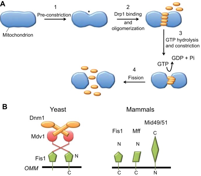
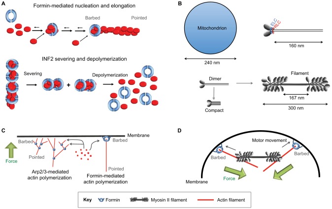
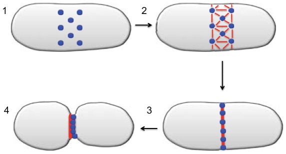
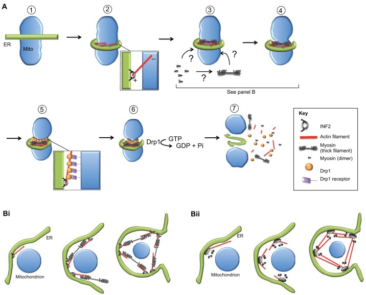
Mitochondrial dynamics, including fusion, fission and translocation, are crucial to cellular homeostasis, with roles in cellular polarity, stress response and apoptosis. Mitochondrial fission has received particular attention, owing to links with several neurodegenerative diseases. A central player in fission is the cytoplasmic dynamin-related GTPase Drp1, which oligomerizes at the fission site and hydrolyzes GTP to drive membrane ingression. Drp1 recruitment to the outer mitochondrial membrane (OMM) is a key regulatory event, which appears to require a pre-constriction step in which the endoplasmic reticulum (ER) and mitochondrion interact extensively, a process termed ERMD (ER-associated mitochondrial division). It is unclear how ER-mitochondrial contact generates the force required for pre-constriction or why pre-constriction leads to Drp1 recruitment. Recent results, however, show that ERMD might be an actin-based process in mammals that requires the ER-associated formin INF2 upstream of Drp1, and that myosin II and other actin-binding proteins might be involved. In this Commentary, we present a mechanistic model for mitochondrial fission in which actin and myosin contribute in two ways; firstly, by supplying the force for pre-constriction and secondly, by serving as a coincidence detector for Drp1 binding. In addition, we discuss the possibility that multiple fission mechanisms exist in mammals.

Keywords: Actin; Mitochondrial fission; Myosin.


Figures

Fig. 1.
Fig. 1.
Role of Drp1 in mitochondrial fission. (A) Schematic illustration of the general steps involved in mitochondrial fission. Step 1: a fission site is marked by an unknown mechanism, and this site undergoes ‘pre-constriction’ (asterisk) prior to the arrival of Drp1 (orange). Step 2: Drp1 binds to the pre-constriction site and oligomerizes. Step 3: GTP hydrolysis by the oligomerized Drp1 causes constriction of the fission site. Step 4: fission occurs by an unknown mechanism. (B) Dnm1/Drp1 receptors in budding yeast (left) and in mammals (right). In budding yeast, the OMM protein Fis1 binds to the dimeric adaptor protein Mdv1, which in turn binds to the Dnm1 dimer (its GTPase domain is represented by the oval). A second adaptor protein, Caf4, can act in place of Mdv1. In mammals, four possible OMM proteins have been postulated to act as Drp1 receptors – Fis1, Mff, MiD49 and MiD51. No adaptor proteins that are homologous to Mdv1 or Caf4 have been identified in mammals.
Fig. 2.
Fig. 2.
Mechanisms of actin-based membrane constriction. (A) The effects of formins on actin. Upper panel, a formin dimer (blue) can enhance the nucleation of actin monomers (red). Subsequently, it remains at the barbed end of the elongating filament, regulating elongation rate by controlling monomer addition. For more detail regarding formin biochemistry, see Higgs, 2005 and Chesarone et al., 2010. Lower panel, INF2 is additionally able to sever filaments and enhance their depolymerization through its ability to bind to the sides of filaments by encircling them. Upon ATP hydrolysis and phosphate release from actin subunits in the filament, INF2 severs the filament and subsequently enhances depolymerization. For further details regarding INF2 biochemistry, see Gurel et al., 2014. (B) Non-muscle myosin II. Upper panel, the fundamental unit of myosin II is a multi-protein complex of two heavy chains, two essential light chains (ELC) and two regulatory light chains (RLC). The heavy chains are tightly dimerized in parallel by their coiled-coil tails of ∼160 nm length. We refer to this fundamental unit as the ‘dimer’ because the dimerized heavy chains dominate the structure. Lower panel, non-muscle myosin II can oligomerize further to create a bipolar filament with the motor domain heads at each end and a ‘bare zone’ in the center. In the presence of ATP, myosin II assumes a ‘10S’ compact structure, presumably by folding the tail into three segments. RLC phosphorylation allows for bipolar filament assembly from the compact structure in the presence of ATP. The latter is a biochemical observation and has not been documented in non-muscle cells. A ‘typical’ mitochondrial diameter is also shown for size comparison. (C) Actin-polymerization-based membrane deformation by monomer addition to membrane-abutting filament barbed ends. Left, the classic example of Arp2/3-complex-based dendritic nucleation is shown. Right, an example of a formin tethered to the membrane directing monomer addition to the barbed end. The Arp2/3 complex and formin are shown in blue. (D) Myosin-II-based membrane deformation. Motor activity of the bipolar myosin II filament on anti-parallel membrane-attached actin filaments causes the deformation and thus constriction of the membrane. In the example shown here, the barbed ends of the filament are tethered to the membrane by a formin.
Fig. 3.
Fig. 3.
Model for fission yeast cytokinesis. Step 1: at the presumptive cleavage site on the plasma membrane, ∼130 ‘nodes’ (blue) assemble the cytokinetic components; each node contains two formin dimers and eight myosin II dimers. Step 2: the formins nucleate actin filaments (red), which remain attached to the formin at their barbed ends. The pointed end of the actin filament interacts with myosin II on a neighboring node by a ‘search and capture’ mechanism. Step 3: myosin II activity along the actin filaments causes the nodes to condense into a compact ring structure. Step 4: further myosin II activity causes the ring to further condense, thereby causing constriction of the plasma membrane. Additional force-generating mechanisms might exist that become more important at subsequent stages of constriction and fission (Proctor et al., 2012).
Fig. 4.
Fig. 4.
The mitokinesis model of mitochondrial fission. (A) Step 1: The ER (green) and mitochondrion (blue) interact at the future site of mitochondrial fission. Step 2: actin filaments (red) are nucleated and elongate with INF2 (gray donut) at their barbed ends, thus staying tethered to the ER through INF2. Step 3: Myosin II is recruited to the fission site. The recruited myosin might come from a cytosolic pool of free dimers or from pre-assembled bipolar filaments. Step 4 (pre-constriction): myosin II activity on anti-parallel actin filaments causes deformation of the network, resulting in constriction of both the surrounding ER and the underlying mitochondrion. Step 5 (coincidence detection): Drp1 (brown circles) binds and oligomerizes at the pre-constriction site, owing to coincidence detection of two signals – an OMM-bound Drp1 receptor (purple) and actin filaments. Step 6: GTPase activity of Drp1 causes increased mitochondrial constriction. Step 7: The actual membrane fission process occurs, and components of the fission complex (actin, myosin, Drp1 oligomer) disassemble. (B) Two possible models for myosin II arrangement during mitokinesis. Side views of the enlarged fission site at Step 3 and 4 of the model depicted in A are shown. In both models, ER is not required to completely encircle the mitochondrion, but encircles sufficiently to allow a continuous actomyosin ring around the mitochondrion. (Bi) Bipolar arrangement. Myosin II is assembled in the form of bipolar filaments around the mitochondrion and acts on actin filaments that are bound by INF2 at the ER membrane to constrict both ER and the mitochondrion. (Bii) Nodal arrangement. Several nodes assemble on the ER membrane and each contains INF2 and myosin II. Actin filaments that are assembled at one node bind to myosin II at neighboring nodes. Myosin II activity then pulls the nodes together, in turn constricting both the ER and mitochondrial membranes. In this model, similar to models of fission yeast cytokinesis (see Fig. 3), the myosin II is organized as individual dimers that are attached to the nodes by their tails. Possibly, the tails are also folded so that the myosin II dimer is shorter than its extended length of 175 nm.

Similar articles

Cited by

References

    1. Anand R., Wai T., Baker M. J., Kladt N., Schauss A. C., Rugarli E., Langer T. (2014). The i-AAA protease YME1L and OMA1 cleave OPA1 to balance mitochondrial fusion and fission. J. Cell Biol. 204, 919–929. 10.1083/jcb.201308006 - DOI - PMC - PubMed
    1. Archer S. L. (2013). Mitochondrial dynamics – mitochondrial fission and fusion in human diseases. N. Engl. J. Med. 369, 2236–2251. 10.1056/NEJMra1215233 - DOI - PubMed
    1. Beck H., Flynn K., Lindenberg K. S., Schwarz H., Bradke F., Di Giovanni S., Knöll B. (2012). Serum Response Factor (SRF)-cofilin-actin signaling axis modulates mitochondrial dynamics. Proc. Natl. Acad. Sci. USA 109, E2523–E2532. 10.1073/pnas.1208141109 - DOI - PMC - PubMed
    1. Belin B. J., Mullins R. D. (2013). What we talk about when we talk about nuclear actin. Nucleus 4, 291–297. 10.4161/nucl.25960 - DOI - PMC - PubMed
    1. Benda C. (1898). Arch. Anat. Physiol. 73, 393–398

Publication types