. 2014 Nov 7;289(45):31253-61.
doi: 10.1074/jbc.M114.581918. Epub 2014 Sep 12.

Neurogenin 2 mediates amyloid-β precursor protein-stimulated neurogenesis

Affiliations

Neurogenin 2 mediates amyloid-β precursor protein-stimulated neurogenesis

Marta Bolós et al. J Biol Chem. .

Abstract

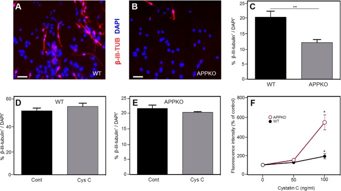
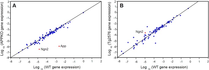
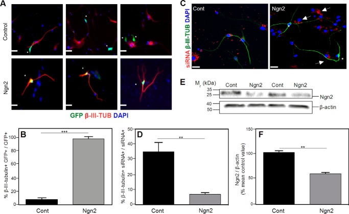
Amyloid-β precursor protein (APP) is well studied for its role in Alzheimer disease, although its normal function remains uncertain. It has been reported that APP stimulates the proliferation and neuronal differentiation of neural stem/progenitor cells (NSPCs). In this study we examined the role of APP in NSPC differentiation. To identify proteins that may mediate the effect of APP on NSPC differentiation, we used a gene array approach to find genes whose expression correlated with APP-induced neurogenesis. We found that the expression of neurogenin 2 (Ngn2), a basic helix-loop-helix transcription factor, was significantly down-regulated in NSPCs from APP knock-out mice (APPKO) and increased in APP transgenic (Tg2576) mice. Ngn2 overexpression in APPKO NSPCs promoted neuronal differentiation, whereas siRNA knockdown of Ngn2 expression in wild-type NSPCs decreased neuronal differentiation. The results demonstrate that APP-stimulated neuronal differentiation of NSPCs is mediated by Ngn2.

Keywords: Amyloid Precursor Protein (APP); Basic Helix-loop-helix Transcription Factor (bHLH); Neurogenesis; RNA Interference (RNAi); Transgenic Mice.


Figures

FIGURE 1.
FIGURE 1.
Neuronal differentiation of NSPCs from WT and APPKO mice. Panels A and B show representative immunofluorescence images demonstrating that there are more β-III-tubulin+ (TUB) cells in WT cultures (A) than in APPKO cultures (B) when NSPCs are incubated for 5 days in differentiation conditions. β-III-Tubulin staining (red) and DAPI staining (blue) are shown. C, quantification of results from immunofluorescence analysis. The panel shows % β-III-tubulin+/DAPI+ of ∼300 cells per group in three independent experiments. Bars show the means ± S.E. D and E, percentage of β-III-tubulin+ cells (β-III-tubulin+/DAPI+ × 100%) after cystatin C treatment (100 ng/ml) in WT cultures (D) and APPKO cultures (E). F, effect of cystatin C on NSPC proliferation. Dissociated neurosphere-derived cells were incubated for 5 days in proliferation medium. The number of viable cells was calculated from the fluorescence intensity in an Alamar Blue assay. Values are the means ± S.E. (n = 4) and shown as the percentage of the control value (no added cystatin C) (* = p < 0.05 as assessed by analysis of variance with post hoc Tukey's test; ** = p < 0.01 as assessed by χ2 test). Scale bars = 30 μm. Cont, control; Cys C, cystatin C.
FIGURE 2.
FIGURE 2.
Two-dimensional plot of gene expression levels in APPKO NSPCs (A) or Tg2576 NSPCs (B) versus WT NSPCs. Gene expression was calculated relative to the expression in the corresponding background strain (Tables 1 and 2) and is shown on a log10 scale. Eighty-four genes were analyzed. Gene expression relative to β-actin (reference gene) is shown. Analysis of gene expression from n = 3 independent experiments was carried out using the RT2 profiler PCR array data analysis v3.5 provided by Qiagen.
FIGURE 3.
FIGURE 3.
Western blotting analysis of Ngn2 in NSPC culture and brain cortex derived from WT and APPKO mice. The figure shows western blots (A, B, and E) and the corresponding quantification of immunoreactivity (C, D, and F). NSPCs from WT and APPKO mice were incubated in proliferation medium (A and C) or differentiation medium (B and D). Cells were lysed after 5 days, and the lysates were analyzed for Ngn2 by western blotting and for β-actin (loading control). E and F, analysis of in vivo expression. Brain cortices from WT and APPKO mice were homogenized, and the lysates were analyzed for Ngn2 by western blotting and for β-actin (loading control). Bars show the means ± S.E. (* = p < 0.05; ** p < 0.01 as determined by Student's t test).
FIGURE 4.
FIGURE 4.
Transfection of NSPCs with a Ngn2 cDNA induces neuronal differentiation. Cells were cultured for 5 days in differentiation medium. A, NSPCs derived from APPKO mice were transfected either with empty vector pCAG-IRES-GFP (Control) or with pCAG-Ngn2-IRES-GFP (Ngn2). The figure shows 3 independent representative immunofluorescence images for each group. Transfected cells (green) are marked with asterisks. Cells were stained for β-III-tubulin (red). No double-stained GFP+ β-III-tubulin+ cells were observed in empty vector incubation, indicating that cells transfected with the empty vector are not neurons. In cells transfected with the Ngn2 plasmid, there was co-staining of GFP and β-III-tubulin, indicating that each cell transfected with Ngn2 was neuronally differentiated. B, quantification of the results in panel A shows the % of β-III-tubulin+ GFP+ cells to total GFP+ cells. C, representative immunofluorescence images of NSPCs derived from WT mice transfected with siRNA-control-Cy3 vector or siRNA-Ngn2-Cy3 vector (Ngn2). The figure shows siRNA (red), β-III-tubulin (green), and DAPI (blue) staining. Cells transfected with the control plasmid (61) showed co-staining of siRNA and β-III-tubulin. Cells transfected with the Ngn2 siRNA (Ngn2), marked with arrows, were not β-III-tubulin+ (asterisk). D, quantification of the results in C showing the % β-III-tubulin+ siRNA+ cells to total siRNA+ cells. F, western blotting analysis of Ngn2 levels. The figure shows a representative western blot (E) and quantification (F). Decreased expression of Ngn2 was observed in NSPCs after siRNA interference. Cells were cultured for 5 days in differentiation medium. β-Actin was used as a control for protein loading. Scale bars = 50 μm. Data are expressed as the means ± S.E. (** = p < 0.001; *** = p < 0.0001 as determined by Student's t test). Cont, control; Ngn2, neurogenin 2.
FIGURE 5.
FIGURE 5.
Neuronal differentiation of NSPCs is not increased by APP cleavage products. Panels A–D show representative immunofluorescence images of NSPCs stained for β-III-tubulin (green) and DAPI (blue). The cultures were either untreated (Control, panel A) or treated with 10 nm sAPPα (B), 1 μm Aβ 1–40 (C), and 1 μm Aβ 1–42 (D). E, quantification of results from immunofluorescence analysis showing % β-III-tubulin+/DAPI+. Approximately 300 cells per group in 3 independent experiments were counted. Bars show means ± S.E. Scale bars = 30 μm.

Similar articles

Cited by

References

    1. Wang Z., Wang B., Yang L., Guo Q., Aithmitti N., Songyang Z., Zheng H. (2009) Presynaptic and postsynaptic interaction of the amyloid precursor protein promotes peripheral and central synaptogenesis. J. Neurosci. 29, 10788–10801 - PMC - PubMed
    1. Tyan S. H., Shih A. Y., Walsh J. J., Maruyama H., Sarsoza F., Ku L., Eggert S., Hof P. R., Koo E. H., Dickstein D. L. (2012) Amyloid precursor protein (APP) regulates synaptic structure and function. Mol. Cell Neurosci. 51, 43–52 - PMC - PubMed
    1. Laßek M., Weingarten J., Einsfelder U., Brendel P., Müller U., Volknandt W. (2013) Amyloid precursor proteins are constituents of the presynaptic active zone. J. Neurochem. 127, 48–56 - PubMed
    1. Young-Pearse T. L., Chen A. C., Chang R., Marquez C., Selkoe D. J. (2008) Secreted APP regulates the function of full-length APP in neurite outgrowth through interaction with integrin β1. Neural Dev. 3, 15. - PMC - PubMed
    1. Small D. H., Clarris H. L., Williamson T. G., Reed G., Key B., Mok S. S., Beyreuther K., Masters C. L., Nurcombe V. (1999) Neurite-outgrowth regulating functions of the amyloid protein precursor of Alzheimer's disease. J. Alzheimers Dis. 1, 275–285 - PubMed

Publication types

Substances

LinkOut - more resources