. 2014 Oct 24;289(43):30144-60.
doi: 10.1074/jbc.M114.587345. Epub 2014 Sep 12.

A C-terminal membrane anchor affects the interactions of prion proteins with lipid membranes

Affiliations

A C-terminal membrane anchor affects the interactions of prion proteins with lipid membranes

Nam K Chu et al. J Biol Chem. .

Abstract

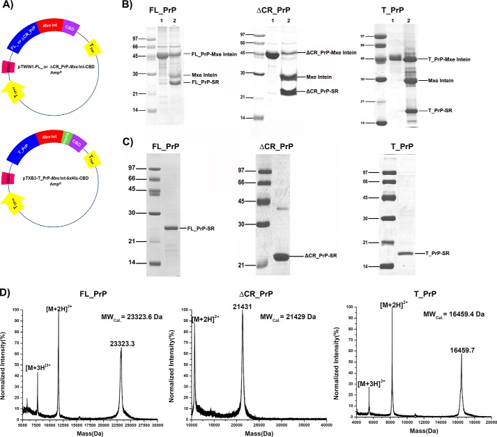
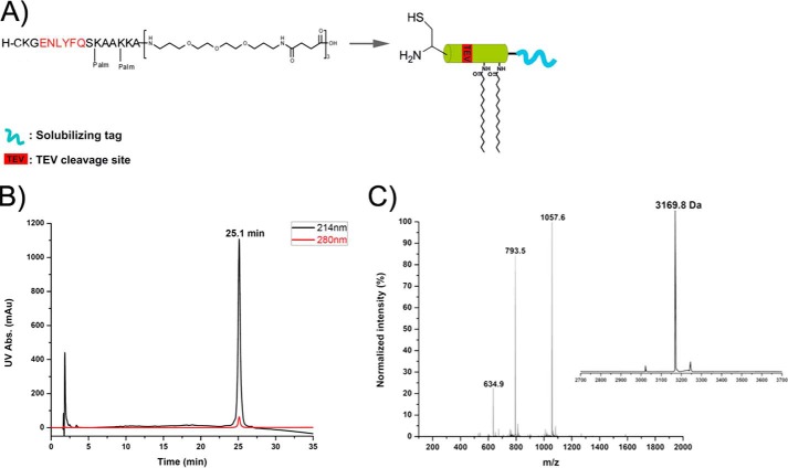
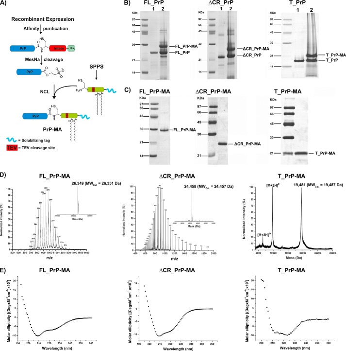
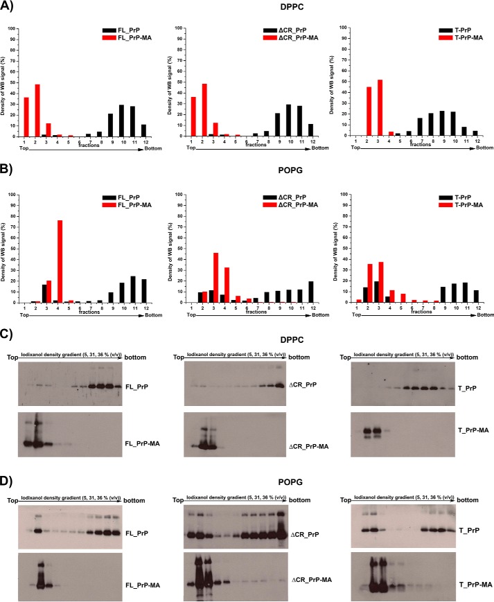
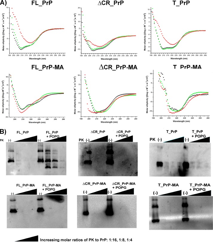
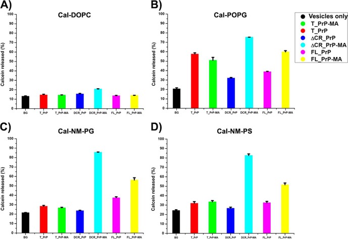
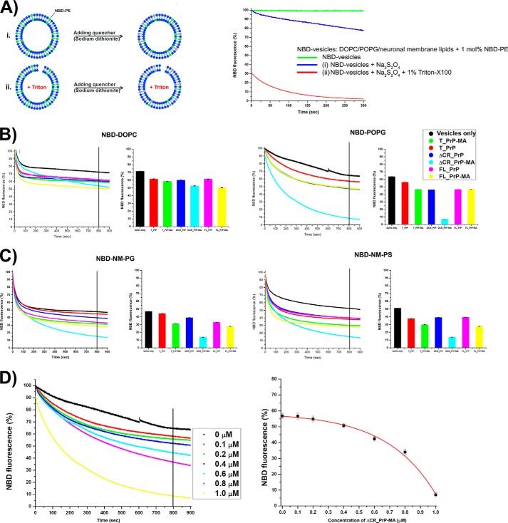
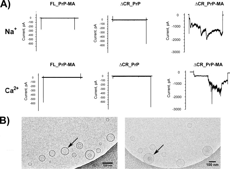
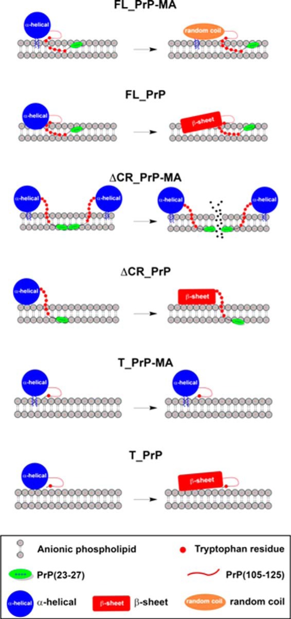
Membrane attachment via a C-terminal glycosylphosphatidylinositol anchor is critical for conversion of PrP(C) into pathogenic PrP(Sc). Therefore the effects of the anchor on PrP structure and function need to be deciphered. Three PrP variants, including full-length PrP (residues 23-231, FL_PrP), N-terminally truncated PrP (residues 90-231, T_PrP), and PrP missing its central hydrophobic region (Δ105-125, ΔCR_PrP), were equipped with a C-terminal membrane anchor via a semisynthesis strategy. Analyses of the interactions of lipidated PrPs with phospholipid membranes demonstrated that C-terminal membrane attachment induces a different binding mode of PrP to membranes, distinct from that of non-lipidated PrPs, and influences the biochemical and conformational properties of PrPs. Additionally, fluorescence-based assays indicated pore formation by lipidated ΔCR_PrP, a variant that is known to be highly neurotoxic in transgenic mice. This finding was supported by using patch clamp electrophysiological measurements of cultured cells. These results provide new evidence for the role of the membrane anchor in PrP-lipid interactions, highlighting the importance of the N-terminal and the central hydrophobic domain in these interactions.

Keywords: Liposome; Membrane Anchor; Pore Formation; Prion; Protein Aggregation; Protein Chemistry; Protein Conformation; Protein Semisynthesis; lipid-Protein Interactions.


Figures

FIGURE 1.
FIGURE 1.
Preparation of PrP constructs containing C-terminal α-thioesters for expressed protein ligation. A, plasmid maps of FL_PrP or ΔCR_PrP (top) and T_PrP (bottom) C-terminally fused to Mxe intein. B, cleavage reactions of PrP-intein fusion proteins. C and D, SDS-PAGE (C) and MALDI-MS (D) analyses of purified PrP-Cα-thioesters.
FIGURE 2.
FIGURE 2.
Chemical synthesis of the GPI anchor-mimicking peptide (membrane anchor) using Fmoc-based solid phase peptide synthesis. peptide carries two palmitoyl moieties, a polymer tag to enhance solubility, and an N-terminal cysteine for the NCL reaction. A, structure and representation of membrane anchor. B, analytical RP-HPLC chromatogram. C, ESI-MS spectrum and deconvoluted mass of the purified membrane anchor.
FIGURE 3.
FIGURE 3.
Semisynthesis of PrP variants with membrane anchors. A, scheme of the semisynthesis strategy. B and C, SDS-PAGE analyses of ligation reactions (B) and purified PrP constructs (C) carrying MAs. D and E, MS analysis (D) and CD measurements (E) of purified PrPs with membrane anchor.
FIGURE 4.
FIGURE 4.
Strong and specific binding of PrPs to liposomes via their C-terminal membrane anchors. The interactions of anchorless PrPs (black) and PrPs with a membrane anchor (red) with DPPC liposomes (A) and POPG liposomes (B) were analyzed by flotation assays in combination with Western blots. C and D, The original Western blots are depicted in C for experiments with DPPC liposomes and in D for POPG liposomes. POPG liposomes loaded with PrP variants were treated with a solution of 10 mm NaOH to disrupt electrostatic interactions. Quantification of all blots was performed by using ImageJ software.
FIGURE 5.
FIGURE 5.
Biochemical and structural properties of PrP constructs within membrane environments. A, CD analyses of PrPs without membrane anchor (top row) and PrPs with membrane anchor (bottom row) in solution (black) or in the presence of DOPC (green) and POPG (red) liposomes. B, PK digestions of FL_PrP constructs after incubation at 37°C for 1 h with buffer alone (left) or POPG liposomes (right) are shown in each panel; the results of PrPs with and without a membrane anchor are presented in the left- and right-hand panels, respectively. The PK-to-PrP molar ratios were 1:16, 1:8, and 1:4.
FIGURE 6.
FIGURE 6.
Stern-Volmer plots of tryptophan fluorescence quenching. A, FL_PrP without a membrane anchor in a phosphate buffer (red) or in the presence of 8 m urea (black). B–D, PrPs with and without a membrane anchor in solution (black and green) or bound to POPG liposomes (red and blue) or DOPC liposomes (cyan and magenta) for T_PrP, ΔCR_PrP and FL_PrP, respectively.
FIGURE 7.
FIGURE 7.
Calein release assays. The influence of PrP-lipid interaction on the integrity of lipid membranes was analyzed by calcein release assays using concentrations of 100 nm of different PrP variants and 200 μm of calcein-loaded vesicles DOPC (A) and POPG (B) and phospholipid mixtures mimicking neuronal membranes with anionic phospholipids POPG (NM-PG) (C) and POPS (NM-PS) (D). One sample containing only calcein-loaded vesicles was included as the background control (BG).
FIGURE 8.
FIGURE 8.
Fluorescence quenching assays for PrP variants mixed with different NBD-labeled vesicles. A, explains the principle setup and control experiments with vesicles in the absence of protein. The green trace indicates stable fluorescence during measurements. i, dithionite quenching of NBD lipids on the outside of intact vesicles (blue trace, negative control); ii, dithionite quenching of NBD fluorescence on triton-disrupted vesicles (red trace, positive control). B, results for PrP variants on NBD-DOPC (left) and NBD-POPG vesicles (right). C, results for PrP variants on vesicles mimicking neuronal membranes NBD-NM-PG (left) and NBD-NM-PS (right). Graphs on the left show NBD fluorescence decay recorded for 900 s. After 800 s, NBD fluorescence was averaged and summarized in a bar graph shown on the right. D, ΔCR_PrP-MA causes the influx of the quencher dithionite into NBD-POPG vesicles in a concentration-dependent manner. Concentrations of ΔCR_PrP-MA from 0 to 1.0 μm were mixed with NBD-POPG vesicles (200 μm lipids), and 10 mm dithionite was added (left side). After 800 s (vertical line) the remaining fluorescence was measured. Averages of two independent measurements were plotted (right panel).
FIGURE 9.
FIGURE 9.
Electrophysiological and cryo-EM analysis. A, ΔCR_PrP with a membrane anchor induces spontaneous inward currents in HEK293 cells. Representative whole-cell patch clamp recordings are shown. HEK293 cells were clamped from a holding potential of 0 mV to a test potential of −100 mV for 10 s every 30 s. After a control phase, the indicated proteins were added directly to the bath solution. In the presence of FL_PrP-MA (left) and ΔCR_PrP without a membrane anchor (middle), no or only weak pore forming activity was observed, whereas ΔCR_PrP-MA induced large sodium (top right) and calcium (bottom right) inward currents. B, cryo-EM images of POPG vesicles (arrows) mixed with ΔCR_PrP-MA (left) and POPG vesicles alone (right). The scale bar is 100 nm.
SCHEME 1.
SCHEME 1.
Schematic illustration of how the PrP variants FL_PrP, ΔCR_PrP, and T_PrP with or without an MA may interact with anionic phospholipid membranes. The scheme illustrates a distinct binding mode of predominantly α-helical PrP constructs (large blue circle) carrying a membrane anchor to POPG vesicles when compared with PrP constructs without a membrane anchor. Tryptophan residues (small red circles), the N-terminal polybasic region (residues 23–27, green oval) and central hydrophobic region (residues 105–125, red line) are indicated. The position of the latter region has not been determined by our experiments and is therefore not accurately depicted in this scheme. In addition, the conformational changes of the globular domain of PrP (residues 125–231) from mainly α-helical (large blue circle) to random coil (orange oval) or β-sheet (red rectangle) are highlighted.

Similar articles

Cited by

References

    1. Prusiner S. B. (1998) Prions. Proc. Natl. Acad. Sci. U.S.A. 95, 13363–13383 - PMC - PubMed
    1. Cobb N. J., Surewicz W. K. (2009) Prion diseases and their biochemical mechanisms. Biochemistry 48, 2574–2585 - PMC - PubMed
    1. Caughey B., Baron G. S., Chesebro B., Jeffrey M. (2009) Getting a grip on prions: oligomers, amyloids, and pathological membrane interactions. Annu. Rev. Biochem. 78, 177–204 - PMC - PubMed
    1. Aguzzi A., Heppner F. L. (2000) Pathogenesis of prion diseases: a progress report. Cell Death Differ. 7, 889–902 - PubMed
    1. Linden R., Martins V. R., Prado M. A., Cammarota M., Izquierdo I., Brentani R. R. (2008) Physiology of the prion protein. Physiol. Rev. 88, 673–728 - PubMed

Publication types

MeSH terms

LinkOut - more resources