. 2014 Nov 15;307(10):L765-74.
doi: 10.1152/ajplung.00100.2014. Epub 2014 Sep 12.

Chronic exposure to perfluorinated compounds: Impact on airway hyperresponsiveness and inflammation

Affiliations

Chronic exposure to perfluorinated compounds: Impact on airway hyperresponsiveness and inflammation

Min H Ryu et al. Am J Physiol Lung Cell Mol Physiol. .

Abstract

Emerging epidemiological evidence reveals a link between lung disease and exposure to indoor pollutants such as perfluorinated compounds (PFCs). PFC exposure during critical developmental stages may increase asthma susceptibility. Thus, in a murine model, we tested the hypothesis that early life and continued exposure to two ubiquitous household PFCs, perfluorooctanoic acid (PFOA) and perflurooctanesulfonic acid (PFOS), can induce lung dysfunction that exacerbates allergen-induced airway hyperresponsiveness (AHR) and inflammation. Balb/c mice were exposed to PFOA or PFOS (4 mg/kg chow) from gestation day 2 to 12 wk of age by feeding pregnant and nursing dams, and weaned pups. Some pups were also sensitized and challenged with ovalbumin (OVA). We assessed lung function and inflammatory cell and cytokine expression in the lung and examined bronchial goblet cell number. PFOA, but not PFOS, without the OVA sensitization/challenge induced AHR concomitant with a 25-fold increase of lung macrophages. PFOA exposure did not affect OVA-induced lung inflammatory cell number. In contrast, PFOS exposure inhibited OVA-induced lung inflammation, decreasing total cell number in lung lavage by 68.7%. Interferon-γ mRNA in the lung was elevated in all PFC-exposed groups. Despite these effects, neither PFOA nor PFOS affected OVA-induced AHR. Our data do not reveal PFOA or PFOS exposure as a risk factor for more severe allergic asthma-like symptoms, but PFOA alone can induce airway inflammation and alter airway function.

Keywords: asthma; environmental pollutant; interferon-γ; perfluorooctanoic acid; perflurooctanesulfonic acid.


Figures

Fig. 1.
Fig. 1.
Schematic diagram showing perfluorooctanoic acid (PFOA) and perflurooctanesulfonic acid (PFOS) exposure timeline and ovalbumin (OVA) protocol. A: timed-pregnant dams were fed PFOA- or PFOS-enriched diet ad libitum beginning on gestation day (GD) 2. The PFC-enriched diet was prepared by mixing in 4 mg of PFOA or PFOS (dissolved in methanol)/1 kg of diet. Dams were fed PFC-enriched diet throughout pregnancy and the lactation period. Postweaning, the pups were maintained on the PFC-enriched diet until 12 wk of age. B: subgroups of PFC-exposed mice were subjected to OVA sensitization and challenge: beginning at 8–9 wk of age, mice were sensitized by ip (OVA IP) injection (2 μg OVA adsorbed to 2 mg of alum in 0.5 ml saline). Fifteen days later (day 15) a second OVA IP was administered along with an initial intranasal OVA challenge (IN OVA): 50 μg OVA in 50 μl saline applied directly to nostrils. IN OVA was repeated for two more consecutive days (days 16 and 17 of the OVA protocol). On day 19, 48 h after the final OVA IN challenge, lung function was assessed using a flexiVENT; thereafter, we collected bronchoalveolar lavage fluid (BALF), blood, and lung specimens.
Fig. 2.
Fig. 2.
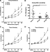
Early life and continued exposure of mice to PFOA alone induces airway hyperresponsiveness. Lung function was assessed in mice that were exposed to PFOA or PFOS. Airway mechanics were assessed after inhalation of nebulized saline and increasing concentrations of methacholine (MCh, 3–50 mg/ml). MCh dose-response curve for Newtonian resistance (Rn) (A), tissue damping (G, C), and tissue elastance (H, D) are plotted for each group. B: central or conducting airway sensitivity to MCh was measured by calculating provocative concentration of MCh needed to elicit 100% increase in Rn (PC100). Significant difference from the chemical-naïve was determined by 2-way ANOVA, and statistical difference compared with the chemical-naïve control was denoted P < 0.05 (*), P < 0.01(**), and P < 0.001(***). Error bars represent means ± SE. The no. of animals studied in each group was 8–10.
Fig. 3.
Fig. 3.
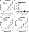
Exposure of mice to dietary PFOA or PFOS does not affect allergen-induced airway hyperresponsiveness. Lung function was assessed in mice that were sensitized and challenged with OVA in addition to PFOA or PFOS exposure. Airway mechanics were measured after inhalation of nebulized saline and increasing concentrations of MCh (3–50 mg/ml). MCh dose-response curves for Rn (A), G (C), and H (D) are plotted for each group. B: conducting airway sensitivity to MCh was measured by calculating PC100. OVA-alone challenge consistently induced AHR to MCh, as indicated by a 93% increase in maximum Rn and 82% decrease in PC100. Statistical comparisons between OVA-alone and chemical-naïve control were compared by ANOVA, and statistical difference between OVA-alone and the chemical-naïve control was denoted P < 0.05 (*), P < 0.01(**), and P < 0.001(***). No statistical difference was found between PFOA + OVA, PFOS + OVA, and OVA-alone group (ANOVA, P > 0.05). Error bars represent means ± SE. The no. of animals studied in each group was 8–10.
Fig. 4.
Fig. 4.
Inflammatory cell counts in BALF are altered by exposure to PFOA or PFOS. BALF was collected after mice were anesthetized and mechanically ventilated to assess respiratory mechanics. A: total cell counts in BALF were estimated using a hemocytometer. Values are expressed as no. of cells/ml of BALF. One-way ANOVA was used to compare the total cell counts. Cell distribution was analyzed by manual counting of macrophages (B), eosinophils (C), and neutrophils (D) in 200 μl of BALF after cytospin, fixation, and modified Wright-Giemsa staining. Cell counts were obtained in 6 random fields examined from a light microscope (×40 magnification). Statistical difference in the differential counts was determined by ANOVA. Means were compared with that of the naïve and are denoted with P < 0.01(**) and P < 0.001(***) for significant difference. #Significant difference compared with the OVA-only group (ANOVA, P < 0.05). As expected, OVA-alone challenge induced a significant increase in total, macrophage, and eosinophil count. In OVA-challenged mice, PFOS decreased the total leukocyte count in BALF by 68.7% compared with OVA alone. This decrease in the total cell count was associated with a blunting of macrophage nos. by 64.8% compared with OVA-only exposed mice. In contrast, PFOA had no detectable impact on the allergen-induced leukocyte infiltration observed. Data points represent counts from 6 to 8 mice in each group (n = 4 for PFOS). Mean values for each group are indicated with a horizontal bar, and error bars for means ± SE are shown.
Fig. 5.
Fig. 5.
Relative mRNA abundance of inflammatory cytokines in mouse lungs is altered by exposure to PFOA and PFOS. mRNA abundance was measured using quantitative RT-PCR, with normalization to an internal standard, ribosomal subunit 18S. Relative mRNA abundances for eotaxin (A), tumor necrosis factor-α (TNF-α, B), interferon-γ (INF-γ, C), and interleukin-13 (IL-13, D) are presented. Relative expression between groups was compared based on ΔΔCt values for PFOA and PFOS exposure alone, OVA sensitization and challenge, or OVA sensitization and challenge in combination with PFOA or PFOS exposure. All data are plotted relative to the abundance of mRNA for each target that was detected in the OVA-only exposed mice. Statistical comparison of mean ΔΔCt was carried out using ANOVA included in SPSS 20, and significant differences from the chemical-naïve mice are denoted with * when P < 0.05. Error bars represent the means ± SE. Data are from at least 2 replicates from 3–4 mice in each group. ND, not detected (Ct >35).
Fig. 6.
Fig. 6.
Semiquantitative analysis of goblet cell and mucus abundance in airways of mice exposed to PFOA or PFOS revealed no impact on the OVA-induced goblet cell hyperplasia and mucus production by PFOA or PFOS exposure. Histological images of murine airways from chemical-naïve and no OVA challenge mice (A), PFOA-only exposed mice (B), PFOS-only exposed mice (C), chemical-naïve and OVA-sensitized/challenged mice (D), PFOA-exposed and OVA-sensitized/challenged mice (E), and PFOA-exposed and OVA-sensitized/challenged mice (F) are presented. Arrows in each panel indicate areas of positive staining for mucous and goblet cells in the airway epithelium. G: scores from semiquantitative assessment of goblet cell and mucus scores are plotted. To obtain a mean score, 12 medium-sized airways (cross-sectional epithelial basement membrane length >1,000 μm) from 4 independent animals were scored two times by 2 blinded observers. Goblet cell hyperplasia and mucus abundance were scored from 0 to 3; 0 = no presence of goblet cell; 1 = mild hyperplasia with occupation of <1/3 of the epithelial area by goblet cells with mild mucus, 2 = moderate hyperplasia with occupation of ≥1/3 to ≤2/3 of the epithelial area with mucus and goblet cells, and 3 = severe goblet cell hyperplasia with >2/3 of the airway epithelial with mucus and goblet cells. Kruskal-Wallis test along with Dunn's Multiple Comparison were used to compare the means. ***Significant statistical difference compared with the naïve mice (P < 0.001).

References

    1. Anderson-Mahoney P, Kotlerman J, Takhar H, Gray D, Dahlgren J. Self-reported health effects among community residents exposed to perfluorooctanoate. J Environ Occup Health Policy 18: 129–143, 2008. - PubMed
    1. Apelberg BJ, Witter FR, Herbstman JB, Calafat AM, Halden RU, Needham LL, Goldman LR. Cord serum concentrations of perfluorooctane sulfonate (PFOS) and perfluorooctanoate (PFOA) in relation to weight and size at birth. Environ Health Perspect 115: 1670–1676, 2007. - PMC - PubMed
    1. Arai N, Kondo M, Izumo T, Tamaoki J, Nagai A. Inhibition of neutrophil elastase-induced goblet cell metaplasia by tiotropium in mice. Eur Resp J 35: 1164–1171, 2010. - PubMed
    1. Belvisi MG, Mitchell JA. Targeting PPAR receptors in the airway for the treatment of inflammatory lung disease (Abstract). Br J Pharmacol 158: 809164, 2009. - PMC - PubMed
    1. Chan E, Burstyn I, Cherry N, Bamforth F, Martin JW. Perfluorinated acids and hypothyroxinemia in pregnant women. Environ Res 111: 559–564, 2011. - PubMed

Publication types