. 2014 Nov:468-470:293-302.
doi: 10.1016/j.virol.2014.08.017. Epub 2014 Sep 15.

Deletions in the fifth alpha helix of HIV-1 matrix block virus release

Affiliations

Deletions in the fifth alpha helix of HIV-1 matrix block virus release

Bridget Sanford et al. Virology. 2014 Nov.

Abstract

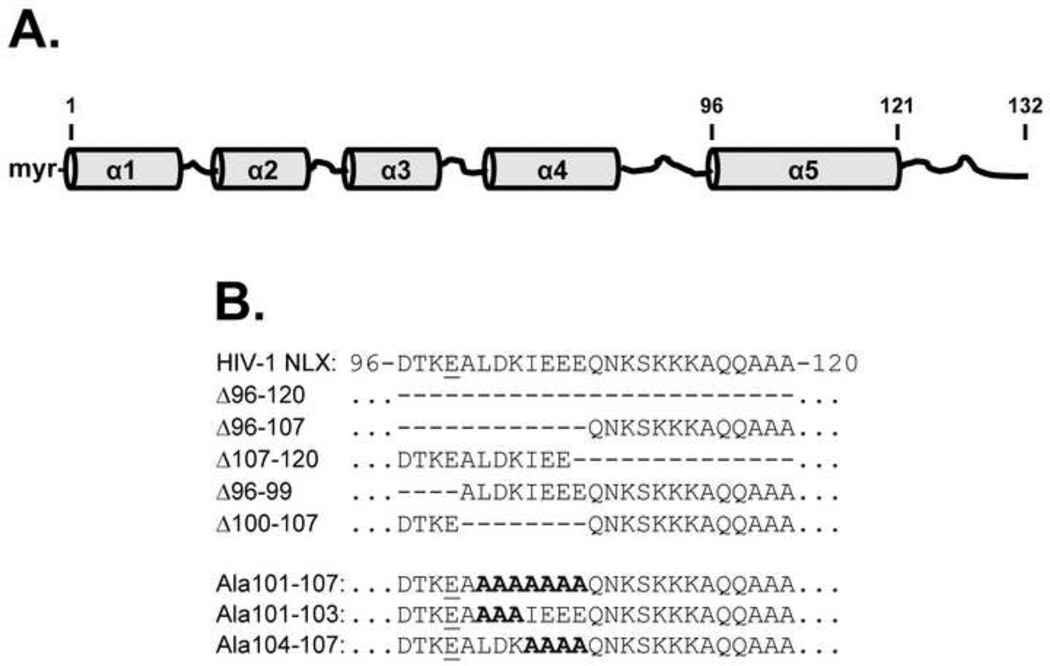
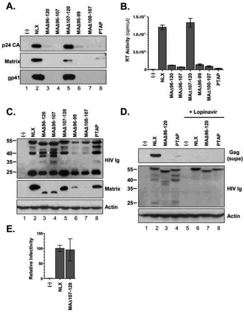
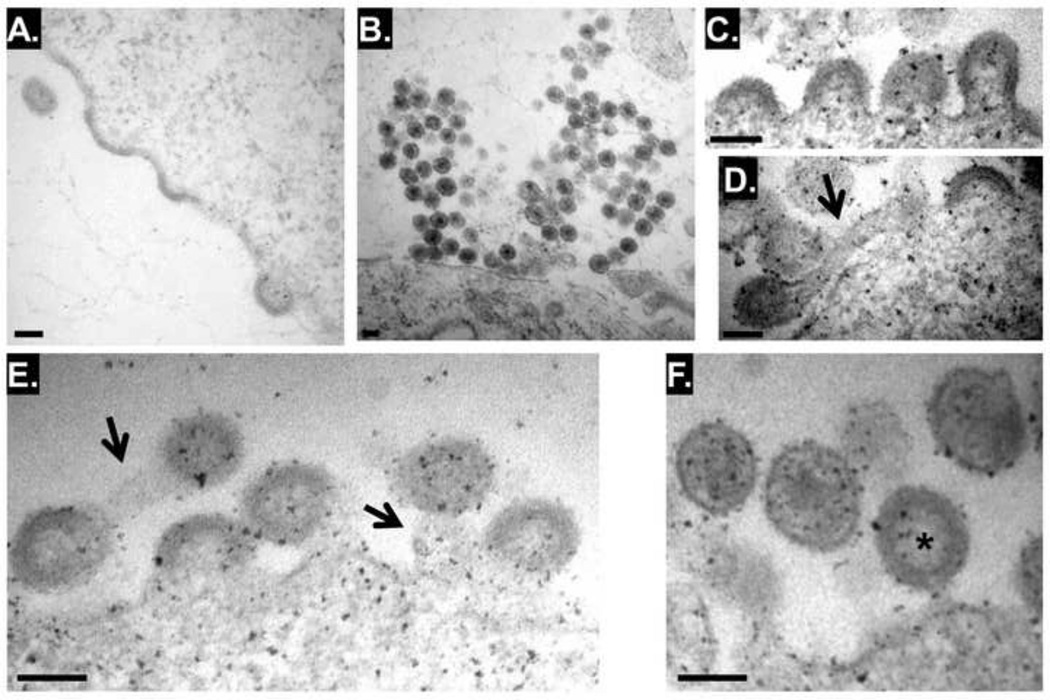
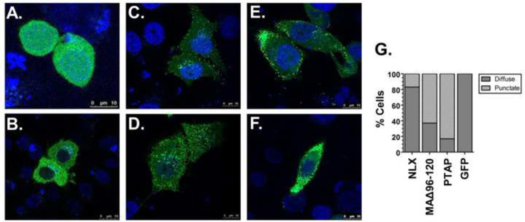
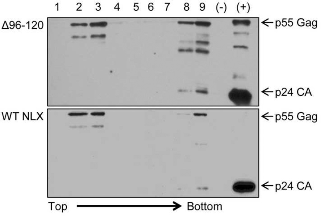
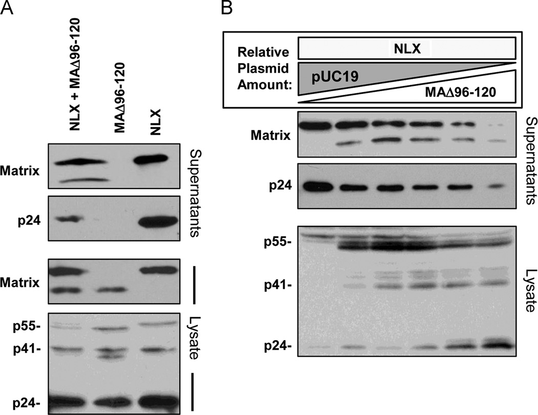
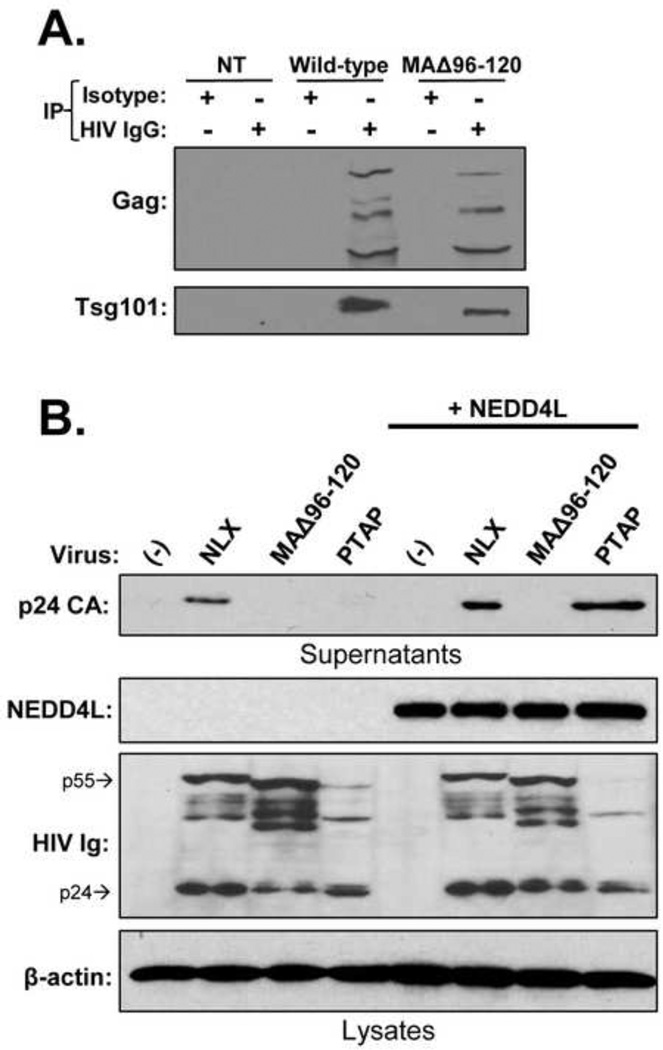
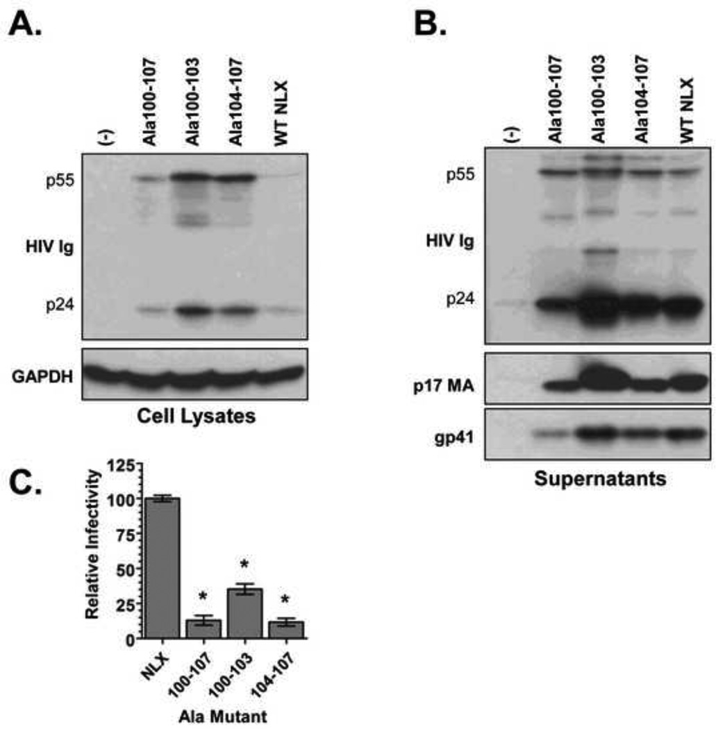
The matrix (MA) protein of HIV-1 is the N-terminal component of the Gag structural protein and is critical for the early and late stages of viral replication. MA contains five α-helices (α1-α5). Deletions in the N-terminus of α5 as small as three amino acids impaired virus release. Electron microscopy of one deletion mutant (MA∆96-120) showed that its particles were tethered to the surface of cells by membranous stalks. Immunoblots indicated all mutants were processed completely, but mutants with large deletions had alternative processing intermediates. Consistent with the EM data, MA∆96-120 retained membrane association and multimerization capability. Co-expression of this mutant inhibited wild type particle release. Alanine scanning mutation in this region did not affect virus release, although the progeny virions were poorly infectious. Combined, these data demonstrate that structural ablation of the α5 of MA inhibits virus release.

Keywords: Deletion mutagenesis; HIV-1 assembly; HIV-1 budding; Matrix; NEDD4L.


Figures

FIG 1
FIG 1
MA deletion mutants used in this study. (A) Cartoon depiction of the α-helicies in MA. The position of the amino acids flanking α5 are indicated. (B) Mutants used in this study. The sequence of amino acids 96-120 of the NLX molecular clone is shown. Names of mutants used in the study are indicated on the left and the amino acids deleted or mutated are shown on right. Deleted amino acids are denoted with a dash.
FIG 2
FIG 2
Expression and release of MA deletion mutants. (A) Virus release. Supernatants were collected from 293T cells transiently transfected with the HIV-1 clones indicated at the top and directly resolved by SDS-PAGE. The indicated viral proteins detected by western blot. All blots are representative of at least three independent experiments. (B) Reverse transcriptase activity released from cells. Supernatants were assayed by in vitro [32P]-TTP incorporation assay. Error bars show standard error of triplicate experiments. (C) Immunoblot of cell lysates. Gag products were detected with HIV Ig (top panel). Aberrant processing intermediates are indicated with a (*). (D) Effects of Lopinavir treatment. Supernatants were collected from 293T cells transiently transfected with the HIV-1 clones and directly resolved by SDS-PAGE and immunoblotted to detect release (top panel). Lower panels show immunoblots of cell lysates. (E) Infectivity of MAΔ107-120. Virus stocks were normalized by RT activity and infectivity measured using TZM-bl indicator cells. The results were normalized relative to wild-type NLX. Data represents the mean of three independent experiments and the error bars denote the combined SEM of all experiments.
FIG 3
FIG 3
Transmission electron microscopic images showing viral particles in the surface regions of cells expressing wild-type NLX (A) and MAΔ96-120 (B–F). Arrows indicate visible membrane stalks. Scale bars: 0.1 µm.
FIG 4
FIG 4
Localization of Gag-GFP constructs. (A) Confocal microscopy of HeLa cells transiently transfected with wild-type (a, b), MAΔ96-120 (c, d), or PTAP mutant Gag-GFP (e, f) constructs. (G) Graphical representation of observed localizations of proteins.
FIG 5
FIG 5
Membrane floatation assay showing MAΔ96-120 is associated with membranes. 293T cells were transfected with either MAΔ96-120 (top panel) or wild-type NLX (bottom panel) for 24 hours. Cells were lysed by hypotonic swelling/dounce homogenization and ultracentrifuged on a sucrose step gradient. Fractions were collect from the top and analyzed by SDS-PAGE and western blot with anti-Gag antiserum. Data is representative of two independent experiments.
FIG 6
FIG 6
Co-expression of MAΔ96-120 and NLX. (A) MAΔ96-120 is capable of multimerization with wild-type NLX. 293T cells were transfected with both NLX and MAΔ96-120, or either clone alone as indicated at the top. (B) MAΔ96-120 trans-dominantly inhibits wild-type virus release. Cells were transfected transiently with a constant amount of NLX plasmid, increasing amounts of MAΔ96-120, and pUC19 to normalize the overall amount of plasmid DNA. Supernatant and lysate samples were isolated and the indicated viral proteins detected as described in the Fig. 2 legend. Panels are a representative of three independent experiments.
FIG 7
FIG 7
The MAΔ96-120 phenotype is distinct from late domain mutants. (A) Co-immunoprecipiation assay showing MAΔ96-120 Gag interacts with TSG101. Cells transfected with wild-type or MAΔ96-120 Gag-GFP were lysed, immunoprecipitated with HIV IgG, and immmunoblotted for TSG101 and Gag as indicated. (B) NEDD4L over-expression does not rescue MAΔ96-120 release. The indicated molecular clones were co-transfected with a NEDD4L expression construct. Supernatants and lysates were tested for the indicated proteins as described in Fig. 2. β-actin was detected as a control. Data is representative of two independent experiments.
FIG 8
FIG 8
Alanine scanning mutagenesis of MA aa 100–107. Virus expression (A) and release (B) from 293T cells. Proteins were resolved by SDS-PAGE and the indicated viral proteins detected by western blot. (C) Virus infection of Ala mutants measured on TZM-bl indicator cells. Data is a combination of two independent experiments with infections performed in triplicate. Error bars denote standard error of the mean; *p<0.001 by unpaired two-tail T-test.

Similar articles

Cited by

References

    1. Abacioglu YH, Fouts TR, Laman JD, Claassen E, Pincus SH, Moore JP, Roby CA, Kamin-Lewis R, Lewis GK. Epitope mapping and topology of baculovirus-expressed HIV-1 gp160 determined with a panel of murine monoclonal antibodies. AIDS Res Hum Retroviruses. 1994;10(4):371–381. - PubMed
    1. Babe LM, Craik CS. Constitutive production of nonenveloped human immunodeficiency virus type 1 particles by a mammalian cell line and effects of a protease inhibitor on particle maturation. Antimicrobial agents and chemotherapy. 1994;38(10):2430–2439. - PMC - PubMed
    1. Balasubramaniam M, Freed EO. New insights into HIV assembly and trafficking. Physiology (Bethesda) 2011;26(4):236–251. - PMC - PubMed
    1. Belshan M, Schweitzer CJ, Donnellan MR, Lu R, Engelman A. In vivo biotinylation and capture of HIV-1 matrix and integrase proteins. J Virol Methods. 2009;159(2):178–184. - PMC - PubMed
    1. Bhatia AK, Campbell N, Panganiban A, Ratner L. Characterization of replication defects induced by mutations in the basic domain and C-terminus of HIV-1 matrix. Virology. 2007;369(1):47–54. - PMC - PubMed

Publication types

Substances

LinkOut - more resources