

The great divide: septation and malformation of the cloaca, and its implications for surgeons
- PMID: 25217828
- PMCID: PMC4302733
- DOI: 10.1007/s00383-014-3593-8
The great divide: septation and malformation of the cloaca, and its implications for surgeons
Abstract
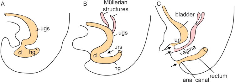
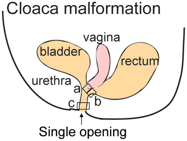
The anorectal and urogenital systems arise from a common embryonic structure termed cloaca. Subsequent development leads to the division/septation of the cloaca into the urethra, urinary bladder, vagina, anal canal, and rectum. Defective cloacal development and the resulting anorectal and urogenital malformations are some of the most severe congenital anomalies encountered in children. In the most severe form in females, the rectum, vagina, and urethra fail to develop separately and drain via a single common channel known as a cloaca into the perineum. In this review, we summarize our current knowledge of embryonic cloaca development and malformation, and compare them to what has already been described in the literature. We describe the use of mouse models of cloaca malformation to understand which signaling pathways and cellular mechanisms are involved in the process of normal cloaca development. We also discuss the embryological correlation of the epithelial and stromal histology found in step sections of the common channel in 14 human cloaca malformations. Finally, we highlight the significance of these findings, compare them to prior studies, and discuss their implications for the pediatric surgeons. Understanding and identifying the molecular basis for cloaca malformation could provide foundation for tissue engineering efforts that in the future would reflect better surgical reconstruction and improved quality of life for patients.
Conflict of interest statement
Figures
Similar articles
-
Embryology of the hindgut.Semin Pediatr Surg. 2011 Aug;20(3):152-60. doi: 10.1053/j.sempedsurg.2011.03.002. Semin Pediatr Surg. 2011. PMID: 21708335
-
The embryology of persistent cloaca and urogenital sinus malformations.Asian J Androl. 2020 Mar-Apr;22(2):124-128. doi: 10.4103/aja.aja_72_19. Asian J Androl. 2020. PMID: 31322137 Free PMC article. Review.
-
Clarification of mammalian cloacal morphogenesis using high-resolution episcopic microscopy.Dev Biol. 2016 Jan 1;409(1):106-113. doi: 10.1016/j.ydbio.2015.10.018. Epub 2015 Oct 17. Dev Biol. 2016. PMID: 26485363 Free PMC article.
-
Defining the molecular pathologies in cloaca malformation: similarities between mouse and human.Dis Model Mech. 2014 Apr;7(4):483-93. doi: 10.1242/dmm.014530. Epub 2014 Feb 13. Dis Model Mech. 2014. PMID: 24524909 Free PMC article.
-
Pathology of cloaca anomalies with case correlation.Semin Pediatr Surg. 2016 Apr;25(2):66-70. doi: 10.1053/j.sempedsurg.2015.11.003. Epub 2015 Nov 10. Semin Pediatr Surg. 2016. PMID: 26969228 Review.
Cited by
-
Host-derived protein profiles of human neonatal meconium across gestational ages.Nat Commun. 2024 Jul 17;15(1):5543. doi: 10.1038/s41467-024-49805-w. Nat Commun. 2024. PMID: 39019879 Free PMC article.
-
The role of ultrasonography in detecting and classifying anorectal malformations in neonates: a retrospective study of 30 cases.Pediatr Surg Int. 2025 May 28;41(1):148. doi: 10.1007/s00383-025-06054-2. Pediatr Surg Int. 2025. PMID: 40437069
-
Cloacal Dysgenesis Sequence in a Preterm Neonate.Am J Case Rep. 2024 Feb 27;25:e942203. doi: 10.12659/AJCR.942203. Am J Case Rep. 2024. PMID: 38412145 Free PMC article.
-
Prenatal detection of chromosomal abnormalities and copy number variants in fetuses with congenital gastrointestinal obstruction.BMC Pregnancy Childbirth. 2022 Jan 19;22(1):50. doi: 10.1186/s12884-022-04401-y. BMC Pregnancy Childbirth. 2022. PMID: 35045821 Free PMC article.
-
Cloacal Malformation in Female Children: Outcome of Initial Management.Pak J Med Sci. 2020 Jan-Feb;36(2):187-191. doi: 10.12669/pjms.36.2.1095. Pak J Med Sci. 2020. PMID: 32063957 Free PMC article.
References
-
- Fritsch H, Aigner F, Ludwikowski B, Reinstadler-Zankl S, Illig R, Urbas D, Schwarzer C, Longato S. Epithelial and muscular regionalization of the human developing anorectum. Anat Rec (Hoboken) 2007;290:1449–58. - PubMed
-
- Kluth D. Embryology of anorectal malformations. Semin Pediatr Surg. 2010;19:201–8. - PubMed
-
- Warne SA, Hiorns MP, Curry J, Mushtaq I. Understanding cloacal anomalies. Arch Dis Child 2011 - PubMed
-
- Robboy SJ, Bentley RC. In: Vagina Histology for Pathologists. 3. Mills SE, editor. Philadelphia: Lippincott Williams &Wilkins; 2011. pp. 999–1010.
Publication types
MeSH terms
Grants and funding
LinkOut - more resources
Full Text Sources
Other Literature Sources
