

Gene structure, regulatory control, and evolution of black widow venom latrotoxins
- PMID: 25217831
- PMCID: PMC4253598
- DOI: 10.1016/j.febslet.2014.08.034
Gene structure, regulatory control, and evolution of black widow venom latrotoxins
Abstract
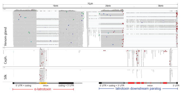
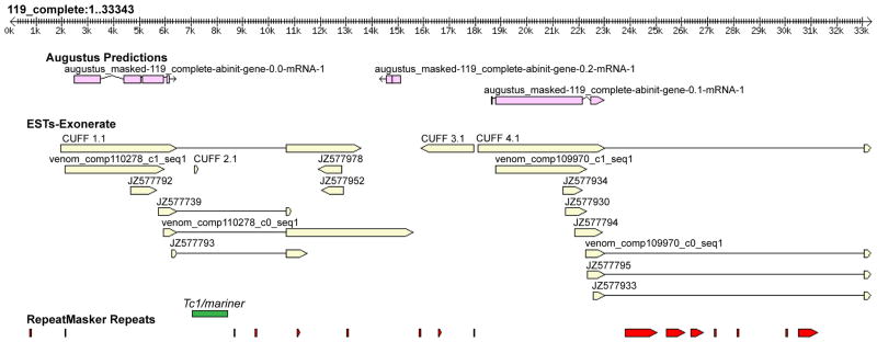
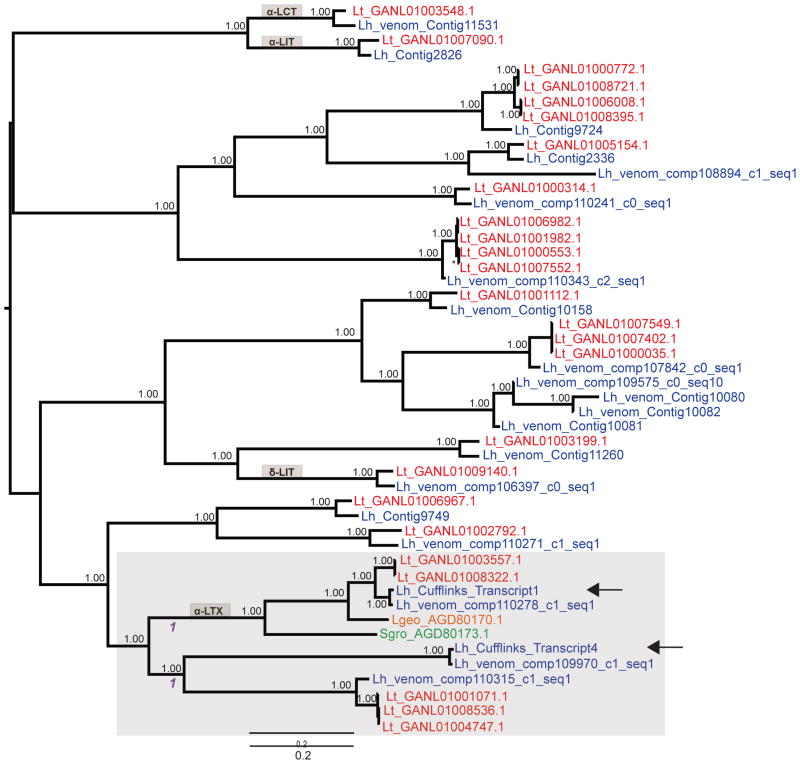
Black widow venom contains α-latrotoxin, infamous for causing intense pain. Combining 33 kb of Latrodectus hesperus genomic DNA with RNA-Seq, we characterized the α-latrotoxin gene and discovered a paralog, 4.5 kb downstream. Both paralogs exhibit venom gland specific transcription, and may be regulated post-transcriptionally via musashi-like proteins. A 4 kb intron interrupts the α-latrotoxin coding sequence, while a 10 kb intron in the 3' UTR of the paralog may cause non-sense-mediated decay. Phylogenetic analysis confirms these divergent latrotoxins diversified through recent tandem gene duplications. Thus, latrotoxin genes have more complex structures, regulatory controls, and sequence diversity than previously proposed.
Keywords: Genomics; Latrodectus; Molecular evolution; Neurosecretion; Venom; α-Latrotoxin.
Copyright © 2014 Federation of European Biochemical Societies. Published by Elsevier B.V. All rights reserved.
Figures
Similar articles
-
Recent Advances in Research on Widow Spider Venoms and Toxins.Toxins (Basel). 2015 Nov 27;7(12):5055-67. doi: 10.3390/toxins7124862. Toxins (Basel). 2015. PMID: 26633495 Free PMC article. Review.
-
House spider genome uncovers evolutionary shifts in the diversity and expression of black widow venom proteins associated with extreme toxicity.BMC Genomics. 2017 Feb 16;18(1):178. doi: 10.1186/s12864-017-3551-7. BMC Genomics. 2017. PMID: 28209133 Free PMC article.
-
Dramatic expansion of the black widow toxin arsenal uncovered by multi-tissue transcriptomics and venom proteomics.BMC Genomics. 2014 Jun 11;15(1):366. doi: 10.1186/1471-2164-15-366. BMC Genomics. 2014. PMID: 24916504 Free PMC article.
-
Molecular evolution of α-latrotoxin, the exceptionally potent vertebrate neurotoxin in black widow spider venom.Mol Biol Evol. 2013 May;30(5):999-1014. doi: 10.1093/molbev/mst011. Epub 2013 Jan 21. Mol Biol Evol. 2013. PMID: 23339183 Free PMC article.
-
The multiple actions of black widow spider toxins and their selective use in neurosecretion studies.Toxicon. 2004 Apr;43(5):527-42. doi: 10.1016/j.toxicon.2004.02.008. Toxicon. 2004. PMID: 15066411 Review.
Cited by
-
Eukaryotic association module in phage WO genomes from Wolbachia.Nat Commun. 2016 Oct 11;7:13155. doi: 10.1038/ncomms13155. Nat Commun. 2016. PMID: 27727237 Free PMC article.
-
Venomix: a simple bioinformatic pipeline for identifying and characterizing toxin gene candidates from transcriptomic data.PeerJ. 2018 Jul 31;6:e5361. doi: 10.7717/peerj.5361. eCollection 2018. PeerJ. 2018. PMID: 30083468 Free PMC article.
-
Recent Advances in Research on Widow Spider Venoms and Toxins.Toxins (Basel). 2015 Nov 27;7(12):5055-67. doi: 10.3390/toxins7124862. Toxins (Basel). 2015. PMID: 26633495 Free PMC article. Review.
-
Gene sequence analysis of toxins from the spider Phoneutria nigriventer revealed an intronless feature.J Venom Anim Toxins Incl Trop Dis. 2020 Apr 30;26:e20190075. doi: 10.1590/1678-9199-JVATITD-2019-0075. eCollection 2020. J Venom Anim Toxins Incl Trop Dis. 2020. PMID: 32395122 Free PMC article.
-
Chromosome-level genome assembly of the black widow spider Latrodectus elegans illuminates composition and evolution of venom and silk proteins.Gigascience. 2022 May 25;11:giac049. doi: 10.1093/gigascience/giac049. Gigascience. 2022. PMID: 35639632 Free PMC article.
References
-
- Moura-da-Silva A, Paine MI, Diniz MV, Theakston RD, Crampton J. The molecular cloning of a phospholipase A2 from Bothrops jararacussu snake venom: evolution of venom group II phospholipase A2’s may imply gene duplications. [Accessed 4 April 2014];J Mol Evol. 1995 41:174–179. http://link.springer.com/10.1007/BF00170670. - DOI - PubMed
Publication types
MeSH terms
Substances
Grants and funding
LinkOut - more resources
Full Text Sources
Other Literature Sources
