. 2014 Sep;16(1):61-8.
doi: 10.1016/j.gep.2014.09.001. Epub 2014 Sep 10.

Glial enriched gene expression profiling identifies novel factors regulating the proliferation of specific glial subtypes in the Drosophila brain

Affiliations

Glial enriched gene expression profiling identifies novel factors regulating the proliferation of specific glial subtypes in the Drosophila brain

Amélie Avet-Rochex et al. Gene Expr Patterns. 2014 Sep.

Abstract

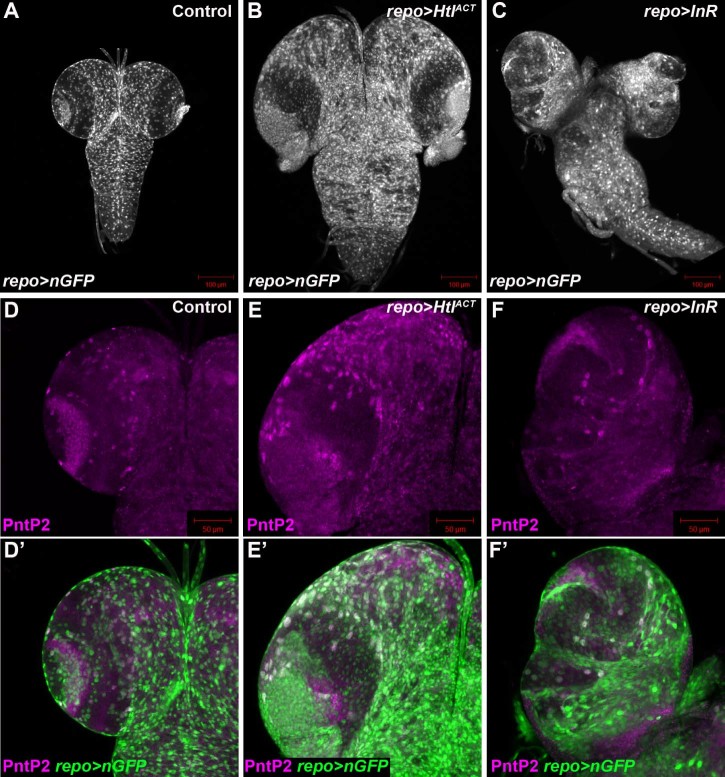
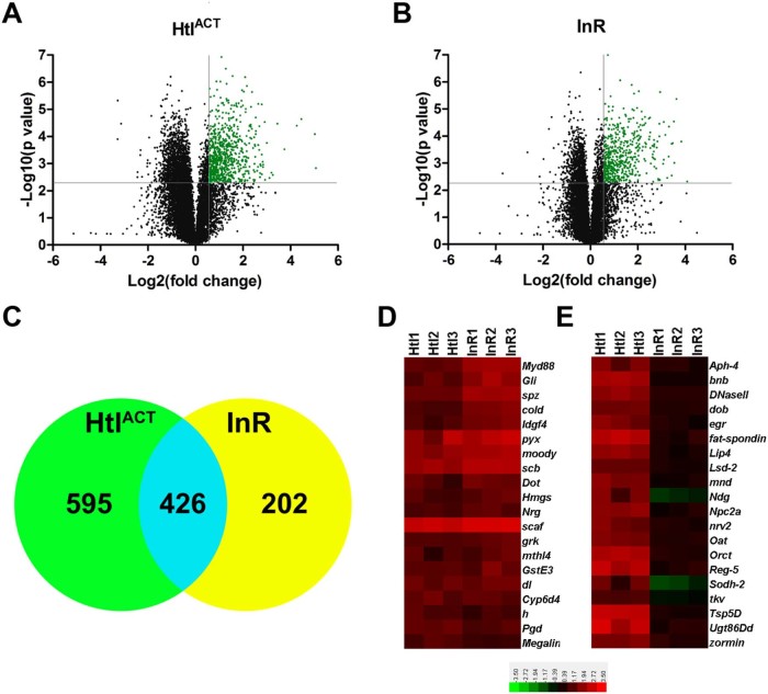
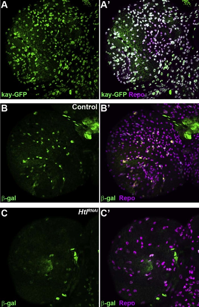
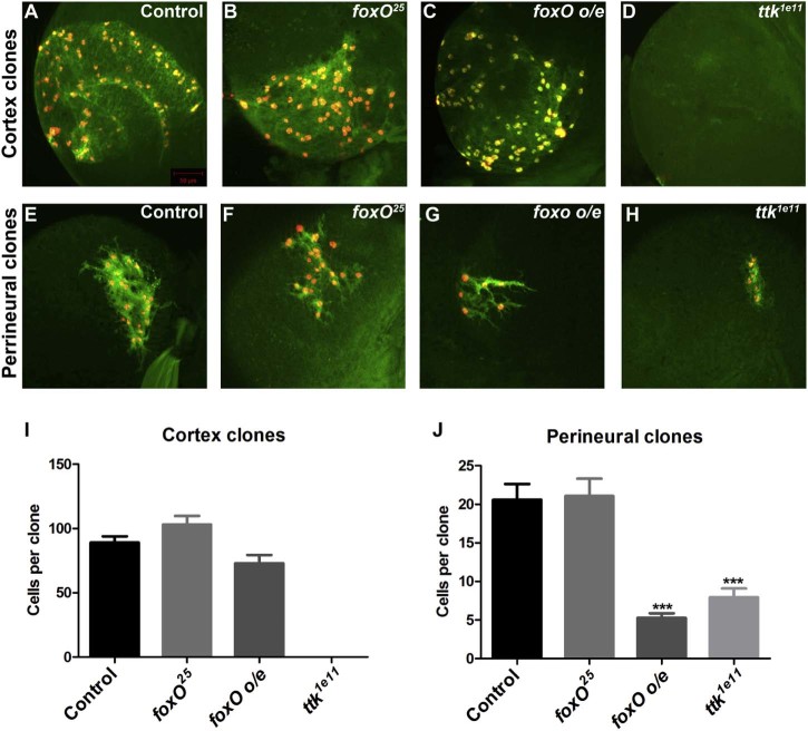
Glial cells constitute a large proportion of the central nervous system (CNS) and are critical for the correct development and function of the adult CNS. Recent studies have shown that specific subtypes of glia are generated through the proliferation of differentiated glial cells in both the developing invertebrate and vertebrate nervous systems. However, the factors that regulate glial proliferation in specific glial subtypes are poorly understood. To address this we have performed global gene expression analysis of Drosophila post-embryonic CNS tissue enriched in glial cells, through glial specific overexpression of either the FGF or insulin receptor. Analysis of the differentially regulated genes in these tissues shows that the expression of known glial genes is significantly increased in both cases. Conversely, the expression of neuronal genes is significantly decreased. FGF and insulin signalling drive the expression of overlapping sets of genes in glial cells that then activate proliferation. We then used these data to identify novel transcription factors that are expressed in glia in the brain. We show that two of the transcription factors identified in the glial enriched gene expression profiles, foxO and tramtrack69, have novel roles in regulating the proliferation of cortex and perineurial glia. These studies provide new insight into the genes and molecular pathways that regulate the proliferation of specific glial subtypes in the Drosophila post-embryonic brain.

Keywords: Cortex; Drosophila; Glia; Perineurial; Tramtrack; foxO.


Figures

Fig. 1
Fig. 1
Generation of larval CNS tissue enriched in glia. (A) Late third instar larval CNS expressing nuclear GFP in glia using repo-Gal4 (repo>nGFP). (B,C) Overexpression of HtlACT (B), or the InR (C) in glia using repo-Gal4 causes glial overproliferation. Glia are marked by the expression of nuclear GFP as in A. (D–F') Overexpression of HtlACT (E), but not the InR (F), in glia using repo-Gal4 causes overproliferation of PntP2 expressing cortex glia. PntP2 expression shown in magenta (D–F') and glia (green in D–E') are marked by the expression of nuclear GFP as in (A–C).
Fig. 2
Fig. 2
Glial enriched larval CNS gene expression profiles. (A,B) Volcano plots of transcript expression levels from larval CNS tissue overexpressing HtlACT (A), or the InR (B) in glia using repo-Gal4. Transcripts whose expression increased ≥1.5 fold with a p value ≤0.05 are shown in green. (C) Venn diagram showing the numbers of genes whose expression was significantly increased ≥1.5 fold in either HtlACT overexpressing CNS tissue (green circle), InR overexpressing CNS tissue (yellow circle), or in both conditions (blue overlap). (D,E) Heat maps representing expression levels (log2) of 20 genes whose expression was similar (D), or significantly different (E) in HtlACT (Htl1-3) and InR (InR1-3) overexpressing CNS tissue.
Fig. 3
Fig. 3
 kayak and hairy are expressed in glia in the brain. (A,A') Superficial layer of a late third instar larval brain expressing kayak-GFP (kay-GFP) stained for GFP (green) and Repo (magenta) expression. (B,B') β-Galactosidase expression (green) in the superficial layer of a control brain from a hE11 enhancer trap larva, co-stained for Repo expression (magenta). (C,C') β-Galactosidase expression (green) in the superficial layer of a repo-Gal4>htlRNAi third instar larval brain carrying the hE11 enhancer trap, co-stained for Repo expression (magenta).
Fig. 4
Fig. 4
 foxO and ttk69 regulate glial proliferation in the postembryonic brain. (A–D) Representative repo-MARCM cortex clones marked with GFP (green) and nuclear-RFP (red) expression. (E–H) Representative repo-MARCM perineurial clones marked with GFP (green) and nuclear-RFP (red) expression. (I) Quantification of cortex repo-MARCM clone sizes. Average clone size of FRT82B control clones (n = 10), foxO25 (n = 9), foxO overexpression (o/e) (n = 8) and ttk1e11 clones (no cortex clones were observed in >50 brains). (J) Quantification of perineurial repo-MARCM clone sizes. Average clone size of FRT82B control clones (n = 34), foxO25 (n = 49), foxO overexpression (o/e) (n = 24) and ttk1e11 clones (n = 24). Data are represented as mean +/− SEM. ***p < 0.001.

References

    1. Altenhein B., Becker A., Busold C., Beckmann B., Hoheisel J.D., Technau G.M. Expression profiling of glial genes during Drosophila embryogenesis. Dev. Biol. 2006;296:545–560. - PubMed
    1. Auld V.J., Fetter R.D., Broadie K., Goodman C.S. Gliotactin, a novel transmembrane protein on peripheral glia, is required to form the blood-nerve barrier in Drosophila. Cell. 1995;81:757–767. - PubMed
    1. Avet-Rochex A., Kaul A.K., Gatt A.P., McNeill H., Bateman J.M. Concerted control of gliogenesis by InR/TOR and FGF signalling in the Drosophila post-embryonic brain. Development. 2012;139:2763–2772. - PMC - PubMed
    1. Awasaki T., Lai S.L., Ito K., Lee T. Organization and postembryonic development of glial cells in the adult central brain of Drosophila. J. Neurosci. 2008;28:13742–13753. - PMC - PubMed
    1. Azevedo F.A., Carvalho L.R., Grinberg L.T., Farfel J.M., Ferretti R.E., Leite R.E. Equal numbers of neuronal and nonneuronal cells make the human brain an isometrically scaled-up primate brain. J. Comp. Neurol. 2009;513:532–541. - PubMed

Publication types