Case Reports
. 2014 Oct;46(10):1135-1139.
doi: 10.1038/ng.3066. Epub 2014 Sep 14.

Mutation of NLRC4 causes a syndrome of enterocolitis and autoinflammation

Affiliations
Case Reports

Mutation of NLRC4 causes a syndrome of enterocolitis and autoinflammation

Neil Romberg et al. Nat Genet. 2014 Oct.

Abstract

Upon detection of pathogen-associated molecular patterns, innate immune receptors initiate inflammatory responses. These receptors include cytoplasmic NOD-like receptors (NLRs) whose stimulation recruits and proteolytically activates caspase-1 within the inflammasome, a multiprotein complex. Caspase-1 mediates the production of interleukin-1 family cytokines (IL1FCs), leading to fever and inflammatory cell death (pyroptosis). Mutations that constitutively activate these pathways underlie several autoinflammatory diseases with diverse clinical features. We describe a family with a previously unreported syndrome featuring neonatal-onset enterocolitis, periodic fever, and fatal or near-fatal episodes of autoinflammation. We show that the disease is caused by a de novo gain-of-function mutation in NLRC4 encoding a p.Val341Ala substitution in the HD1 domain of the protein that cosegregates with disease. Mutant NLRC4 causes constitutive IL1FC production and macrophage cell death. Infected macrophages from affected individuals are polarized toward pyroptosis and exhibit abnormal staining for inflammasome components. These findings identify and describe the cause of a life-threatening but treatable autoinflammatory disease that underscores the divergent roles of the NLRC4 inflammasome.


Figures

Figure 1
Figure 1
Kindred with syndrome of infantile enterocolitis and autoinflammation caused by mutation of NLRC4. (a) Kindred structure. Affected members are denoted by filled symbols. Genotypes at the NLRC4 locus are indicated. (b) Hematoxylin and eosin staining of duodenal tissue obtained by autopsy (upper panel, patient III.3) or biopsy (lower panel, patient III.2) demonstrating villous blunting and intraepithelial lymphocytes. Original magnification 200x. Scale bars, 500 μm (c) Hematoxylin and eosin staining of bone marrow from patient II.3 during an episode of acute autoinflammation demonstrating hemo and myelo-phagocytosis. Original magnification 1000x. Scale bar, 20 μm. (d) Elevated serum ferritin concentrations in three affected patients. Immune modulatory treatments used in patient II.3 (blue) are indicated. Institutional normal range is indicated in gray. T=0 represents the birth date of subject III.3. The father (II.3) presented with acute illness at day 28. Subject III.2 has been interictal during laboratory assessments and without severe febrile episodes throughout the observed interval. (e) Elevated plasma IL-18 in patients II.3 and III.2. Normal range, indicated in gray, was determined by testing plasma of four related (three adult, one pediatric) and 18 un-related (13 adult, five pediatric) healthy controls.
Figure 2
Figure 2
The V341A amino acid substitution is positioned within the HD1 domain of NLRC4. (a) A schematic representation of the NLRC4 protein with individual domains colored as follows: CARD in black, NBD in blue, HD1 in cyan, WHD in pink, HD2 in green, LRR in lilac. The location of the V341A substitution is displayed (b) Mapping of Val-341 onto the crystal structure of murine NLRC4 in the ADP-bound state (PDB accession code 4KXF),. The ribbon diagram excludes the N-terminal CARD domain which was not included in its crystal structure. ADP is drawn as sticks, and the position of Val-341 is indicated with red spheres. The zoomed-in region (structure rotated 90° toward the reader) shows the position of Val-341 on α–helix 12. Neighboring hydrophobic residues within the HD1 (black outlines) and adjacent α-helices are numbered.
Figure 3
Figure 3
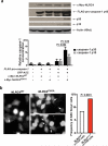
NLRC4V341A promotes spontaneous cleavage of pro-caspase-1 and ASC multimerization in HEK293 cells. (a) Increased cleavage of FLAG-procaspase-1 in HEK293 cells expressing NLRC4V341A versus NLRC4WT. Western blot at top of figure shows results of blotting for NLRC4, pro-caspase-1 (p45) and its p35 and p10 cleavage products as well as actin controls in cells expressing constructs shown below, as described in Methods. Levels of p35 and p10 are normalized in each case to the level of pro-caspase-1. Alternate analysis normalizing p35 and p10 to levels of actin and NLRC4 also yielded statistically significant differences between lines transfected with NLRC4WT and NLRC4V341A. Mean and standard deviation of four independent transfections is shown. A two-sided Student's t-test was used to determine statistical significance. (b) Spontaneous ASC multimerization (white arrows) in HEK293 cells expressing GFP-ASC and either NLRC4WT (left panel) or NLRC4V341A (right panels) using epifluorescent microscopy. A total of 1422 cells transfected with wildtype NLRC4 and 1155 cells transfected with mutant NLRC4 were scored. * the frequency of ASC puncta+ cells in lines transfected with NLRC4WT is significantly different (P<0.0001) from lines transfected with NLRC4V341A (chi-square testing). Scale bars, 20 μm.
Figure 4
Figure 4
Increased production of IL-1β,IL-18 and increased cell death in macrophages harboring NLRC4V341A. Monocyte-derived macrophages from SCAN4 patients (II.3 and III.2) and WT controls (one related and four unrelated) were cultured for 18 hours in media containing low-dose LPS (1 ng/ml) followed by measurement of (a) IL-1-β(b) IL-18, and (c) LDH as described in Methods. LDH release is reported relative to result following total lysis by TritonX-100 (0.1%). Cytokine secretion and cell death results were similar in macrophages from patients (II.3 and III.2). Bar graphs show mean ± S.E.M from three separate experiments. Significance by unpaired Student's t-test is indicated.
Figure 5
Figure 5
SCAN4 macrophages infected with S. typhimurium strain SL1344 show abnormal ASC staining and limited activation of caspase-1. (a) The majority of S. typhimurium infected monocyte-derived macrophages from a representative healthy control (HD II.2, the mother of SCAN4 patient III.2) display features of conventional macrophage activation including biotin-YVAD-CMK staining (green cytoplasm) and a limited number (1-3) of ASC foci (red puncta) per cell (left panel). A subset of S. typhimurium infected macrophages (white arrows) from SCAN4 patients II.3 and III.2 display numerous (>6) ASC foci but more limited staining for biotin-YVADCMK (right panel). Scale bars, 20 μm (b) Quantitation of ASC foci in 2499 SL1344 infected healthy control macrophages and 1224 S. typhimurium infected patient macrophages. (c) Representative biotin-YVAD-CMK staining in S. typhimurium infected healthy control or SCAN4 macrophages from groups with either 1-3, 4-6 or >6 ASC foci/cell. Pie charts display the frequency of biotin-YVAD-CMK positive cells per group. Total cell number analyzed within each group listed within parentheses. Scale bars, 20 μm. (d) Diameter of ASC foci in biotin-YVADCMK positive macrophages (filled green circles) and biotin-YVAD-CMK negative macrophages (unfilled circles) from healthy control and SCAN4 patients II.3 and III.2. Mean foci diameter displayed as white bars. * patient cell distribution is significantly different (P<0.0001) from the healthy control cell distribution by chi-square statistical testing (for b and c). Significance by unpaired Student's t-test is indicated (for d).

Comment in

References

    1. Mariathasan S, Monack DM. Inflammasome adaptors and sensors: intracellular regulators of infection and inflammation. Nat Rev Immunol. 2007;7:31–40. - PubMed
    1. Dinarello CA. Immunological and inflammatory functions of the interleukin-1 family. Annu Rev Immunol. 2009;27:519–50. - PubMed
    1. Ozen S, Bilginer Y. A clinical guide to autoinflammatory diseases: familial Mediterranean fever and next-of-kin. Nat Rev Rheumatol. 2014;10:135–47. - PubMed
    1. Thornberry NA, et al. A novel heterodimeric cysteine protease is required for interleukin-1 beta processing in monocytes. Nature. 1992;356:768–74. - PubMed
    1. Miao EA, et al. Cytoplasmic flagellin activates caspase-1 and secretion of interleukin 1beta via Ipaf. Nat Immunol. 2006;7:569–75. - PubMed

Publication types

Substances