. 2014 Sep 14;15(1):112.
doi: 10.1186/s12931-014-0112-3.

The role of bronchial epithelial cells in the pathogenesis of COPD in Z-alpha-1 antitrypsin deficiency

The role of bronchial epithelial cells in the pathogenesis of COPD in Z-alpha-1 antitrypsin deficiency

Laura Pini et al. Respir Res. .

Abstract

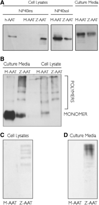
Background: Alpha-1 antitrypsin is the main inhibitor of neutrophil elastase in the lung. Although it is principally synthesized by hepatocytes, alpha-1 antitrypsin is also secreted by bronchial epithelial cells. Gene mutations can lead to alpha-1 antitrypsin deficiency, with the Z variant being the most clinically relevant due to its propensity to polymerize. The ability of bronchial epithelial cells to produce Z-variant protein and its polymers is unknown.

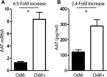
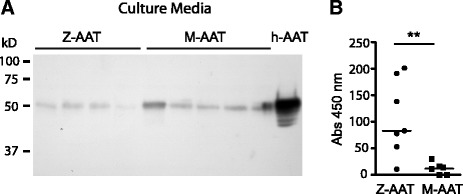
Methods: Experiments using a conformation-specific antibody were carried out on M- and Z-variant-transfected 16HBE cells and on bronchial biopsies and ex vivo bronchial epithelial cells from Z and M homozygous patients. In addition, the effect of an inflammatory stimulus on Z-variant polymer formation, elicited by Oncostatin M, was investigated. Comparisons of groups were performed using t-test or ANOVA. Non-normally distributed data were assessed by Mann-Whitney U test or the Kruskal-Wallis test, where appropriate. A P value of < 0.05 was considered to be significant.

Results: Alpha-1 antitrypsin polymers were found at a higher concentration in the culture medium of ex vivo bronchial epithelial cells from Z-variant homozygotes, compared with M-variant homozygotes (P < 0.01), and detected in the bronchial epithelial cells and submucosa of patient biopsies. Oncostatin M significantly increased the expression of alpha-1 antitrypsin mRNA and protein (P < 0.05), and the presence of Z-variant polymers in ex vivo cells (P < 0.01).

Conclusions: Polymers of Z-alpha-1 antitrypsin form in bronchial epithelial cells, suggesting that these cells may be involved in the pathogenesis of lung emphysema and in bronchial epithelial cell dysfunction.


Figures

Figure 1
Figure 1
Expression of M- or Z-AAT in the 16HBE cell line. ( A ) 16HBE cells expressing M- or Z-AAT were assayed for AAT expression by SDS PAGE and Western blot analysis. 24 hours after transfection, proteins from 50 μl of cell culture media or from 10 μg or 10 μl of NP40-soluble and insoluble cell lysates respectively were separated by SDS-PAGE, transferred to PVDF membranes, and hybridized with anti-AAT antibody to detect total AAT protein. Purified human serum AAT (hAAT) was used as positive control. Typical fluorograms from three independent experiments, which gave superimposable results, are shown. ( B ) Nondenaturing PAGE and Western blot analysis using anti-AAT antibody to detect total AAT in 10 μg of NP40-soluble cell lysate and 10 μl of cell culture media from 16HBE cells expressing M- or Z-AAT. The migration of polymeric and monomeric AAT species is indicated. Typical fluorograms from three independent experiments, which gave superimposable results, are shown. ( C ) Nondenaturing PAGE and Western blot analysis using the antipolymeric Z-AAT antibody ATZ11 in NP40-soluble cell lysate fraction (10 μg) and ( D ) cell culture media (50 μl)from 16HBE cells expressing M- or Z-AAT. Typical fluorograms from three independent experiments, which gave superimposable results, are shown. Definition of abbreviations: AAT = alpha-1 antitrypsin; h-AAT = human serum AAT; PAGE = polyacrylamide gel electrophoresis; PVDF = polyvinylidene fluoride; SDS-PAGE = sodium dodecyl sulphate polyacrylamide gel electrophoresis.
Figure 2
Figure 2
Effects of Oncostatin M on expression of endogenous AAT in the 16HBE cell line. Nontransfected 16HBE cells were cultured in the absence (OsM-) or presence (OsM+) of 50 ng/ml Oncostatin M for 24 hours, and AAT mRNA expression and AAT protein secretion were analyzed. ( A ) Mean AAT mRNA expression levels, normalized for β-actin expression levels (×103), as measured by real-time PCR analysis from total cellular RNA. ( B ) AAT protein levels as measured by ELISA for total AAT in cell culture media. The fold increase in expression of Oncostatin M-treated versus untreated cells is indicated. Results are expressed as mean ± SEM (n = 3–4). *P < 0.05 Student’s t-test. Definition of abbreviations: AAT = alpha-1 antitrypsin; ELISA = enzyme-linked immunosorbent assay; mRNA = messenger ribonucleic acid; Real-time PCR = reverse transcription real-time polymerase chain reaction; RNA = ribonucleic acid.
Figure 3
Figure 3
Immunohistochemistry of bronchial epithelial biopsy. Endobronchial biopsy sections taken from patients homozygous for M-AAT or Z-AAT were fixed and stained with the ATZ11 antibody. Evidence of polymers of Z-AAT was confirmed in the epithelial cells and submucosa of Z-AAT homozygote tissue. Definition of abbreviation: AAT = alpha-1 antitrypsin.
Figure 4
Figure 4
Detection of AAT in ex-vivo cultured BECs. ( A ) SDS-PAGE and Western blot analysis using an antibody to detect total AAT in 20 μl of 10x concentrated culture media from a 72-hour culture of ex vivo BECs from five patients homozygous for M-AAT and four patients homozygous for Z-AAT. Purified human serum AAT (h-AAT) served as positive control. A typical fluorogram from three independent experiments, which gave superimposable results, is shown. ( B ) Cell culture media from a 72-hour culture of ex vivo BECs from six patients homozygous for M-AAT and seven patients homozygous for Z-AAT were concentrated 10 times, opportunely diluted to a final concentration of 10 μg/ml of AAT as assessed by an ELISA for total AAT, and were analyzed by ELISA using the antipolymeric AAT antibody ATZ11. . * P = 0.01 Mann–Whitney U test. Definition of abbreviations: AAT = alpha-1 antitrypsin; BEC = bronchial epithelial cell; h-AAT = human serum AAT; PAGE = polyacrylamide gel electrophoresis; ELISA = enzyme-linked immunosorbent assay.
Figure 5
Figure 5
The effect of Oncostatin M on Z-AAT expression in ex-vivo cultured BECs. ( A ) Real-time PCR analysis for AAT mRNA in ex vivo cultured BECs from patients homozygous for M-AAT or Z-AAT. Cells from four patients homozygous for Z-AAT and five patients homozygous for M-AAT were incubated in the absence (OsM-) or presence (OsM+) of Oncostatin M (50 ng/ml) for 24 hours. After treatment cells from M-AAT and Z-AAT patients were pooled and AAT mRNA levels were measured. The fold increase in expression of Oncostatin M-treated versus control cells is indicated. Results are expressed as mean ± SEM (n = 3–4). *P < 0.05 Student’s t-test. ( B ) ELISA for total AAT protein in cell lysates and ( C ) cell culture media of ex vivo cultured BECs from four Z-AAT and five M-AAT homozygous patients. Cells were incubated in the absence (OsM-) or presence (OsM+) of Oncostatin M (50 ng/ml) for 24 hours and the intracellular AAT accumulation and secretion were measured for lysates of pooled cells and 10x concentrated cell culture media, respectively. Results are expressed as mean ± SEM (n = 3–4). *P < 0.05 ANOVA followed by Bonferroni t-test, Oncostatin M-treated versus untreated cells; +P < 0.05 ANOVA followed by Bonferroni t-test, M-AAT– versus Z-AAT–expressing cells. SDS-PAGE and Western blot analysis using an antibody to detect total AAT was carried out on the cell culture media of ex vivo cultured BECs. Cells from five patients homozygous for Z-AAT ( D ) and five patients homozygous for M-AAT ( E ) were cultured in the absence (OsM-) or presence (OsM+) of 50 ng/ml of Oncostatin M. At 24 hours after treatment cell media were harvested and concentrated 10x. Typical fluorograms from three independent experiments, which gave superimposable results, are shown.
Figure 6
Figure 6
Effect of OsM on AAT polymers formation in ex-vivo cultured BECs. ELISA for polymeric AAT protein, using the ATZ11 antibody, in cell culture media and cell lysates of ex vivo cultured BECs from Z-AAT homozygous patients. Cells from five patients were incubated in the absence (OsM-) and presence (OsM+) of Oncostatin M (50 ng/ml) for 24 hours, and AAT secretion and intracellular accumulation were measured in the 10x concentrated cell medium. Results are expressed as mean absorbance ± SEM (n = 3–4), measured at 450 nm. *P = < 0.01 Student’s t-test. Definition of abbreviations: AAT = alpha-1 antitrypsin; BEC = bronchial epithelial cell; ELISA = enzyme-linked immunosorbent assay; SEM = standard error of mean.

References

    1. Brantly M, Nukiwa T, Crystal RG. Molecular basis of alpha-1-antitrypsin deficiency. Am J Med. 1988;84:13–31. doi: 10.1016/0002-9343(88)90154-4. - DOI - PubMed
    1. Johnson D, Travis J. Structural evidence for methionine at the reactive site of human alpha-1-proteinase inhibitor. J Biol Chem. 1978;253:7142–7144. - PubMed
    1. Cichy J, Potempa J, Travis J. Biosynthesis of alpha1-proteinase inhibitor by human lung-derived epithelial cells. J Biol Chem. 1997;272:8250–8255. doi: 10.1074/jbc.272.13.8250. - DOI - PubMed
    1. Du Bois RM, Bernaudin JF, Paakko P, Hubbard R, Takahashi H, Ferrans V, Crystal RG. Human neutrophils express the alpha 1-antitrypsin gene and produce alpha 1-antitrypsin. Blood. 1991;77:2724–2730. - PubMed
    1. Mornex JF, Chytil-Weir A, Martinet Y, Courtney M, LeCocq JP, Crystal RG. Expression of the alpha-1-antitrypsin gene in mononuclear phagocytes of normal and alpha-1-antitrypsin-deficient individuals. J Clin Invest. 1986;77:1952–1961. doi: 10.1172/JCI112524. - DOI - PMC - PubMed

Publication types

MeSH terms

Substances