

Regional wall function before and after acute myocardial infarction; an experimental study in pigs
- PMID: 25218585
- PMCID: PMC4169797
- DOI: 10.1186/1471-2261-14-118
Regional wall function before and after acute myocardial infarction; an experimental study in pigs
Abstract
Background: Left ventricular function is altered during and after AMI. Regional function can be determined by cardiac magnetic resonance (CMR) wall thickening, and velocity encoded (VE) strain analysis. The aims of this study were to investigate how regional myocardial wall function, assessed by CMR VE-strain and regional wall thickening, changes after acute myocardial infarction, and to determine if we could differentiate between ischemic, adjacent and remote segments of the left ventricle.
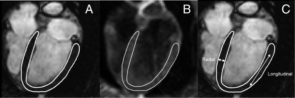
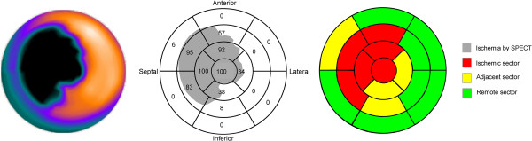
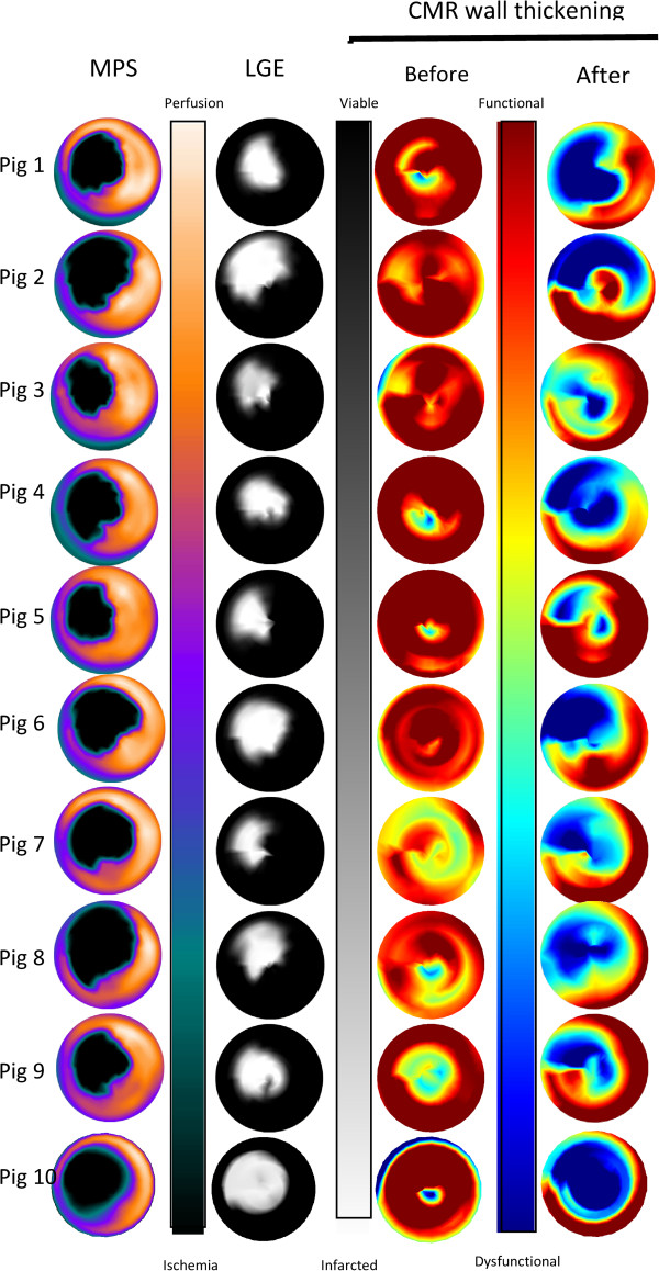
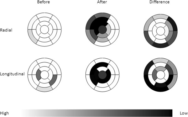
Methods: Ten pigs underwent baseline CMR study for assessment of wall thickening and VE-strain. Ischemia was then induced for 40-minutes by intracoronary balloon inflation in the left anterior descending coronary artery. During occlusion, (99m)Tc tetrofosmin was administered intravenously and myocardial perfusion SPECT (MPS) was performed for determination of the ischemic area, followed by a second CMR study. Based on ischemia seen on MPS, the 17 AHA segments of the left ventricle was divided into 3 different categories (ischemic, adjacent and remote). Regional wall function measured by wall thickening and VE-strain analysis was determined before and after ischemia.
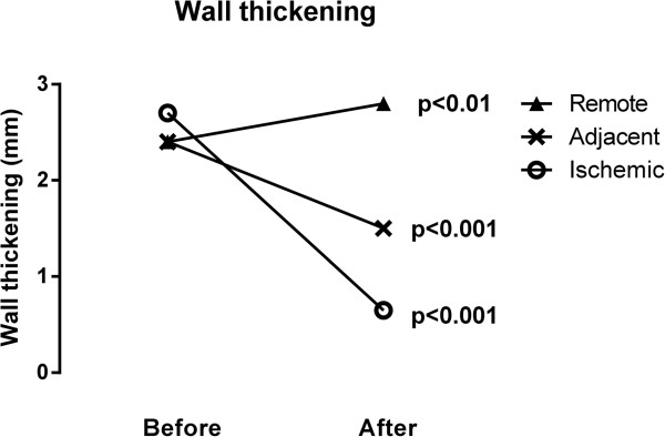
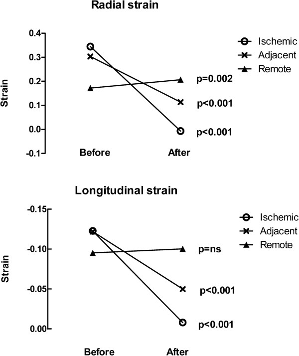
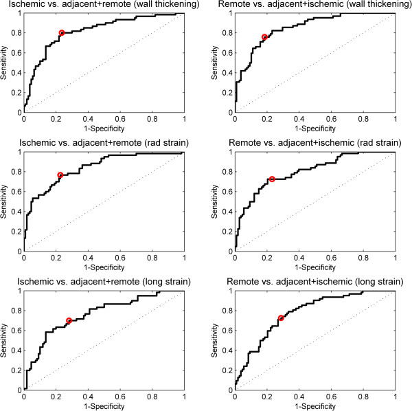
Results: Mean wall thickening decreased significantly in the ischemic (from 2.7 mm to 0.65 mm, p < 0.001) and adjacent segments (from 2.4 to 1.5 mm p < 0.001). In remote segments, wall thickening increased significantly (from 2.4 mm to 2.8 mm, p < 0.01). In ischemic and adjacent segments, both radial and longitudinal strain was significantly decreased after ischemia (p < 0.001). In remote segments there was a significant increase in radial strain (p = 0.002) while there was no difference in longitudinal strain (p = 0.69). ROC analysis was performed to determine thresholds distinguishing between the different regions. Sensitivity for determining ischemic segments ranged from 70-80%, and specificity from 72%-77%. There was a 9% increase in left ventricular mass after ischemia.
Conclusion: Differentiation thresholds for wall thickening and VE-strain could be established to distinguish between ischemic, adjacent and remote segments but will, have limited applicability due to low sensitivity and specificity. There is a slight increase in radial strain in remote segments after ischemia. Edema was present mainly in the ischemic region but also in the combined adjacent and remote segments.
Figures
Similar articles
-
Correspondence between gated SPECT and cardiac magnetic resonance quantified myocardial wall thickening.Int J Cardiovasc Imaging. 2011 Oct;27(7):1095-104. doi: 10.1007/s10554-010-9746-5. Epub 2010 Nov 26. Int J Cardiovasc Imaging. 2011. PMID: 21110103
-
Three-dimensional regional strain analysis in porcine myocardial infarction: a 3T magnetic resonance tagging study.J Cardiovasc Magn Reson. 2012 Dec 13;14(1):85. doi: 10.1186/1532-429X-14-85. J Cardiovasc Magn Reson. 2012. PMID: 23237210 Free PMC article.
-
Perfusion and functional abnormalities outside the septal region in patients with left bundle branch block assessed with gated SPECT.Q J Nucl Med. 2001 Mar;45(1):108-14. Q J Nucl Med. 2001. PMID: 11456369
-
Quantification of myocardial segmental function in acute and chronic ischemic heart disease and implications for cardiovascular cell therapy trials: a review from the NHLBI-Cardiovascular Cell Therapy Research Network.JACC Cardiovasc Imaging. 2011 Jun;4(6):671-9. doi: 10.1016/j.jcmg.2011.02.015. JACC Cardiovasc Imaging. 2011. PMID: 21679903 Free PMC article. Review.
-
CMR for myocardial characterization in ischemic heart disease: state-of-the-art and future developments.Eur Radiol Exp. 2021 Mar 25;5(1):14. doi: 10.1186/s41747-021-00208-2. Eur Radiol Exp. 2021. PMID: 33763757 Free PMC article. Review.
Cited by
-
Evolution of left ventricular function among subjects with ST-elevation myocardial infarction after percutaneous coronary intervention.BMC Cardiovasc Disord. 2020 Jun 29;20(1):309. doi: 10.1186/s12872-020-01540-y. BMC Cardiovasc Disord. 2020. PMID: 32600336 Free PMC article. Clinical Trial.
-
Necropsy Validation of a Novel Method for Left Ventricular Mass Quantification in Porcine Transthoracic and Transdiaphragmal Echocardiography.Front Cardiovasc Med. 2022 May 3;9:868603. doi: 10.3389/fcvm.2022.868603. eCollection 2022. Front Cardiovasc Med. 2022. PMID: 35592401 Free PMC article.
-
Acute phase segmental radial strain correlates with recovery and late gadolinium extent in ST-elevation myocardial infarction (STEMI): analysis of the abciximab intracoronary versus intravenously drug application in STEMI substudy.Quant Imaging Med Surg. 2021 Aug;11(8):3595-3603. doi: 10.21037/qims-21-56. Quant Imaging Med Surg. 2021. PMID: 34341734 Free PMC article.
-
Precision imaging of cardiac function and scar size in acute and chronic porcine myocardial infarction using ultrahigh-field MRI.Commun Med (Lond). 2024 Jul 18;4(1):146. doi: 10.1038/s43856-024-00559-y. Commun Med (Lond). 2024. PMID: 39026075 Free PMC article.
-
Injectable Myocardial Matrix Hydrogel Mitigates Negative Left Ventricular Remodeling in a Chronic Myocardial Infarction Model.JACC Basic Transl Sci. 2021 Mar 10;6(4):350-361. doi: 10.1016/j.jacbts.2021.01.003. eCollection 2021 Apr. JACC Basic Transl Sci. 2021. PMID: 33997521 Free PMC article.
References
-
- Go AS, Mozaffarian D, Roger VL, Benjamin EJ, Berry JD, Borden WB, Bravata DM, Dai S, Ford ES, Fox CS, Franco S, Fullerton HJ, Gillespie C, Hailpern SM, Heit JA, Howard VJ, Huffman MD, Kissels BM, Kittner SJ, Lackland DT, Lichtman JH, Lisabeth JH, Magid D, Marcus GM, Marelli A, Matchar DB, McGuire DK, Mohler ER, Moy CS, Mussolino ME. Heart disease and stroke statistics–2013 update: a report from the American Heart Association. Circulation. 2013;127(1):e6–e245. doi: 10.1161/CIR.0b013e31828124ad. - DOI - PMC - PubMed
-
- Shimkunas R, Zhang Z, Wenk JF, Soleimani M, Khazalpour M, Acevedo-Bolton G, Wang G, Saloner D, Mishra R, Wallace AW, Ge L, Baker AJ, Guccione JM, Ratcliffe MB. Left ventricular myocardial contractility is depressed in the borderzone after posterolateral myocardial infarction. Ann Thorac Surg. 2013;95(5):1619–1625. doi: 10.1016/j.athoracsur.2013.02.005. - DOI - PMC - PubMed
Pre-publication history
-
- The pre-publication history for this paper can be accessed here:http://www.biomedcentral.com/1471-2261/14/118/prepub
Publication types
MeSH terms
Substances
LinkOut - more resources
Full Text Sources
Other Literature Sources
Medical
