Case Reports
. 2015 Feb;114(2):233-241.
doi: 10.1016/j.ymgme.2014.08.011. Epub 2014 Sep 2.

CNS, lung, and lymph node involvement in Gaucher disease type 3 after 11 years of therapy: clinical, histopathologic, and biochemical findings

Affiliations
Case Reports

CNS, lung, and lymph node involvement in Gaucher disease type 3 after 11 years of therapy: clinical, histopathologic, and biochemical findings

Thomas A Burrow et al. Mol Genet Metab. 2015 Feb.

Abstract

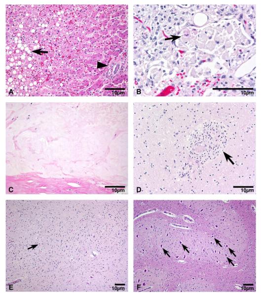
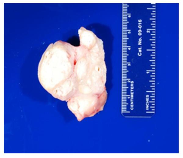
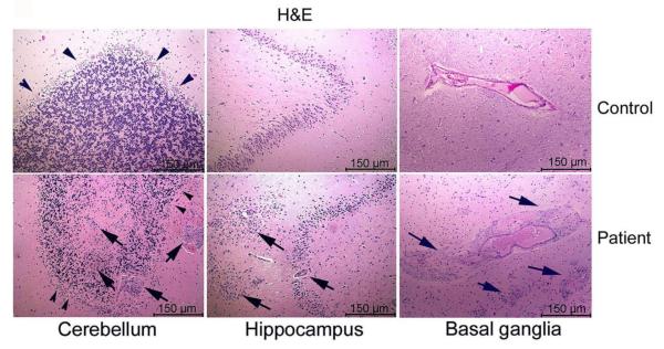
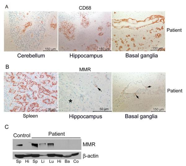
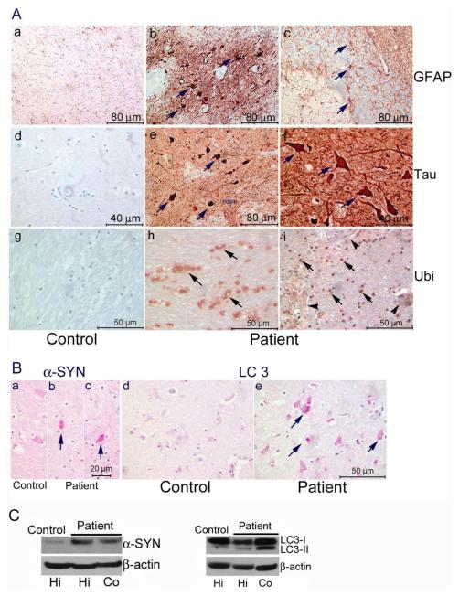
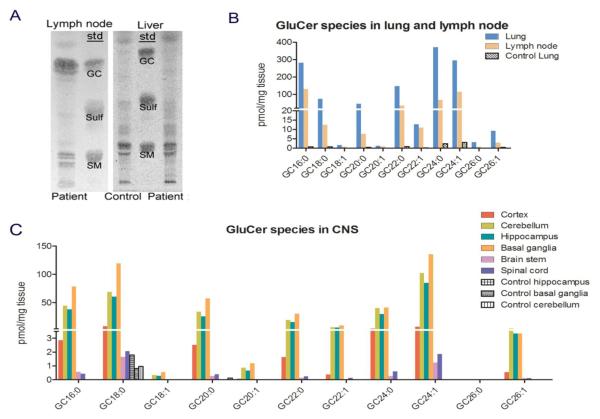
A Caucasian male with Gaucher disease type 3, treated with continuous enzyme therapy (ET) for 11 years, experienced progressive mesenteric and retroperitoneal lymphadenopathy, lung disease, and neurological involvement leading to death at an age of 12.5 years. Autopsy showed significant pathology of the brain, lymph nodes, and lungs. Liver and spleen glucosylceramide (GluCer) and glucosylsphingosine (GluS) levels were nearly normal and storage cells were cleared. Clusters of macrophages and very elevated GluCer and GluS levels were in the lungs, and brain parenchymal and perivascular regions. Compared to normal brain GluCer (GC 18:0), GluCer species with long fatty acid acyl chains were increased in the patient's brain. This profile was similar to that in the patient's lungs, suggesting that these lipids were present in brain perivascular macrophages. In the patient's brain, generalized astrogliosis, and enhanced LC3, ubiquitin, and Tau signals were identified in the regions surrounding macrophage clusters, indicating proinflammation, altered autophagy, and neurodegeneration. These findings highlight the altered phenotypes resulting from increased longevity due to ET, as well as those in poorly accessible compartments of brain and lung, which manifested progressive disease involvement despite ET.

Keywords: Enzyme replacement therapy; Gaucher disease; Lymphadenopathy; Lysosomal storage disease; Pathology.


Figures

Figure 1
Figure 1. Pathological findings by H&E
A) Liver histology: Moderate steatosis (arrow) and mild portal fibrosis (arrow ahead) were noted. B) Lung histology: Extensive accumulation of storage cells in alveolar spaces (arrow). C) Mesenteric lymph node histology: The lymphoid architecture was significantly replaced with an acellular material with areas of calcification. D) Pathology of cerebrum: The storage cells (arrow) consisted of large histiocytes with vacuolated cytoplasm and containing typical inclusions for Gaucher disease frequently compressed the lumen of the arterioles. E) Decreased neurons (arrow) in patient cerebellar dentate nucleus. F) Neurons (arrows) in unaffected age matched human control cerebellar dentate nucleus.
Figure 2
Figure 2. Mesenteric lymph node
Cut section of Mesenteric lymph node showing replacement of gross structure with a white, chalky material.
Figure 3
Figure 3. H&E staining of paraffin brain sections
The storage cells (arrows) were in parenchyma of cerebellum and hippocampus and lining around perivascular region in basal ganglia from the patient. Normal Purkinje cell (arrowheads) layer was in control cerebellum. Loss of Purkinje cells (arrowheads) was prominent in patient cerebellum.
Figure 4
Figure 4. Storage cells and Macrophage mannose receptor (MMR) expression
(A) The storage cells were positive for CD68 antibody staining (brown), a macrophage marker, and were present in patient’s cerebellum, hippocampus, basal ganglia. (B) MMR positive staining (brown) was in patient spleen. A few MMR positive cells (arrows) were around vessels in the basal ganglia and hippocampus. The storage cells (star) in the hippocampus did not stain positive for MMR. The nuclei were counter stained with hematoxicillin (blue). Scales are indicated in the images. (C) Immunoblot of MMR. Control and patient spleen (Sp), liver (Li) and lung (Lu) expressed MMR protein. Control and patient brain samples had no detectable MMR protein. Hi, hippocampus; Ba, basal ganglia; Co, cerebral cortex. β-actin was used as loading control.
Figure 5
Figure 5. Brain pathology
(A) Astrogliosis, Tau and Ubiquitin (Ubi) pathology. (a-c) Astrogliosis (brown) was determined by anti-GFAP staining. The control human basal ganglia (a) had low level GFAP signal. The engorged astrocytes (arrows) were detected in patient hippocampus (b) and cerebellum (c). (d-f) Tau pathology was assessed by anti-phospho-Tau antibody. No phospho-Tau signal was detected in control human basal ganglia (d). Strong phospho-Tau signals (brown) were in the cells (arrows) of patient basal ganglia (e) and hippocampus (f). (g-i) Ubi signals (brown) were detected in white matter areas of patient basal ganglia (h) and around storage cells (arrowhead) in hippocampus (i). No positive Ubi staining was observed in basal ganglia white matter in control (g). (B) α-Synuclein (α-SYN) and LC3 detection. Low level of α-SYN signals were detected in the cells of control human hippocampus (a). Enhanced α-SYN signals were found in a few cells in the patient’s cerebral cortex (b) and hippocampus (c). In comparison to control hippocampus (d), LC3 signals were shown in patient’s hippocampus (e). Scales are indicated in the images. Nuclei were counter stained with hematoxylin. C. Immunoblots. Left, α-SYN levels were increased in patient hippocampus (Hi) and cortex (Co) compared to control hippocampus. Right, Increased LC3-II was detected in patient hippocampus (Hi) and cortex (Co). β-actin was used as a loading control.
Figure 6
Figure 6. Glycosphingolipid analyses
(A) TLC analyses of lymph node and liver. Patient lymph node had accumulated glucosylceramide (GluCer, GC). GC in patient liver was at control (normal) level. Lipids standards (std) on TLC: GC, glucosylceramide; Sulf, sulfatide; and SM, sphingomyelin. (B) GluCer species profile in visceral tissues. Patient lung and lymph node had accumulation of short and long chain GluCer species compared to control lung. (C) GluCer species profile in patient CNS. GC18:0 is the major species in control brains (black bars). In addition to GC18:0, GC16:0 and various long fatty acid acyl chain GluCer species were detected and increased in patient cortex, cerebellum, hippocampus, basal ganglion, brain stem and spinal cord as shown in colored bars. More GluCer species were accumulated in cortex, cerebellum, hippocampus, basal ganglion than that in brain stem and spinal cord. GluCer species were analyzed by LC/MS and normalized by mg of tissue weight.

Similar articles

Cited by

References

    1. Grabowski G, Petsko G, Kolodny E. Gaucher Disease. In: Valle D, Beaudet A, Vogelstein B, Kinzler K, Antonarakis S, Ballabio A, Gibson K, Mitchell G, editors. The Online Metabolic and Molecular Bases of Inherited Disease. The McGraw-Hill Companies, Inc.; New York, NY: [Accessed on; 06/23/14]. 2010. Chapt 146.
    1. Altarescu G, Hill S, Wiggs E, Jeffries N, Kreps C, Parker CC, Brady RO, Barton NW, Schiffmann R. The efficacy of enzyme replacement therapy in patients with chronic neuronopathic Gaucher’s disease. J Pediatr. 2001;138:539–547. - PubMed
    1. Burrow TA, Cohen MB, Bokulic R, Deutsch G, Choudhary A, Falcone RA, Jr., Grabowski GA. Gaucher disease: progressive mesenteric and mediastinal lymphadenopathy despite enzyme therapy. J Pediatr. 2007;150:202–206. - PubMed
    1. Grabowski GA, Leslie N, Wenstrup R. Enzyme therapy for Gaucher disease: the first 5 years. Blood Rev. 1998;12:115–133. - PubMed
    1. Zhao H, Bailey LA, Elsas LJ, 2nd, Grinzaid KA, Grabowski GA. Gaucher disease: in vivo evidence for allele dose leading to neuronopathic and nonneuronopathic phenotypes. Am J Med Genet A. 2003;116A:52–56. - PubMed

Publication types

MeSH terms