

Neural crest development in fetal alcohol syndrome
- PMID: 25219761
- PMCID: PMC4827602
- DOI: 10.1002/bdrc.21078
Neural crest development in fetal alcohol syndrome
Abstract
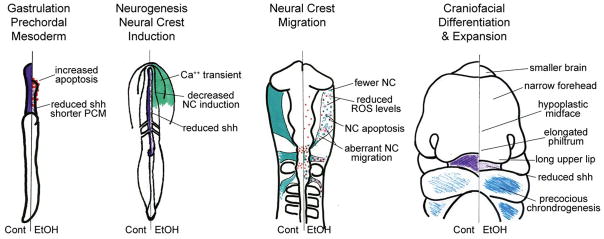
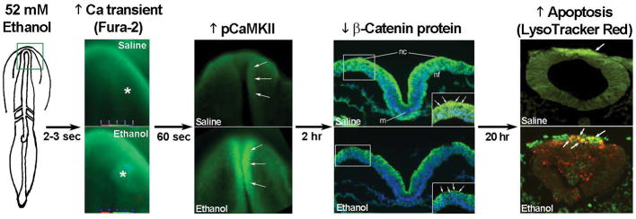
Fetal alcohol spectrum disorder (FASD) is a leading cause of neurodevelopmental disability. Some affected individuals possess distinctive craniofacial deficits, but many more lack overt facial changes. An understanding of the mechanisms underlying these deficits would inform their diagnostic utility. Our understanding of these mechanisms is challenged because ethanol lacks a single receptor when redirecting cellular activity. This review summarizes our current understanding of how ethanol alters neural crest development. Ample evidence shows that ethanol causes the "classic" fetal alcohol syndrome (FAS) face (short palpebral fissures, elongated upper lip, deficient philtrum) because it suppresses prechordal plate outgrowth, thereby reducing neuroectoderm and neural crest induction and causing holoprosencephaly. Prenatal alcohol exposure (PAE) at premigratory stages elicits a different facial appearance, indicating FASD may represent a spectrum of facial outcomes. PAE at this premigratory period initiates a calcium transient that activates CaMKII and destabilizes transcriptionally active β-catenin, thereby initiating apoptosis within neural crest populations. Contributing to neural crest vulnerability are their low antioxidant responses. Ethanol-treated neural crest produce reactive oxygen species and free radical scavengers attenuate their production and prevent apoptosis. Ethanol also significantly impairs neural crest migration, causing cytoskeletal rearrangements that destabilize focal adhesion formation; their directional migratory capacity is also lost. Genetic factors further modify vulnerability to ethanol-induced craniofacial dysmorphology and include genes important for neural crest development, including shh signaling, PDFGA, vangl2, and ribosomal biogenesis. Because facial and brain development are mechanistically and functionally linked, research into ethanol's effects on neural crest also informs our understanding of ethanol's CNS pathologies.
Keywords: apoptosis; calcium signaling; craniofacial; fetal alcohol spectrum disorders; neural crest; sonic hedgehog.
© 2014 Wiley Periodicals, Inc.
Figures
References
-
- Ahlgren SC, Bronner-Fraser M. Inhibition of sonic hedgehog signaling in vivo results in craniofacial neural crest cell death. Curr Biol. 1999;9:1304–1314. - PubMed
-
- Aoto K, Shikata Y, Higashiyama D, Shiota K, Motoyama J. Fetal ethanol exposure activates protein kinase A and impairs Shh expression in prechordal mesendoderm cells in the pathogenesis of holoprosencephaly. Birth Defects Res A Clin Mol Teratol. 2008;82:224–231. - PubMed
-
- Blader P, Strahle U. Ethanol impairs migration of the prechordal plate in the zebrafish embryo. Dev Biol. 1998;201:185–201. - PubMed
Publication types
MeSH terms
Substances
Grants and funding
LinkOut - more resources
Full Text Sources
Other Literature Sources
Medical
Research Materials
Miscellaneous
