. 2014 Oct 23;514(7523):490-3.
doi: 10.1038/nature13723. Epub 2014 Sep 14.

A Hox regulatory network of hindbrain segmentation is conserved to the base of vertebrates

Affiliations

A Hox regulatory network of hindbrain segmentation is conserved to the base of vertebrates

Hugo J Parker et al. Nature. .

Abstract

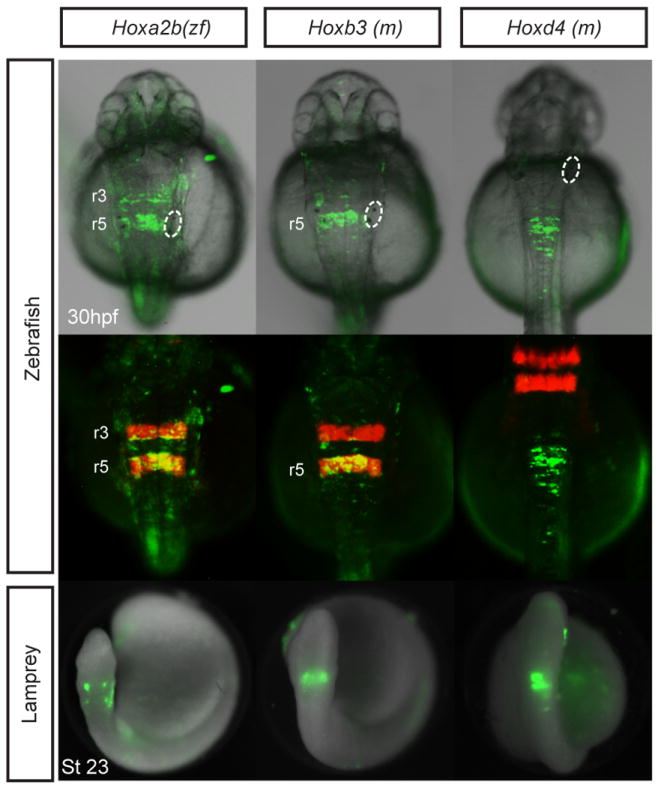
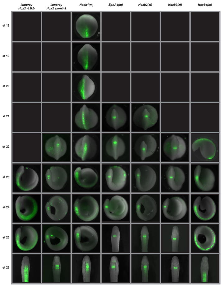
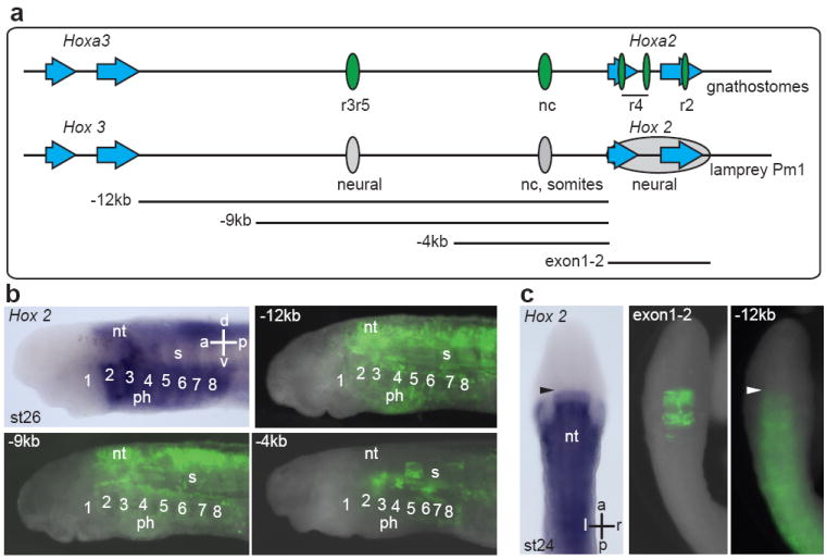
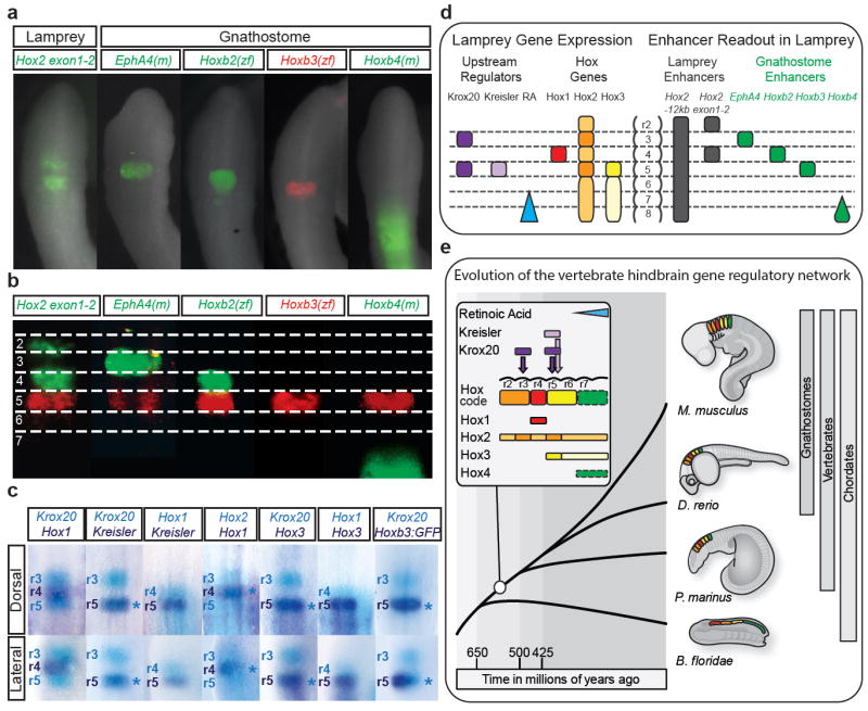
A defining feature governing head patterning of jawed vertebrates is a highly conserved gene regulatory network that integrates hindbrain segmentation with segmentally restricted domains of Hox gene expression. Although non-vertebrate chordates display nested domains of axial Hox expression, they lack hindbrain segmentation. The sea lamprey, a jawless fish, can provide unique insights into vertebrate origins owing to its phylogenetic position at the base of the vertebrate tree. It has been suggested that lamprey may represent an intermediate state where nested Hox expression has not been coupled to the process of hindbrain segmentation. However, little is known about the regulatory network underlying Hox expression in lamprey or its relationship to hindbrain segmentation. Here, using a novel tool that allows cross-species comparisons of regulatory elements between jawed and jawless vertebrates, we report deep conservation of both upstream regulators and segmental activity of enhancer elements across these distant species. Regulatory regions from diverse gnathostomes drive segmental reporter expression in the lamprey hindbrain and require the same transcriptional inputs (for example, Kreisler (also known as Mafba), Krox20 (also known as Egr2a)) in both lamprey and zebrafish. We find that lamprey hox genes display dynamic segmentally restricted domains of expression; we also isolated a conserved exonic hox2 enhancer from lamprey that drives segmental expression in rhombomeres 2 and 4. Our results show that coupling of Hox gene expression to segmentation of the hindbrain is an ancient trait with origin at the base of vertebrates that probably led to the formation of rhombomeric compartments with an underlying Hox code.


Conflict of interest statement

The authors declare no competing financial interests.

Figures

Extended Data Figure 1
Extended Data Figure 1. Gnathostome enhancer elements selected for reporter analysis
Schematic diagrams depicting the gnathostome enhancer elements assayed for activity in zebrafish and lamprey embryos in this study. The endogenous genomic positions of the enhancer elements (green boxes) are shown relative to the genes that they regulate. Known trans-acting factors are listed above the elements, whilst the corresponding regulatory modules and their combined activity domains are detailed below the elements. For each element, the species from which it was cloned are listed on the right. Figure based on Fig. 4.2 from Tumpel et al. (2009).
Extended Data Figure 2
Extended Data Figure 2. Segmental activity of additional jawed vertebrate enhancers in zebrafish and lamprey
GFP reporter expression mediated by gnathostome enhancer elements in zebrafish and lamprey embryos. Dorsal views are shown, with anterior to the top. For zebrafish, two images of the same embryo are shown, presenting GFP plus brightfield (top) and GFP plus endogenous r3r5mCherry (middle) signals. The zebrafish otic vesicle is circled. Abbreviations: m, mouse; r, rhombomere; zf, zebrafish.
Extended Data Figure 3
Extended Data Figure 3. Segmental patterns of GFP reporter expression in transgenic zebrafish lines
Lateral (top) and dorsal (middle) views of 30hpf transgenic (F1) zebrafish embryos show combined brightfield illumination and segmental GFP reporter expression in the hindbrain mediated by five different gnathostome enhancer elements. The corresponding transient transgenic GFP expression patterns mediated by these elements are shown in Fig. 1b and Extended Data Figure 2. When available, GFP lines were crossed with endogenous r3r5 mCherry reporter line as a reference (bottom). The otic vesicle is circled. Abbreviations: m, mouse; r, rhombomere; zf, zebrafish.
Extended Data Figure 4
Extended Data Figure 4. Developmental time-course of GFP reporter expression mediated by lamprey and gnathostome regulatory elements in lamprey embryos
Stages 18 to 26 are shown. All embryos are positioned such that the hindbrain is viewed dorsally, with anterior to the top, except for Hoxb4(m) at stage 22, which is viewed laterally with anterior to the left. Black boxes indicate no GFP expression mediated by that element at that developmental stage. In both fish and lamprey, expression driven by the gnathostome Hoxb1 enhancers appear to be temporally dynamic, starting broad and refining with time, likely caused by autoregulation within this element. However, we cannot rule out the possibility that the enhancers used may be missing some repressor elements that are required for fine-tuning.
Figure 1
Figure 1. Conserved segmental activity of jawed vertebrate enhancers in zebrafish and lamprey
a, Schematic depicting components of the GRN for segmental Hox expression in the gnathostome hindbrain. The rhombomeric (r) expression of upstream segmental regulators (Reg: blue) and the activity domains of known enhancer elements they control (Enh: green) are shown. b, GFP reporter expression in dorsal views of zebrafish and lamprey hindbrains mediated by enhancers from panel a. For zebrafish, two images of the same embryo are shown, GFP plus brightfield (top) and GFP plus endogenous r3r5mCherry (middle) signals. The otic vesicle is circled and GFP+ rhombomeres indicated. Letters in parentheses indicate the species of origin of the element: m, mouse; zf, zebrafish. c, The zebrafish Hoxb3 r5 enhancer contains conserved Kreisler (kr: blue) and Krox20 (krox: purple) binding sites (asterisks). Mutations known to influence activity are detailed below the aligned sites. d, GFP reporter expression of wild type and mutated (mut) versions of the r5 enhancer in zebrafish (dorsal views) and lamprey (lateral views) embryos. Numbers (n) denote the proportion of embryos exhibiting segmental reporter expression. Extended Data Tables 1 and 2 provide the number of embryos and efficiency of specific expression for all constructs. Arrowheads indicate segment-like reporter expression in the lamprey hindbrain. Abbreviations: a, anterior; d, dorsal; hpf, hours post fertilization; nc, neural crest; p, posterior; r, rhombomere; st, stage; v, ventral.
Figure 2
Figure 2. Expression of segmental regulators and Hox genes in the lamprey hindbrain
Gene expression visualized by in-situ hybridization in lamprey embryos at developmental stages (st) 19-26. Dorsal views are shown, with anterior to the top. Arrowheads indicate the onset of segmental-like gene expression in the developing hindbrain. Abbreviations: a, anterior; l, left; p, posterior; r, right.
Figure 3
Figure 3. Identification of enhancers from the lamprey Hox2 locus
a, The Hoxa2-Hoxa3 genomic region from gnathostomes and the equivalent region from the lamprey Pm1 Hox cluster. Hox gene exons (blue arrows) and relative positions of previously characterized enhancer elements in gnathostomes (green ovals) are shown. Hox2 enhancers identified in this study are denoted as grey ovals. Fragments of Pm1 tested in lamprey reporter assays are shown below. b, Lateral views of st26 lamprey embryos comparing the endogenous expression of Pm1 Hox2 with GFP reporter expression mediated by fragments of Pm1. Pharyngeal arches are numbered. c, Dorsal views of st24 lamprey embryos showing endogenous expression of Pm1 Hox2 compared to GFP reporter expression. The exon1-2 region mediates two stripes of segmental expression (Extended Data Table2 provides information on number of embryos and efficiency of specific expression for the exon1-2 region). Arrowheads indicate anterior extent of expression in the neural tube. Abbreviations: nc, neural crest; nt, neural tube; ph, pharynx; s, somites.
Figure 4
Figure 4. Comparison of enhancer activity and segmental gene expression in lamprey supports an origin of the hindbrain GRN at the base of vertebrates
a-b, The register of segmental domains of GFP expression mediated by lamprey and gnathostome enhancers in st24 lamprey embryos (a) are mapped to putative rhombomeres (2-7) by direct comparison with a co-injected r5 enhancer from zebrafish Hoxb3 linked to RFP(b). c, Double in-situ hybridization reveals that endogenous Hox gene expression and GFP reporter expression align with segmental regulators in the lamprey hindbrain. Dorsal (top) and lateral (bottom) views of st23-24 embryos are shown with anterior to the top and the inferred rhombomeric expression domains annotated. Asterisks indicate overlapping domains of in-situ signal. d, Schematic summary of segmental gene expression and enhancer activity in the lamprey hindbrain at st23-24. For Hox2 and Hox3, darker colour shades indicate stronger levels of gene expression. e, An evolutionary model based on our data, indicating that the GRN coupling the Hox code in the neural tube to hindbrain segmentation (rhombomeres) via Krox20 and Kreisler evolved prior to the split between jawed and jawless vertebrates.

Similar articles

Cited by

References

    1. Putnam NH, et al. The amphioxus genome and the evolution of the chordate karyotype. Nature. 2008;453:1064–1071. - PubMed
    1. Shimeld SM, Donoghue PC. Evolutionary crossroads in developmental biology: cyclostomes (lamprey and hagfish) Development. 2012;139:2091–2099. - PubMed
    1. Smith JJ, et al. Sequencing of the sea lamprey (Petromyzon marinus) genome provides insights into vertebrate evolution. Nat Genet. 2013;45:415–421. 421e411–412. - PMC - PubMed
    1. Murakami Y, et al. Segmental development of reticulospinal and branchiomotor neurons in lamprey: insights into the evolution of the vertebrate hindbrain. Development. 2004;131:983–995. - PubMed
    1. Takio Y, et al. Evolutionary biology: lamprey Hox genes and the evolution of jaws. Nature. 2004;429:1–262. - PubMed

Publication types

MeSH terms

Associated data

LinkOut - more resources