. 2014 Nov 15;395(2):287-98.
doi: 10.1016/j.ydbio.2014.09.008. Epub 2014 Sep 16.

Chibby functions in Xenopus ciliary assembly, embryonic development, and the regulation of gene expression

Affiliations

Chibby functions in Xenopus ciliary assembly, embryonic development, and the regulation of gene expression

Jianli Shi et al. Dev Biol. .

Abstract

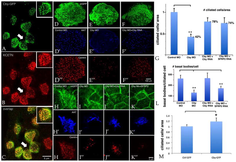
Wnt signaling and ciliogenesis are core features of embryonic development in a range of metazoans. Chibby (Cby), a basal-body associated protein, regulates β-catenin-mediated Wnt signaling in the mouse but not Drosophila. Here we present an analysis of Cby's embryonic expression and morphant phenotypes in Xenopus laevis. Cby RNA is supplied maternally, negatively regulated by Snail2 but not Twist1, preferentially expressed in the neuroectoderm, and regulates β-catenin-mediated gene expression. Reducing Cby levels reduced the density of multiciliated cells, the number of basal bodies per multiciliated cell, and the numbers of neural tube primary cilia; it also led to abnormal development of the neural crest, central nervous system, and pronephros, all defects that were rescued by a Cby-GFP chimera. Reduction of Cby led to an increase in Wnt8a and decreases in Gli2, Gli3, and Shh RNA levels. Many, but not all, morphant phenotypes were significantly reversed by the Wnt inhibitor SFRP2. These observations extend our understanding of Cby's role in mediating the network of interactions between ciliogenesis, signaling systems and tissue patterning.

Keywords: Chibby; Cilia; Hedgehog; Neural crest; Neural plate; Pronephros; Snail2; Wnt signaling; Xenopus laevis.


Conflict of interest statement

Competing Interests: none

Figures

Figure 1
Figure 1
Cby RNA is supplied maternally and its level remains high throughout early Xenopus development (A: based on Xenbase data for X. laevis (red) and X. tropicalis (green); B: RT-PCR analysis in X. laevis of Cby RNA at various developmental stages using ornithine decarboxylase (ODC) RNA as the normalizing control. In situ hybridization studies indicate that Cby is expressed at high levels in the neuroectoderm of stage 18 embryos (C); later in embryogenesis (D) Cby is expressed in a range of tissues including the myotome, pronephros, otic vesicle, central nervous system, migrating neural crest, the eye, and blood islands. Standard (E) and qPCR (F) analyses of ectodermal explants derived from control, Snail2/Slug, or Twist1 morpholino (MO) injected embryos revealed an increase in Cby RNA in response to inhibition of Snail2 expression. G: Ectodermal explants were derived from embryos injected with GR-Snail2 RNA (200 pg/embryo) and either left untreated (−dex) or treated for 2 hours with dexamethasone (+dex), dexamethasone and emetine (+dex, +eme), or emetine alone (+eme). Cby RNA levels were measured by qPCR with the Y-axis corresponding to the change in Cby RNA level with respect to control condition, either control MO injected (F) or in the absence of dexamethasone (G). Student t-test values of < 0.05 are indicated by a “*”, while a p value < 0.01 is indicated by “**” in this and all other figures.
Figure 2
Figure 2
A: The Cby MO aligns with the translation start region of the Cby RNA; this same sequence is present in the Cby-GFP-match RNA. An alternative, rescuing version of the Cby-GFP RNA, Cby-GFP-rescue, has a number of mismatches in the morpholino-binding region. Immunoblot analysis was carried out using either an anti-rabbit Cby antibody (B) or an anti-GFP antibody (C); embryos were injected with RNAs encoding GFP (200 pgs/embryo) and Cby-GFP match (200 pg/side) and either control or Cby morpholino (10 ngs/embryo) and analyzed at stage 11. Cby MO reduced Cby and Cby-GFP protein levels. In this experiment, the blot was first probed with anti-GFP antibody (C), then stripped and probed with the anti-Cby antibody (B). D: Embryos were injected with TOPFLASH and FOPFLASH (control) plasmid DNAs (100 pgs/embryo) together with ΔG-β-catenin RNA (100 pgs/embryo) either alone or together with GFP or Cby-GFP (100 pgs/embryo) RNAs or control or Cby morpholinos (10 ngs/embryo). The Y-axis indicates fold increase relative to the control TOPFLASH/FOPFLASH value (set equal to 1). Comparisons between conditions are marked by horizontal bars; in each case, p-values were < 0.05.
Figure 3
Figure 3
Compared to uninjected (not shown) or control MO injected embryos (A), Cby MO injected embryos typically displayed a noticeable kink (B); injection of Cby-GFP-rescue RNA together with the Cby MO reversed this kink (C,D), while Cby-GFP-rescue RNA alone produced a distinct phenotype (E,F). In situ hybridization studies revealed the loss of the neural and patterning markers Tubb2b (G), Engrailed (H), and Krox20 (I). These phenotypes were rescued by injection of Cby-GFP-rescue RNA. In panels G–I embryos were injected with either control MO (left panel), Cby MO (center panel), or Cby MO together with Cby-GFP RNA (right panel). All embryos were injected with RNA encoding β-galactosidase as a lineage tracer. Quantitation is provided in panel J. Comparisons between conditions are marked by horizontal bars (* for p < 0.05 and ** for p < 0.01).
Figure 4
Figure 4
To examine the effects of morpholino down regulation of Cby, we carried out in situ hybridization of embryos injected in one of two blastomeres. Compared to embryos injected with control MO (A,C), injection of 5ng/blastomere of Cby MO (B,D) had little apparent effect on Sox9 (A,B) or Twist1 (C,D) expression. In contrast to control MO injected embryos (E,H), the injection of 10 ngs/blastomere Cby MO (F, I) produced a dramatic reduction in both Sox9 (E,F) and Twist1 (H,I). These effects were rescued by the co-injection of Cby-GFP-rescue RNA (200 pg/side) with Cby MO (10 ngs/blastomere)(G - Sox9, J - Twist1). A similar effect was seen in later stage embryos; compared to control embryos (K,N), the injection of 10 ngs/blastomere Cby MO (L,O) led to a reduction in Sox9 (K,L) and Twist1 (N,O) expression. This phenotype could be partially rescued by the co-injection of Cby-GFP-rescue RNA (200 pg/side)(M - Sox9, P - Twist1). The results from10 ngs/blastomere injection experiments are quantified in part Q with p-values (* for p < 0.05 and ** for p < 0.01). Alcian Blue staining revealed defects in Cby MO (10 ngs/blastomere) injected embryos (S) compared to control (R) embryos; these defects were ameliorated by co-injection of Cby-GFP-rescue RNA (V). Neural crest transplants from GFP injected embryos migrate normally (U) while the analogous region from Cby morphant embryos (5ngs/embryo) failed to migrate (V).
Figure 5
Figure 5
To characterize the intracellular localization of Cby-GFP, both blastomeres of two-cell stage embryos were injected with Cby-GFP RNA (200 pg/embryo). Ectodermal explants were isolated at stage 9 and fixed at stage 18. Confocal images were taken at 40X magnification. Immunofluorescence staining was performed with an chicken anti-GFP antibody (A) and a rabbit anti-X. laevis Centrin antibody (B), C is the merged image of A and B, inserts in each panel show higher magnification view. In explants from Cby-GFP RNA injected embryos, we did find juxtaposed ciliated cells (arrows)(see below - part M). At higher injected RNA levels, Cby-GFP can also be seen associated with membranes. To examine the effects of reducing Cby levels on the frequency of ciliated cells (D–G) and the number of basal bodies per ciliated cell (H–L) both blastomeres of two-cell embryos were injected with membrane-GFP RNA together with either control MO (D,D′,D″ and H, H′,H″), Cby MO (E, E′, E″ and I, I′, I″), Cby MO plus Cby-GFP RNA (F, F′,F″ and J, J′,J″), or Cby MO plus SFRP2 RNA (K, K′, K″), Membrane-GFP (D–K) was visualized using an anti-GFP antibody, while anti-AAT (D′–K′) and anti-centrin (D″–K″) antibodies were used to visualized ciliated cells and basal bodies, respectively. Confocal images were taken at 10X magnification. Quantitation of the Cby morpholino’s effect on the number of ciliated cells per cap (G)(y-axis corresponds to number of cilia per area, normalized to control morphant explants) and the number of basal bodies per cell (L) are shown. Injection of Cby-GFP RNA (M) led to an increase in the number of ciliated cell per unit area in ectodermal explants. Comparisons between conditions are marked by horizontal bars (* for p < 0.05 and ** for p < 0.01).
Figure 6
Figure 6
Both blastomeres of two cell stage embryos were injected with either control (A,D) or Cby (B,E) morpholino (10 ngs/embryo) and membrane-GFP RNA. The neural tube region of stage 26 embryos (A,B) and the gastrocoele roof plate regions of stage 19 embryos (D,E) were dissected and stained for injected membrane-GFP (green) and AAT (blue). Primary cilia were absent or greatly reduced in Cby morphant neural tubes (A,B). C: To quantitate the effect of the Cby morpholino on primary cilia formation, and the ability of Cby-GFP or SFRP2 RNAs to rescue this effect, 7–10 GFP positive embryos for each group were analyzed. For each embryo, a series of sections were generated and 5 representative images (taken at 40X) were selected and use to calculate mean number of cilia. ”**” indicates a p value < 0.01 compared to control embryos. In contrast to the effect on primary cilia, gastrocoele roof plate cilia were present in Cby morphant gastrocoele roof plate tissue (D,E). In experiments in which fertilized eggs were injected with Cby-GFP; at stage 25 embryos were fixed, sectioned and stained for AAT (F) or GFP (G); H is the overlap of F and G. While cilia are visible (arrows) GFP staining, presumably associated with CbyGFP is not concentrated there. When similar sections from uninjected embryos were stained for centrin (I - magenta) and AAT (blue), centrin was found localized to nuclei (arrows pointing down) but not to the basal body regions of primary cilia (arrows pointing up). Scale bars in part B, E, and I marks 5 μm for parts A and B, D and E, and F–I respectively.
Figure 7
Figure 7
Ectodermal explants derived from control (A) or Cby (B) morpholino injected embryos were stained in situ for Tubb2b RNA; co-injection of RNA encoding Cby-GFP-rescue (C′ and C″) increased the level of Tubb2b RNA staining. D: Control (Ctrl MO) and Cby MO explants were analyzed at stage 18 using RT-PCR; Cby morphant explants displayed decreased levels of BMP4, Noggin, and Tubb2b RNAs, and increased levels of Wnt8a RNA. Levels of FGF8 RNA were unchanged. E: qPCR analyses of control and Cby morphant explants co-injected with Cby-GFP-rescue, Dkk1, or SRFP2 RNAs. Both Cby-GFP and the two Wnt signaling inhibitors returned all RNAs to control levels. Standard (F) and qPCR (G) analyses of control (Ctrl) and Cby morphant ectodermal explants, analyzed at stage 18, revealed a no change in the levels of the ciliogenesis associated transcription factors Multicilin (F and G), Foxjia, Myb, and Rfx2 (G).
Figure 8
Figure 8
Each blastomere of a two cell embryo was injected with either Control or Cby morpholino (10 ngs/embryo total) together with RNA encoding membrane-bound GFP. In rescue studies, embryos were also injected with RNAs (200 pgs/embryo total) encoding either Cby-GFP-rescue or SFRP2. Embryos were analyzed at stage 11 by qPCR. Panel A displays the results for Wnt8a, BMP4, FGF8, Tubb2b, and Noggin, panel B displays the results for Gli1, Gli2, Gli3, Shh, and Patched RNAs. This experiment was carried out two independent times with similar results.
Figure 9
Figure 9
This cartoon summarizes our observations and integrates them into the context of previous studies. Cby is associated with the basal body region of the cilia of multicilated cells. The Cby morpholino (“MO” in red) down regulates Cby protein levels, leading to a reduction in both the number of cilia per cell and the density of multiciliated cells in ectodermal explants; both phenotypes were rescued by the injection of Cby-GFP RNA. Cby inhibits the effects of β-catenin on gene expression, as reflected by the effects of both Cby RNA and MO on the activity of the β-catenin-responsive TOPFLASH reporter. Cby is thought to act on gene expression primarily by driving β-catenin out of the nucleus. In the context of the canonical (β-catenin-dependent) Wnt signaling pathway, this inhibits the interaction between β-catenin and LEF/TCF HMG-box type transcription factors involved in gene activation. We note that β-catenin also interacts with other transcription factors, including a subset of Sox type HMG box transcription factors, so the exact details of the Cby MO’s effects on gene expression in any particular cell type remain to be resolved. We have identified two distinct patterns of altered RNA levels in Cby morphant explants. In the first (indicated by blue circled 1), there is a increase in Wnt8a RNA levels and a decrease in BMP4, Noggin, and Tubb2b RNAs. These changes are largely blocked by expression (from injected RNA) of the secreted Wnt inhibitors SFRP2 and DKK1 (in red). This suggests that these effects are dependent upon a feed-forward Wnt signaling loop, which may be autocrine, juxtacrine, or both. SFRP2 also at least partially reverses the Cby morpholino’s effects on multiciliated cell density in explants and neuroectodermal and neural crest marker expression in embryos, again suggesting that the Cby morphant phenotype is due, at least in part, to enhanced Wnt-signaling effects. Cby MO effects on Gli2, Gli3, and Shh RNA levels (indicated by blue circled 2) are not blocked by SFRP2, and so appear to be direct effects of decreasing Cby levels and not mediated by downstream Wnt signaling. SFRP2 does not reverse Cby MO’s effects on cilia number in multiciliated cells or the neural tube, suggesting that these effects involve distinct Cby-regulated processes.

Similar articles

Cited by

References

    1. Ariizumi T, Takahashi S, Chan TC, Ito Y, Michiue T, Asashima M. Isolation and differentiation of Xenopus animal cap cells. Curr Protoc Stem Cell Biol. 2009;Chapter 1(Unit 1D):5. - PubMed
    1. Avilion AA, Nicolis SK, Pevny LH, Perez L, Vivian N, Lovell-Badge R. Multipotent cell lineages in early mouse development depend on SOX2 function. Genes Dev. 2003;17(1):126–40. - PMC - PubMed
    1. Bradley L, Sun B, Collins-Racie L, LaVallie E, McCoy J, Sive H. Different activities of the frizzled-related proteins frzb2 and sizzled2 during Xenopus anteroposterior patterning. Dev Biol. 2000;227(1):118–32. - PubMed
    1. Carl TF, Dufton C, Hanken J, Klymkowsky MW. Inhibition of Neural Crest Migration in Xenopus Using Antisense Slug RNA. Dev Biol. 1999;213(1):101–115. - PubMed
    1. Cary RB, Klymkowsky MW. Differential organization of desmin and vimentin in muscle is due to differences in their head domains. J Cell Biol. 1994;126(2):445–56. - PMC - PubMed

Publication types

MeSH terms