. 2014 Dec;26(12):2912-20.
doi: 10.1016/j.cellsig.2014.09.005. Epub 2014 Sep 16.

Class I HDAC inhibition stimulates cardiac protein SUMOylation through a post-translational mechanism

Affiliations

Class I HDAC inhibition stimulates cardiac protein SUMOylation through a post-translational mechanism

Weston W Blakeslee et al. Cell Signal. 2014 Dec.

Abstract

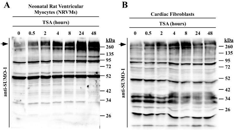
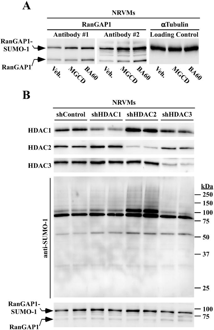
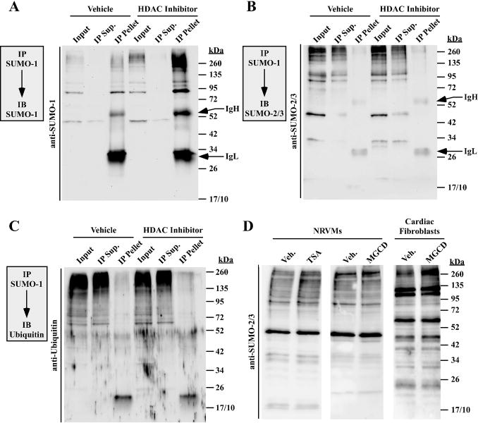
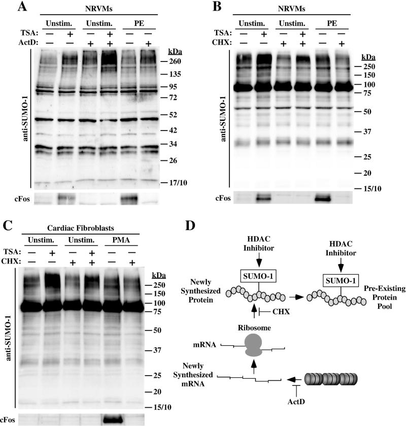
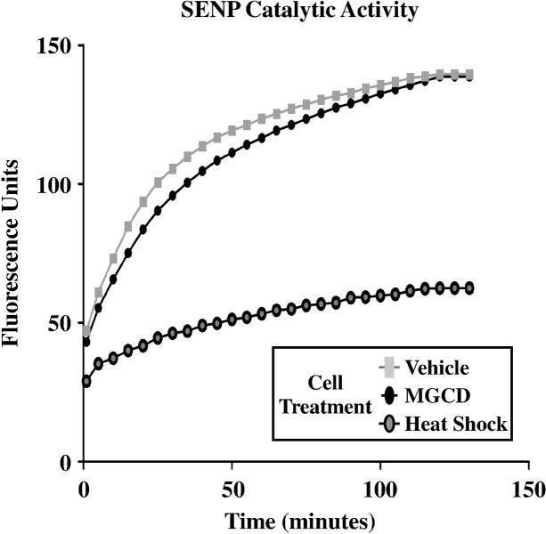
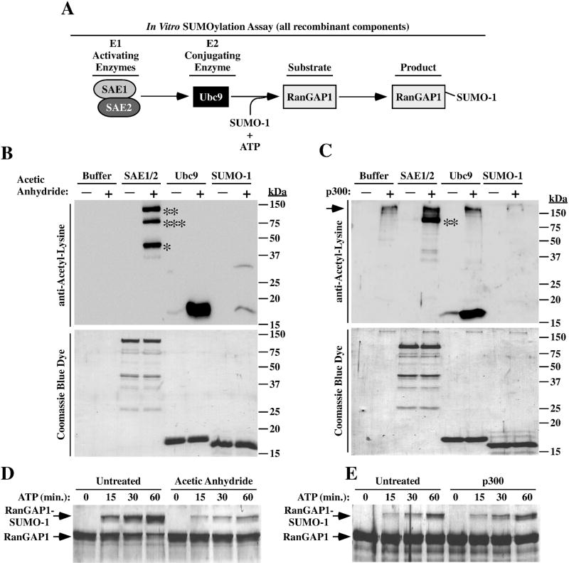
Lysine residues are subject to a multitude of reversible post-translational modifications, including acetylation and SUMOylation. In the heart, enhancement of lysine acetylation or SUMOylation using histone deacetylase (HDAC) inhibitors or SUMO-1 gene transfer, respectively, has been shown to be cardioprotective. Here, we addressed whether there is crosstalk between lysine acetylation and SUMOylation in the heart. Treatment of cardiomyocytes and cardiac fibroblasts with pharmacological inhibitors of HDAC catalytic activity robustly increased conjugation of SUMO-1, but not SUMO-2/3, to several high molecular weight proteins in both cell types. The use of a battery of selective HDAC inhibitors and short hairpin RNAs demonstrated that HDAC2, which is a class I HDAC, is the primary HDAC isoform that controls cardiac protein SUMOylation. HDAC inhibitors stimulated protein SUMOylation in the absence of de novo gene transcription or protein synthesis, revealing a post-translational mechanism of HDAC inhibitor action. HDAC inhibition did not suppress the activity of de-SUMOylating enzymes, suggesting that increased protein SUMOylation in HDAC inhibitor-treated cells is due to stimulation of SUMO-1 conjugation rather than blockade of SUMO-1 cleavage. Consistent with this, multiple components of the SUMO conjugation machinery were capable of being acetylated in vitro. These findings reveal a novel role for reversible lysine acetylation in the control of SUMOylation in the heart, and suggest that cardioprotective actions of HDAC inhibitors are in part due to stimulation of protein SUMO-1-ylation in myocytes and fibroblasts.

Keywords: Acetylation; HDAC; SUMO.


Figures

Fig. 1
Fig. 1
HDAC inhibition stimulates SUMOylation in cardiac cells. (A) Neonatal rat ventricular myocytes (NRVMs) were treated with the pan-HDAC inhibitor trichostatin A (TSA) for the indicated amounts of time. SUMO-1 conjugates were examined by immunoblotting. (B) Primary adult rat cardiac fibroblasts were serum starved for 24 hours prior to treatment with TSA. Arrows indicate the high molecular weight SUMO conjugates that are referenced throughout the text.
Fig. 2
Fig. 2
Selective inhibition of HDAC1 and HDAC2 is sufficient to stimulate SUMOylation in cardiac myocytes and cardiac fibroblasts. (A) Selectivity profiles for the indicated HDAC inhibitors are shown; X = inhibited. (B) NRVMs were stimulated with phenylephrine (PE) in the absence or presence of the indicated HDAC inhibitors for 48 hours; DMSO vehicle (Veh.; 0.1% final concentration) was used as a negative control. SUMO-1 conjugates were examined by immunoblotting. (C) Unstimulated NRVMs were treated for 48 hours with the indicated HDAC inhibitors prior to immunoblotting for SUMO-1 conjugates. (D) Neonatal rat ventricular fibroblasts were serum-starved for 24 hours prior to treatment with the indicated HDAC inhibitors for 48 hours. (E) Independent plates of adult rat ventricular fibroblasts were serum starved for 24 hours prior to treatment with MGCD. SUMO-1 conjugates were resolved on a 7.5% polyacrylamide gel as opposed to (A – D), which employed 10% polyacrylamide gels.
Fig. 3
Fig. 3
HDAC2 regulates cardiac protein SUMOylation. (A) NRVMs were treated with MGCD0103 or BA60 for 48 hours, and protein homogenates were immunoblotted with two distinct antibodies that detect free RanGAP1 (lower band) and SUMO-1-ylated RanGAP1 (upper band). (B) NRVMs were infected with lentiviruses encoding shRNAs directed toward HDAC1, HDAC2 or HDAC3. shControl is an shRNA that is predicted to fail to target any mammalian mRNA transcript. After 96 hours of infection in the presence of PE, cells were lysed and HDAC1, HDAC2, HDAC3, SUMO-1, and RanGAP1 were detected by immunoblotting, as indicated.
Fig. 4
Fig. 4
Inhibition of class I HDACs preferentially enhances SUMO-1 conjugation. SUMO-1 conjugates from DMSO vehicle or HDAC inhibitor-treated cardiac fibroblasts (A) or NRVMs (B and C) were immunoprecipitated (IP) with anti-SUMO-1 antibody-conjugated beads and immunoblotted (IB) with either anti-SUMO-1 antibody (A) anti-SUMO-2/3 antibody (B) or anti-ubiquitin antibody (C). Input = 10% of pre-IP volume; IP supernatant (Sup) = 10% of post-IP volume; pellet = immunoprecipitate. Immunoglobulin heavy (IgH) and light (IgL) chains from the IP antibody are indicated. (D) NRVMs or 24 hour serum-starved adult rat ventricular fibroblasts were treated with DMSO, TSA or MGCD for 48 hours, and SUMO-2/3 conjugates were detected by immunoblotting.
Fig. 5
Fig. 5
Class I HDAC inhibitor-mediated SUMOylation does not require de novo gene transcription or protein synthesis. (A) NRVMs were pre-treated with actinomycin D (ActD) for two hours to block gene transcription, and were subsequently exposed to either vehicle (DMSO; -) or TSA for 4 hours. As controls, some cells received PE for two hours in the absence or presence of ActD. Protein homogenates were immunoblotted with anti-SUMO-1 antibody. In parallel, cFos protein was assessed to confirm that ActD efficiently inhibited gene expression. (B) NRVMs were pre-treated with the protein translation inhibitor cycloheximide (CHX) for 30 minutes, and were subsequently exposed to vehicle (DMSO; -) or TSA for 4 hours. Immunoblotting was performed with anti-SUMO-1 antibody or cFos antibody to confirm CHX efficacy. (C) Adult rat ventricular fibroblasts were serum-starved for 24 hours, pretreated with CHX for 30 minutes, and exposed to DMSO or TSA for 4 hours. As controls, some cells received PMA for 2 hours in the absence or presence of CHX. ActD did not block TSA-mediated SUMO-1-ylation (A), and CHX only partially reduced SUMO-1 conjugation in cardiac myocytes (B) and fibroblasts (C), suggesting that HDAC inhibition stimulates SUMOylation of a pre-existing protein pool (D).
Fig. 6
Fig. 6
Global SENP catalytic activity is not suppressed by a class I HDAC inhibitor. Adult rat ventricular fibroblasts were serum-starved for 24 hours and treated with DMSO vehicle or the class I HDAC inhibitor MGCD for 48 hours. As controls, some cells were subjected to 43°C for 30 minutes prior lysis. Protein homogenates were incubated with a SUMO-1-AMC probe and de-SUMOylase activity (as measured by increased fluorescence) was monitored over 2 hours. Heat shock inactivates SENPs and served as a positive control. For each condition, homogenates from three independent plates of cells were pooled for assessment of SENP activity.
Fig. 7
Fig. 7
Acetylation of SUMO conjugation machinery. (A) Schematic depiction of the in vitro SUMOylation assay. (B) The indicated recombinant proteins were incubated for one hour with acetic anhydride to assess non-enzymatic acetylation of the proteins. Proteins were resolved by SDS-PAGE and either immunoblotted with anti-acetyl-lysine antibody (top panel) or stained with Coomassie blue dye (bottom panel); *SAE1, **SAE2, ***presumed degradation product of SAE2. (C) Recombinant proteins were incubated for one hour with p300 to assess their capacity to be enzymatically acetylated. Samples were analyzed as in (B); **SAE2. Note that p300 (the upper-most acetylated protein) undergoes auto-acetylation (arrow). (D) In vitro SUMOylation assays were performed as outlined in (A) after acetic anhydride treatment (D) or exposure to p300 (E). Reactions were terminated at the indicated times. Proteins were resolved by SDS-PAGE and stained with Coomassie blue dye. The reduced mobility of RanGAP1 is indicative of SUMOylation.

References

    1. Yang XJ, Seto E. Mol Cell. 2008;31:449–461. - PMC - PubMed
    1. Taverna SD, Li H, Ruthenburg AJ, Allis CD, Patel DJ. Nat Struct Mol Biol. 2007;14:1025–1040. - PMC - PubMed
    1. Foster DB, Liu T, Rucker J, O’Meally RN, Devine LR, Cole RN, O’Rourke B. PLoS One. 2013;8:e67513. - PMC - PubMed
    1. Choudhary C, Kumar C, Gnad F, Nielsen ML, Rehman M, Walther TC, Olsen JV, Mann M. Science. 2009;325:834–840. - PubMed
    1. Lundby A, Lage K, Weinert BT, Bekker-Jensen DB, Secher A, Skovgaard T, Kelstrup CD, Dmytriyev A, Choudhary C, Lundby C, Olsen JV. Cell Rep. 2012;2:419–431. - PMC - PubMed

Publication types

LinkOut - more resources