. 2014 Dec;32(12):3266-77.
doi: 10.1002/stem.1847.

Tendon progenitor cells in injured tendons have strong chondrogenic potential: the CD105-negative subpopulation induces chondrogenic degeneration

Affiliations

Tendon progenitor cells in injured tendons have strong chondrogenic potential: the CD105-negative subpopulation induces chondrogenic degeneration

Shuji Asai et al. Stem Cells. 2014 Dec.

Abstract

To study the cellular mechanism of the tendon repair process, we used a mouse Achilles tendon injury model to focus on the cells recruited to the injured site. The cells isolated from injured tendon 1 week after the surgery and uninjured tendons contained the connective tissue progenitor populations as determined by colony-forming capacity, cell surface markers, and multipotency. When the injured tendon-derived progenitor cells (inTPCs) were transplanted into injured Achilles tendons, they were not only integrated in the regenerating area expressing tenogenic phenotype but also trans-differentiated into chondrogenic cells in the degenerative lesion that underwent ectopic endochondral ossification. Surprisingly, the micromass culture of the inTPCs rapidly underwent chondrogenic differentiation even in the absence of exogenous bone morphogenetic proteins or TGFβs. The cells isolated from human ruptured tendon tissues also showed connective tissue progenitor properties and exhibited stronger chondrogenic ability than bone marrow stromal cells. The mouse inTPCs contained two subpopulations one positive and one negative for CD105, a coreceptor of the TGFβ superfamily. The CD105-negative cells showed superior chondrogenic potential in vitro and induced larger chondroid degenerative lesions in mice as compared to the CD105-positive cells. These findings indicate that tendon progenitor cells are recruited to the injured site of tendons and have a strong chondrogenic potential and that the CD105-negative population of these cells would be the cause for chondroid degeneration in injured tendons. The newly identified cells recruited to the injured tendon may provide novel targets to develop therapeutic strategies to facilitate tendon repair.

Keywords: Chondrogenesis; Injury; Progenitor; TGFβ; Tendon.


Figures

Figure 1
Figure 1
Histological changes in the Achilles tendons after injury surgery. Longitudinal sections were prepared from uninjured control Achilles tendons (Control) (A, B, S) or injured Achilles tendons (Injury) 1 (C, D), 2 (E, F), 4 (G, H), 8 (I–P) or 12 (Q–T) weeks after injury, and stained with hematoxylin-eosin (A–J, Q, R) or alcian blue (K, L), toluidine blue (M, N) or anti-collagen 2 antibody (O, P). The images represent the edges (A, C, E, G, I, K, M, O, Q) and the center parts (B, D, F, H, J, L, N, P, R) of the injured tendons. The measurement bars are 200 µm for A–J, Q and R, 50 µm for K–P. S and T, Radiology images of the hind limb. The radiopaque materials were detected at both edges of the injured Achilles tendon (arrows).
Figure 2
Figure 2
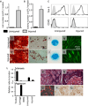
Clonogenicity, cell surface marker expression and multipotency of uninjured and injured tendon derived cells. The cells were isolated from uninjured control (Uninjured) and injured (Injured) Achilles tendons 1 week after the injury surgery. A, The total cell number was counted. B, The number of colonies was counted 10 days after the cells were plated at a low density. C, The cells expressed Sca-1, but not CD45, a hematopoietic stem cell marker. D–J, The cells were cultured under the osteogenic (D, H), adipogenic (E, I) or chondrogenic (F, J) condition, and stained with Alizarin red (D, H), Oil red O (E, I) and alcian blue (F, J), respectively. G and K, The cells were isolated from uninjured and injured tendons of Scx-GFP mice and cultured in the presence of TGFβ1 (10 ng/ml) for 5 days. GFP fluorescence images were taken. L and M, Total RNAs were prepared from Achilles tendons (7 weeks old mice), epiphyseal cartilage (4 days old mice) and monolayer cultures of mouse bone marrow stromal cells (BMSC), uninjured (Uninjured) or injured (Injured) tendon derived cells, and subjected to qPCR analysis for scleraxis and aggrecan (L). The injured tendon-derived cells from RFP-expressing mice were mixed with Matrigel and subcutaneously transplanted into athymic mice, and histologically inspected 10 days after transplantation (M–Q). The frozen sections of the transplant were observed under the fluorescence microscope (O), and the paraffin sections were stained with hematoxylin and eosin (M and N), anti-collagen 2 (P) and collagen 10 (Q) antibodies.
Figure 3
Figure 3
Transplantation of injured tendon derived cells in injured Achilles tendons. The cells were isolated from injured Achilles tendons in the Scx-GFP;RFP (A–C), H2K–GFP (F–I, N, O) or Scx-GFP (J–M) mice 1 week after the injury surgery and transplanted in the injured Achilles tendons in CD1 Nude mice (n=5/group) just after the injury surgery. After 4 weeks (A–E) or 12 weeks (F–O), the injured Achilles tendons that had received cell transplantation (A–C, F, G, J, K) or no transplantation (D, E) were observed under the fluorescence stereomicroscope. The red (A, D) and green (B, E–G, J, K) fluorescence images were superimposed to the brightfield images (BF+RFP or BF+GFP) or each other (RFP+GFP). The sections of the injured tendons that had received HK2-EGFP or Scx-GFP cell transplantation were subjected to the immunohistochemical staining for GFP (H, I, L–O). N and O were the injured Achilles tendons that had no transplantation. The bar is 50 µm for H, I, L–O.
Figure 4
Figure 4
Chondrogenic potential of tendon-derived progenitor cells and bone marrow stromal cells. A–E, Mouse bone marrow stromal cells (A, BMSC), or tendon-derived progenitor cells isolated from mouse uninjured (B, Unnjured) or injured (C, Injured) Achilles tendons were cultured in micromass in 10% FBS-DMEM for 7 days and stained with alcian blue (A–C). Integrated density of the alcian blue-stained cultures (n=4) was measured (D). Aggrecan gene expression was examined by qPCR (E). F, The micromass cultures of the inTPCs were treated with LDN-193189 (BMP inhibitor) at the concentration of 200 nM or SB431542 (TGFβ inhibitor) at the concentration of 2 µM for 7 days, and integrated density of alcian blue-stained cultures (n=4) was measured. G–I, The cells isolated from human bone marrow stromal cells (F, BMSC) or human tendons (H, Tendon) were cultured in micromass in 10% FBS-DMEM for 7 days and stained with alcian blue. Integrated density of the alcian blue-stained cultures (n=3–8) was measured (I). The values are average and SD. *, p<0.05.
Figure 5
Figure 5
Multipotency of CD105-positive and negative injured tendon-derived progenitor cells. A, The inTPCs were sorted to two subpopulations, Sca1+/CD105+ and Scat+/CD105- populations. B–H, The CD105+ and CD105- cells were cultured under the osteogenic, adipogenic or chondrogenic condition and stained with Alizarin red (B, E), Oil red O (C, F) or alcian blue (D, G), respectively. Integrated density of the alcian blue-stained cultures (n=4) was measured (H). I and J, The micromass cultures (I) and TGFβ1-treated monolayer cultures (J) of CD105+ and CD105- cells were subjected to qPCR analysis for aggrecan (I) or scleraxis (J) gene expression, respectively. The values are the average and SD. *, p<0.05.
Figure 6
Figure 6
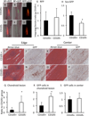
Distribution of transplanted CD105-positive and negative tendon progenitor cells in injured Achilles tendons. The sorted CD105+ and CD105- inTPCs isolated from Scx-GFP;RFP mice (A–H) or H2K-GFP mice (I–S) were transplanted in the injured Achilles tendon of CD1 Nude mice just after the injury surgery. A–H, The injured Achilles tendons that had received cell transplantation were examined under the fluorescence microscope (A–F) 4 weeks after the transplantation. The red (RFP) and green (Scx-GFP) fluorescence images were superimposed to the brightfield images (A/D and B/E, respectively) or each other (C, F). Integrated intensity of RFP (G) and Scx-GFP (H) fluorescence of the samples (n=4) was measured. I–S, The injured Achilles tendons that had received transplantation of H2K-GFP mouse-derived inTPCs were harvested 10 weeks after the transplantation (n=5). Longitudinal sections of the injured tendons were subjected to alcian blue staining (I, K, M, O) and immunohistochemical staining for GFP (J, L, N, P). The chondroid lesion area positive to alcian blue staining at the edges of injured tendons (I, M) was measured (Q). The number of GFP-positive cells was counted at the edges (J, N) and the center parts (L, P) of injured tendons (R and S, respectively). The values are the average and SD. *, p<0.05.
Figure 7
Figure 7
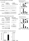
Phosphorylation of smad proteins and chondrogenesis in injured tendon derived progenitor cells. A and B, The sorted CD105-positive and negative inTPCs (A) or CD105-negative inTPCs transduced with adenovirus encoding CD105 or GFP (B) cultures were treated with TGFβ1 (10 ng/ml) for 30–120 min, and subjected to immunoblot with anti-phospho-smad 2, phospho-smad1/5, total smad 2, total smad 1. The immunoblot band intensity was evaluated by Image J and the ratio of P-Smad2 to Smad 2 or P-Smad1/5 to Smad 1 was calculated. The value represents ratio to the value of the non-treated control group. C, CD105-negative inTPCs transduced with adenovirus encoding CD105 or GFP were cultured in 10% FBS-DMEM for 7 days, and gene expression levels of aggrecan and CD105 were examined. The values are the average and SD. *, p<0.05

References

    1. Sharma P, Maffulli N. Tendon injury and tendinopathy: healing and repair. J Bone Joint Surg Am. 2005;87(1):187–202. - PubMed
    1. Rees JD, Wilson AM, Wolman RL. Current concepts in the management of tendon disorders. Rheumatology (Oxford) 2006;45(5):508–521. - PubMed
    1. Aslan H, Kimelman-Bleich N, Pelled G, et al. Molecular targets for tendon neoformation. J Clin Invest. 2008;118(2):439–444. - PMC - PubMed
    1. Tozer S, Duprez D. Tendon and ligament: development, repair and disease. Birth Defects Res C Embryo Today. 2005;75(3):226–236. - PubMed
    1. Manske PR, Gelberman RH, Vande Berg JS, et al. Intrinsic flexor-tendon repair. A morphological study in vitro. J Bone Joint Surg Am. 1984;66(3):385–396. - PubMed

Publication types

Substances