. 2014 Nov;14(11):2491-9.
doi: 10.1111/ajt.12922. Epub 2014 Sep 12.

Critical role of NKT cells in posttransplant alloantibody production

Affiliations

Critical role of NKT cells in posttransplant alloantibody production

J M Zimmerer et al. Am J Transplant. 2014 Nov.

Abstract

We previously reported that posttransplant alloantibody production in CD8-deficient hosts is IL-4+ CD4+ T cell-dependent and IgG1 isotype-dominant. The current studies investigated the hypothesis that IL-4-producing natural killer T cells (NKT cells) contribute to maximal alloantibody production. To investigate this, alloantibody levels were examined in CD8-deficient WT, CD1d KO and Jα18 KO transplant recipients. We found that the magnitude of IgG1 alloantibody production was critically dependent on the presence of type I NKT cells, which are activated by day 1 posttransplant. Unexpectedly, type I NKT cell contribution to enhanced IgG1 alloantibody levels was interferon-γ-dependent and IL-4-independent. Cognate interactions between type I NKT and B cells alone do not stimulate alloantibody production. Instead, NKT cells appear to enhance maturation of IL-4+ CD4+ T cells. To our knowledge, this is the first report to substantiate a critical role for type I NKT cells in enhancing in vivo antibody production in response to endogenous antigenic stimuli.


Figures

Figure 1
Figure 1. Type I NKT cells are critical for maximal alloantibody production following allogeneic hepatocyte transplant
C57BL/6 (wild-type; WT), CD1d KO, and Jα18 KO mice (H-2b) were transplanted with allogeneic FVB/N (H-2q) hepatocytes. Recipients were left untreated or CD8-depleted (day −2,−1). Serum was tested for alloantibody on day 14 posttransplant by two methods- A) percent allogeneic target splenocytes bound and B) by serum dilution to calculate alloantibody titer. Naïve serum and third party B10.BR targets were used as negative controls. Both WT (9.7±2.5%; n=13; titer=90±5 n=5) and CD1d KO (11.0±4.5%; n=4; titer=66.7±10 n=3) recipients produced similar low, but significant alloantibody levels compared to naïve controls (p<0.05). CD8-depleted WT mice (76.9±3.4%; n=7; titer=1800±89 n=5) produced increased alloantibody compared to WT recipients (9.7±2.5%; titer=90±5; p<0.0001). CD8-depleted CD1d KO recipients (34.8±4.0%, n=5; titer=333±39 n=3) produced significantly less alloantibody than CD8-depleted WT recipients (76.9±3.4%, p<0.0001; titer=1800±89, p=0.016; as signified by “*” for both). Jα18 KO recipients exhibited low alloantibody production (2.0±0.4%, n=5; titer=22±3 n=6). CD8-depleted Jα18 KO recipients (6.6±2.4%, n=14; titer=22±2 n=6) produced significantly less alloantibody than CD8-depleted WT recipients (76.9±5.0%, p<0.001; titer=1800±89, p=0.0041; as signified by “†” for both). C) Analysis of alloantibody isotype by titer analysis shows that IgG1 is the dominant posttransplant alloantibody isotype with relatively low contributions from IgG2b, IgG2c, and IgG3.
Figure 2
Figure 2. Type I NKT cell-deficient recipients have fewer antibody producing cells
CD8-depleted wild-type (WT) and Jα18 KO mice were transplanted with FVB/N hepatocytes. ELISPOT was used to determine the number of antibody producing cells on days 4, 8, 11, and 15 posttransplant. A) WT transplant recipients exhibited a significant induction of IgG1-producing cells (spot forming cells, SFC) per 106 cells analyzed on days 8 (288±42) and 11 (266±53) posttransplant, compared to naïve (day 0) WT mice (45±15; p<0.0001 for both comparisons). Jα18 KO recipients exhibited significantly fewer IgG1-producing cells per 106 cells on days 8 (169±28) and 11 (94±30) posttransplant, compared to WT recipients (p<0.006 for both comparisons, as signified by “*”). B) In vitro antibody production, as demonstrated by a side-by-side ELISA, was lower in Jα18 KO splenocytes (day 8= 63.9±17.1, day 11= 58.4±7.2 ng/mL per 106 splenocytes) versus WT splenocytes (day 8= 157.2±29.2, day 11= 106.6±22.0 ng/mL per 106 splenocytes; p<0.02 for both days, as signified by “†”).The experimental results are representative of duplicate experiments. Error bars designate the standard error (based on 4 to 10 wells per data point from 2 mouse spleens).
Figure 3
Figure 3. Type I NKT cell enhanced alloantibody production is IFN-γ-dependent, but IL-4-independent
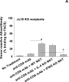
Jα18 KO mice (H-2b) were transplanted with allogeneic FVB/N (H-2q) hepatocytes. Recipients were left untreated or CD8-depleted (day -2,-1). Type I NKT cells were adoptively transferred (AT; 1x10⁶ cells i.v.) into CD8-depleted Jα18 KO mice immediately following allogeneic hepatocyte transplant. Serum alloantibody was tested on day 14 by two methods- A) percent allogeneic target splenocytes bound and B) by serum dilution to calculate alloantibody titer. Naïve serum was used as a negative control. AT of wild-type (WT) NKT cells into CD8-depleted Jα18 KO recipients induced significant enhancement of alloantibody production (33.3±1.7%; n=7; titer=142±11 n=6) compared to CD8-depleted Jα18 KO recipients (6.6±1.2%, n=14, p<0.0001; titer=22±2, n=6; p<0.0031; as signified by “*” for both). Jα18 KO recipients adoptively transferred with IL-4 KO NKT cells (27.0±2.1%; n=5; titer=113±16 n=4) produced similar alloantibody levels compared to recipients which received AT of WT NKT cells (p=ns). CD8-depleted Jα18 KO recipients adoptively transferred with IFN-γ KO NKT cells (4.8±2.3%; n=4; titer=18±1 n=6) produced significantly less alloantibody compared to recipients which received AT of WT NKT cells (, p<0.003 for both methods). C) Analysis of alloantibody isotype by titer analysis shows that IgG1 is the dominant posttransplant alloantibody isotype with relatively low contributions from IgG2b, IgG2c, and IgG3.
Figure 4
Figure 4. NKT cells enhance maturation of IL-4-producing CD4+ T cells following allogeneic hepatocyte transplant
Wild-type (WT) and CD1d KO mice (H-2b) were transplanted with allogeneic FVB/N (H-2q) hepatocytes. Recipients were left untreated or CD8-depleted (day -2,-1). On day 7, splenocytes were harvested and analyzed for intracellular IFN-γ and IL-4 within CD4+ T cells. WT and CD1d KO recipients both exhibited increased percentages of IL-4+CD4+ T cells (1.1%±0.3%, n=3 and 1.5%±0.3%, n=3, respectively) compared to naïve controls (0.2%±0.0%, n=3; p<0.02 for both). CD8-depleted WT recipients exhibited a greater percentage of IL-4+CD4+ T cells (2.6%±0.2%, n=5; p=0.0005, as signified by “*”) compared to CD8-sufficient WT recipients. In contrast, IL-4+CD4+ T cells were significantly lower in CD8-depleted CD1d KO recipients (1.5%±0.2%, n=5; p=0.0028, as signified by “†”) compared to CD8-depleted WT recipients. WT and CD1d KO recipients exhibited a significantly greater percentage of IFN-γ+CD4+ T cells (5.0%±0.4%, n=3 and 5.6%±0.5%, n=3, respectively) compared to naïve controls (0.3%±0.1%, n=3; p<0.01 for both). IFN-γ+CD4+ T cells from CD8-deficient WT and CD1d KO recipients were comparable to each other (5.2%±0.4%, n=5 and 4.3%±0.4%, n=5) and their CD8-sufficient counterparts.

Similar articles

Cited by

References

    1. McKenna RM, Takemoto SK, Terasaki PI. Anti-HLA antibodies after solid organ transplantation. Transplantation. 2000;69(3):319–326. - PubMed
    1. Puttarajappa C, Shapiro R, Tan HP. Antibody-mediated rejection in kidney transplantation: a review. Journal of transplantation. 2012;2012:193724. - PMC - PubMed
    1. Lorenz M, Regele H, Schillinger M, Exner M, Rasoul-Rockenschaub S, Wahrmann M, et al. Risk factors for capillary C4d deposition in kidney allografts: evaluation of a large study cohort. Transplantation. 2004;78(3):447–452. - PubMed
    1. Powell JD, Delgoffe GM. The mammalian target of rapamycin: linking T cell differentiation, function, and metabolism. Immunity. 2010;33(3):301–311. - PMC - PubMed
    1. Strauss G, Osen W, Debatin KM. Induction of apoptosis and modulation of activation and effector function in T cells by immunosuppressive drugs. Clinical and experimental immunology. 2002;128(2):255–266. - PMC - PubMed

Publication types