

Altered functional magnetic resonance imaging responses to nonpainful sensory stimulation in fibromyalgia patients
- PMID: 25220783
- PMCID: PMC4410766
- DOI: 10.1002/art.38781
Altered functional magnetic resonance imaging responses to nonpainful sensory stimulation in fibromyalgia patients
Abstract
Objective: Fibromyalgia (FM) is a disorder characterized by chronic pain and enhanced responses to acute noxious events. However, the sensory systems affected in FM may extend beyond pain itself, as FM patients show reduced tolerance to non-nociceptive sensory stimulation. Characterizing the neural substrates of multisensory hypersensitivity in FM may thus provide important clues about the underlying pathophysiology of the disorder. The aim of this study was to characterize brain responses to non-nociceptive sensory stimulation in FM patients and their relationship to subjective sensory sensitivity and clinical pain severity.
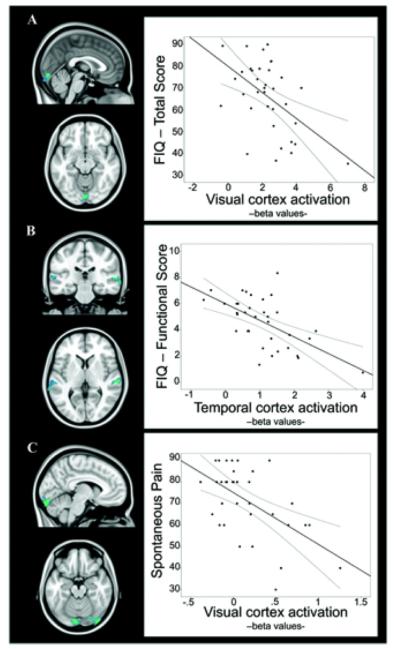
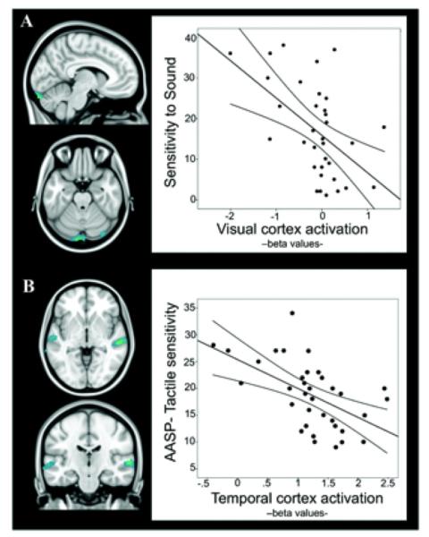
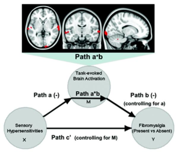
Methods: Functional magnetic resonance imaging (MRI) was used to assess brain response to auditory, visual, and tactile motor stimulation in 35 women with FM and 25 matched controls. Correlation and mediation analyses were performed to establish the relationship between brain responses and 3 types of outcomes: subjective hypersensitivity to daily sensory stimulation, spontaneous pain, and functional disability.
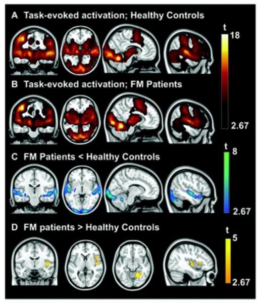
Results: Patients reported increased subjective sensitivity (increased unpleasantness) in response to multisensory stimulation in daily life. Functional MRI revealed that patients showed reduced task-evoked activation in primary/secondary visual and auditory areas and augmented responses in the insula and anterior lingual gyrus. Reduced responses in visual and auditory areas were correlated with subjective sensory hypersensitivity and clinical severity measures.
Conclusion: FM patients showed strong attenuation of brain responses to nonpainful events in early sensory cortices, accompanied by an amplified response at later stages of sensory integration in the insula. These abnormalities are associated with core FM symptoms, suggesting that they may be part of the pathophysiology of the disease.
Copyright © 2014 by the American College of Rheumatology.
Figures
References
-
- Kosek E, Ekholm J, Hansson P. Sensory dysfunction in fibromyalgia patients with implications for pathogenic mechanisms. Pain. 1996;68:375–83. - PubMed
-
- Lautenbacher S, Rollman GB, McCain GA. Multi-method assessment of experimental and clinical pain in patients with fibromyalgia. Pain. 1994;59:45–53. - PubMed
-
- Gibson SJ, Littlejohn GO, Gorman MM, Helme RD, Granges G. Altered heat pain thresholds and cerebral event-related potentials following painful CO2 laser stimulation in subjects with fibromyalgia syndrome. Pain. 1994;58:185–93. - PubMed
-
- Cook DB, Lange G, Ciccone DS, Liu WC, Steffener J, Natelson BH. Functional imaging of pain in patients with primary fibromyalgia. J Rheumatol. 2004;31:364–78. - PubMed
-
- Gracely RH, Petzke F, Wolf JM, Clauw DJ. Functional magnetic resonance imaging evidence of augmented pain processing in fibromyalgia. Arthritis Rheum. 2002;46:1333–43. - PubMed
Publication types
MeSH terms
Grants and funding
LinkOut - more resources
Full Text Sources
Other Literature Sources
Medical
